
Abstract
An innovative technique combining phase transition and microprinting in one step was applied to fabricate the nerve conduits used in peripheral nerve regeneration. The asymmetric microporosity served to generate asymmetric permeability, and the surface microgrooves were introduced to achieve cell alignment in vitro. The symmetric/asymmetric porous poly(D,L-lactide) (PLA) substrates with microgrooves on the surface were tested for their ability to repair 10 mm sciatic nerve transection defects in rats. The in vivo results showed that porous PLA conduits maintained a stable supporting structure during the entire regeneration process. The myelin sheaths of the regenerated nerve in asymmetric conduits were thicker than in symmetric groups at 4 weeks. Moreover, the regenerated nerves in the asymmetric conduits with surface microgrooves had the highest degree of myelination at 4 weeks and the most number of vessels at 6 weeks. The walking track analysis also implied that the asymmetric conduits with surface microgrooves had the highest degree of functional recovery. Based on the study, the combination of microgrooves and asymmetric microporous structure could be employed in the design of nerve conduits for peripheral nerve regeneration in the future.
Introduction
The traditional peripheral nerve conduits are made from silicone rubber, due to the inertness and elastic properties. However, the silicone tube is nonporous and not biodegradable. In contrast to silicone tubes, the permeable conduits have interconnected pores with proper sizes that can increase the nutrient exchange between the lumen and the outer environment, and can prevent the cell infiltration that may impede axon extension. 5 The pore size of permeable conduits determines which molecules pass through the conduit from the surrounding tissue to the regenerating nerve. 6 The bioresorbable nerve conduits with special porous structures to enhance nutrient or transport waste drainage were proposed in many reports.6–8 It has been shown that semipermeable conduits may enhance nerve regeneration over impermeable ones.9–11 In membranes with an asymmetric structure, the mass transfer rates of inflow and outflow for a specific high molecular weight substance are usually different. In our previous work, novel conduits with the asymmetric structure could facilitate peripheral nerve repair, probably through the excellent waste drainage ability in the early stage and inhibition of exogenous cell invasion by the dense skin layer. 6
In addition to the permeability of the conduit, another key factor for nerve regeneration is the directed neuron outgrowth. Extracellular matrices (ECMs) and oriented tissue structures may influence cell migration and axon outgrowth in vivo. 12 The directed growth of Schwann cells on polymer substrates was found to promote neuronal outgrowth in vitro.13,14 Cells could recognize the three-dimensional geometric configurations on the surface of a substrate, and their growth could be controlled and guided through the fabrication of microgrooves or other patterns on the surface.13,15,16 Manufacture of a multichannel conduit is one of the geometric strategies to guide the nerve regeneration between the proximal and distal nerve stumps.17,18 The multiple channels were designed to provide a larger surface area for extending axons to enhance axonal elongation.18,19 However, a unichannel conduit is much easier to fabricate, 20 and the lumen surface can be chemically or physically modified. 21 The unichannel conduit also provides a larger cross-sectional area for nerve tract growth and expansion than the multichannel conduit. The influence of surface texture, for example, hydrophilic or hydrophobic polymer strips on the synthetic nerve guidance channel, has also been investigated. Significantly, more myelinated axons in the hydrophobic tubes than in the hydrophilic tubes were reported. 22 The surface roughness of a guidance channel might, in addition, affect the outcome of peripheral nerve regeneration, potentially by changing the early arrangement of the fibrin matrix and/or inducing different cellular responses. 23
In this study, symmetric/asymmetric porous poly(D,L-lactide) (PLA) substrates with microgrooves on the surface were fabricated. The combination of microgrooves on the surface and asymmetric pore distribution in the cross section were observed by scanning electron microscopy (SEM). Cell alignment on substrates was measured in vitro. Permeability of the substrates was also analyzed. The conduits made of these substrates were tested for their ability to repair 10 mm sciatic nerve defects in rats. Our goal was to combine the advantages from both asymmetric porous structure and microgroove geometry in a biodegradable substrate simultaneously. The design concept in our work can be applied to biomaterial-guided regeneration of other tissues as well.
Materials and Methods
Creation of the micropattern
The pattern was prepared on glass by photolithographical techniques. The negative photoresist (THB-120N; JSR, Tokyo, Japan) was spin coated onto the glass substrate (6 × 5 cm) by two-step spin-coating processes. The wavelength of the exposure system (OAI-500, Milpitas, CA) was 365 nm, and the exposure time was 10 s for the photoresist. To achieve the 3 μm thickness of the photoresist, the coating speed was performed at 1500 rpm for 10 s to form a layer of uniform thickness first, and then performed at 2500 rpm for 25 s to achieve the desired 3 μm thickness. The width/spacing size on the mask was 20 μm/20 μm. The unreacted photoresist was removed to obtain the microgrooves of 3 μm depth on the glass substrate.
Fabrication of the symmetric/asymmetric microporous PLA substrates with or without microgrooved features
The pattern transfer process has been described in our previous study. 24 Briefly, a poly(dimethylsiloxane) (PDMS) submaster mold was created by pouring Dow Corning Silastic MDX4-4210 on the photoresist patterned glass. Ten percent solution of PLA (8300D; Cargill Dow, Minneapolis, MN) in 1,4-dioxane was prepared. The pattern on PLA was transferred from the PDMS mold. The PLA polymer solution was cast on the PDMS mold fit into a 5 cm glass dish. After that, these dishes were placed at room temperature for 24 h in different concentrations of alcohol (95%, 40%, and 20%) that served as the nonsolvent. By the process, the substrates with symmetric or asymmetric porous structure were formed. Then, the PLA substrates were immersed in water for 8 h and air dried in a 40°C oven for 24 h to remove any residue organic solvent. The three types of grooved substrates prepared from the above immersion–precipitation phase inversion process using 95%, 40%, or 20% of alcohol were abbreviated as “95g,” “40g,” and “20g,” respectively. The surface of the substrate that faced the PDMS mold before removal from the mold was defined as the top surface of the substrate.
The PLA substrates without surface grooves as the flat controls were also fabricated using a similar casting method. The three control substrates obtained from 95%, 40%, or 20% of alcohol were denoted as “95f,” “40f,” and “20f,” respectively. In addition, the nonporous PLA films (with or without grooves) were fabricated by solvent casting and air dry without the use of nonsolvent.
Porous structures
The integrity of microgrooves on the substrates and the microporous structure of the substrates were examined by a scanning electron microscope (SEM, ABT-150S; Topcon, Livermore, CA).
Permeability studies
The permeability of the substrates was measured by a method described in an earlier work. 6 The solute was bovine serum albumin (BSA; molecular weight, 62 kDa). BSA was dissolved in the phosphate-buffered saline (PBS; pH 7.5), so that the concentration was 1%. Then, 1.5 mL of BSA solution was added to a 2 mL tube, the top of which was sealed using the test substrate with a 0.785 cm2 diffusion area. The sealed tube, containing the solution, was immersed in a vial containing 3 mL of PBS. Twenty microliters of solution was retrieved from the PBS vial and analyzed after 3, 24, and 48 h. Solute concentrations were measured using a UV/VIS spectrophotometer (Hitachi U-2000, Tokyo, Japan) at the wavelength of 240 nm. Four samples were tested for each group. The procedure was repeated on the reverse side of the substrates to determine the directional permeability, that is, inflow and outflow rates of diffusion, where the inflow defined as the direction of solute movement from bottom toward the top surface.
Cell alignment
Cells from a glioma cell line BCRC-60046 (C6 cells) were used for cell alignment studies. The grooved PLA substrates were sterilized by 70% ethanol for 30 min, cut as 15 mm disks, and placed into the bottom of the well in a 24-well tissue culture plate. C6 cells at a density of 1 × 10⁴ per well were seeded on these substrates. The culture medium was high-glucose Dulbecco's modified Eagle's medium (H-DMEM; Gibco, Los Angeles, CA) containing 10% fetal bovine serum (Gibco), 1% penicillin–streptomycin–amphotericin B solution, and 44 mM sodium bicarbonate. Cell alignment was evaluated at 24 and 72 h after seeding. To evaluate cell alignment, 25 the cells were stained with methylene blue. The orientation angle was determined by examining the cells in nine different areas for three samples under a reflective microscope (Nikon TE-300, Tokyo, Japan) using the ImageProPlus software. The angle created by the major axis and the direction of grooves was defined as the orientation angle. The data were imported into Microsoft Excel to create the distribution of the angle (frequency plot). Those cells with an orientation angle between −10° and 10° were identified as aligned cells. The percent cell alignment was defined as the percentage of the aligned cells (±10°) over the whole population in the frequency distribution. 25 In addition, the cells on the substrates at 72 h were fixed with the glutaraldehyde solution, dehydrated in a series of ethanol aqueous solutions, and critical-point dried for observation by SEM.
Animal implantation of nerve conduits
The polymer substrates were rolled into conduits using a 1.5-mm-diameter mandrel, and the edges were adhered tightly by a small amount of solvent. After rolling, the top surface of the substrates became the internal surface of the conduits. Conduits were checked for their dimensional fidelity and sectioned into 12 mm segments before implantation. Before implantation, the conduits were dried under vacuum overnight to remove any residual solvent.
Sixty male Sprague-Dawley rats weighing 250–300 g were used for the preliminary animal studies. They were divided into four experimental groups and one control group (n = 12 for each group) with two time points. Each of the four experimental groups received 95g, 40g, 95f, and 40f conduits (1.53 mm ID, 0.21 mm in wall thickness, and 12 mm long). The control group received reversed autografts into their left sciatic nerves.
Animals were deeply anesthetized with isoflurane (Halocarbon, River Edge, NJ) throughout the surgical procedures. Surgery was conducted on the left hind leg for each rat under aseptic conditions. After an incision had been made in the skin, the sciatic nerve was exposed by making a muscle splitting incision. A 10 mm nerve segment was excised with microscissors. The conduit was interposed into the 10 mm nerve defect, respectively. The proximal nerve was anchored in the conduit by 7-0 nylon microsutures. The distal end was then sutured into the other end of the conduit. Nerve stumps at both ends were sutured into the conduit to a length of approximately 1 mm. The wound was then closed in layers using 3-0 Dexon sutures. The animals were housed in temperature-controlled (25°C) and humidity-controlled (45%) rooms with 12 h light cycles. All procedures followed the guidelines of the animal care and use committee of our university.
Histological analysis
Four and 6 weeks after implantation, animals were euthanized by CO2 overdose treatment. The implanted grafts were harvested and histologically analyzed. Grafts were immediately fixed in cold-buffered 3% glutaraldehyde solution. After 2 days, the nerve conduits were cut open longitudinally. The specimens were then washed in PBS and transected to proximal, medium, and distal three segments. These samples were then postfixed in 1% osmium tetroxide (Polysciences, Warrington, PA), dehydrated in a graded series of ethanol solutions, and finally embedded. The embedded samples were cut into 3-μm-thick sections, and then stained with 1% toluidine blue, which did not stain PLA. All nerve sections were observed under the optical microscope, and photographs were taken using a digital camera (Nikon H666L). Analyses were conducted using an image analysis system (Image-Pro Lite; Media Cybernetics, Silver Spring, MD) to determine the number of individual myelinated axons and blood vessels as well as the regenerated area of individual samples. All myelinated axons and blood vessels in histological sections were counted.
Functional assessment
Walking track analysis was performed on all animals weekly before the animals were sacrificed. This time schedule was chosen to allow adequate time for nerve regeneration, considering the length of the autograft/conduits. Preoperatively, the animals were trained to walk down a 150 × 8 cm track in a darkened enclosure. The sciatic functional index (SFI) that assessed the functional muscle reinnervation was calculated based on the walking track analysis using the equation SFI = −38.3 (PLF) + 109.5 (TSF)+ 13.3 (ITF) − 8.8, where PLF (print length function) = (experimental PL − normal PL)/normal PL; TSF (toe spread function) = (experimental TS − normal TS)/normal TS (first to fifth toe); and ITF (intermedian toe spread function) = (experimental IT − normal IT)/normal IT (second to fourth toe). 26
Statistical analysis
Data from the experiments were expressed as mean ± standard deviation. Statistical differences were analyzed by one-way analysis of variance (ANOVA). p < 0.05 was considered as statistically significant.
Results
The groove features on symmetric/asymmetric porous biodegradable polymer substrates
A technique to combine microgrooves and each different type of microporous structure on biodegradable PLA substrates was developed in this study using transprinting and phase transition techniques at the same time. The microporous PLA substrates with or without microgrooves could be further rolled into nerve conduits for animal implantation. The fabrication process in this study was reproducible. Figure 1 shows the SEM images of the top surfaces and the cross sections of the grooved/flat substrates made from alcohol in three different concentrations or without alcohol (i.e., nonporous control). On the nonporous PLA substrate (Fig. 1k), there was nearly no pore to possibly interfere with the integrity of the microgrooves. For the microporous substrates, the average pore size on the top surface increased and the integrity of the microgrooves decreased with the decreasing alcohol concentration. There was a skin layer on the top surfaces of 95f and 95g (Fig. 1a, c). The microgrooves on 95g were well integrated, as on the nonporous substrate. On the other hand, there were small pores on the top surfaces of 40f and 40g (Fig. 1e, g). The integrity of microgrooves on top of 40g substrate was disturbed by these small pores. There were many large pores on 20f and 20g. The large and plenty pores destroyed the microgrooves on the 20g surface (Fig. 1i).

SEM images of the symmetric microporous PLA substrates with or without microgrooves. (
Because of the fragile porous structure under microgrooves, it was not easy to measure the depth of groove by physics contact instrument. From SEM pictures, the integrity and precision of the microgrooves on the top surface of various substrates followed the order of nonporous >95g > 40g > 20g. The cross-sectional thickness of 95g and 40g was close, each about 65 and 70 μm. The thickness of 20g, however, increased to about 120 μm.
The micropores in the cross sections of 95f and 95g substrates were uniform in size (Fig. 1b, d). The pore size was about 5 μm. The micropores in the cross sections of 40f and 40g substrates were asymmetric in size (Fig. 1f, h). The pore near the top surface was about 5 μm in size, close to that in symmetric groups. The pores near the bottom surface (the surface facing the nonsolvent) were larger and in elliptical shape. The average length of the long axis of the pores was 10 μm, and the average length of the short axis of the pores was 5 μm. The pores in the cross sections of 20f and 20g substrates were also asymmetric in size, as in 40f and 40g substrates. However, the ellipse-shaped pores in 20f and 20g were larger than those in 40f and 40g. The average length of the long axis of the pores was 20 μm, and the average length of the short axis of the pores was 10 μm.
Permeability studies
The inflow and the outflow permeabilities of various substrates are shown in Figure 2. BSA could penetrate through all three microporous PLA substrates. Notably, 40f substrate demonstrated clearly directional selectivity; that is, the transport was different between the two flow directions, and in this case, high outflow and low inflow. 95f substrate had nearly identical inflow and outflow rates. The difference between inflow and outflow rates for 20f, however, was not as significant as in the 40f substrate. Especially, the inflow permeability of 20f was much higher than that of 40f or 95f. The lowest BSA permeability was detected in the nonporous PLA substrate. These results indicated that the directional permeability of PLA substrates was strongly dependent on the microporous structures. The introduction of microgrooves did not change the permeability of the substrates significantly.

Concentrations of permeated solute for various microporous PLA substrates measured from two different directions, inflow and outflow.
Cell alignment
Cell alignment on grooved or flat surfaces was analyzed by SEM. In Figure 3, it was apparent that more cells were oriented on 95g than on 40g substrates (Fig. 3c, d). Random orientation was observed in flat control groups. Percent cell alignment is quantified in Figure 4. Cells of the highest percentage were aligned on the nonporous grooved substrate. The degree of cell alignment decreased with the decreasing integrity of the microgrooves. On 20g substrate, the lowest percent cell alignment was found. For this reason, only 95g and 40g (vs. the control groups 95f and 40f) were evaluated in the animal studies.

SEM images of the C6 cells cultured on the surface of (
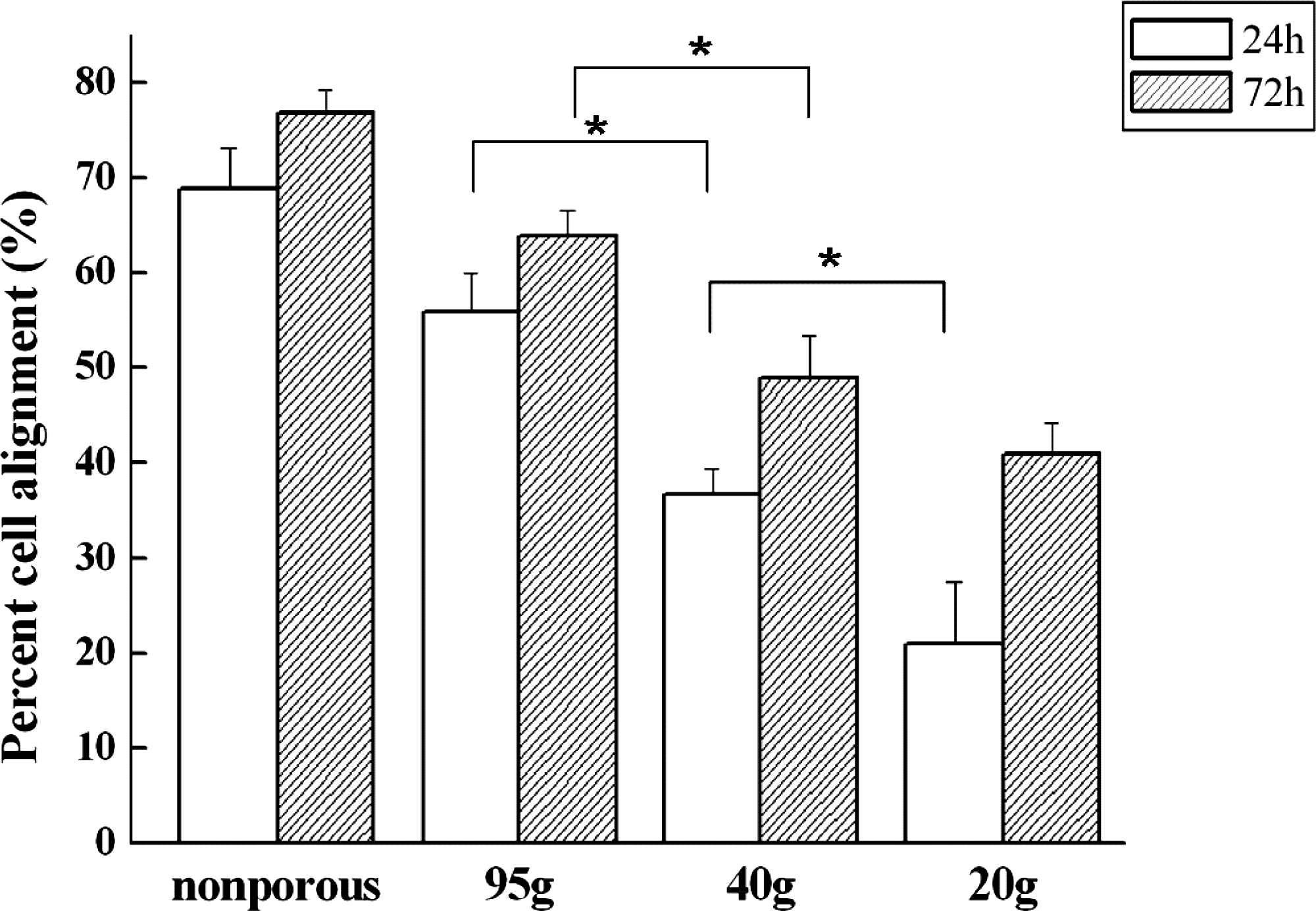
Percentage of C6 cell alignment on different microgrooved substrates. *p < 0.05.
Animal implantation
After the nerve conduit was cut open vertically, the newly regenerated nerve in the form of thin white tubular substance that connected the two ends, as well as a gel-like substance that filled the space in the conduit, was observed in all groups of implantation after 4 and 6 weeks. Histological data showing the primary myelin sheaths and the regenerated blood vessels at the midconduit are displayed in Figures 5 and 6. The quantitative data are summarized in Figure 7.

Histology of the nerve sections at the midconduit at 4 weeks for (

Histology of the nerve sections at the midconduit at 6 weeks for (
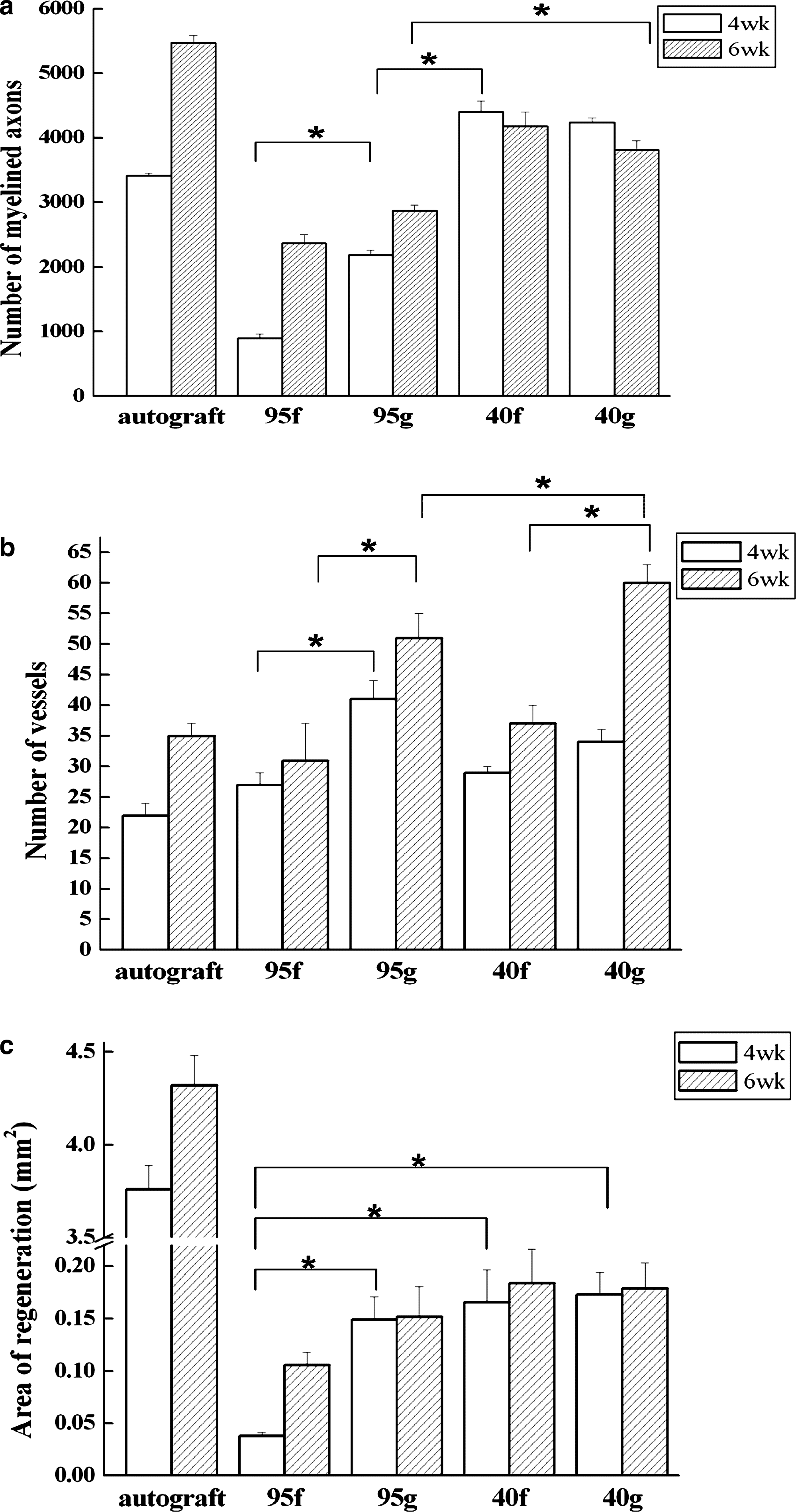
Quantitative data from histological analyses of different conduits. (
When the two experimental groups with surface groove features were compared, the larger and more deeply colored myelin sheaths were observed in the asymmetric conduits (40g); that is, the degree of myelination of the regenerated nerve in the asymmetric conduits (40g) (Figs. 5d and 6d) was higher than in the symmetric conduits (95g) (Figs. 5c and 6c). A similar trend was found in the two nongrooved groups, that is, 40f > 95f in degree of myelination (Figs. 5a, b, and 6a, b). On the other hand, when the two experimental groups with the same microporous structure were compared, the larger and more deeply colored myelin sheaths were found in the grooved conduits than in the nongrooved conduits, that is, 95g > 95f (Fig. 5a, c), 40g > 40f (Fig. 5b, d), 95g > 95f (Fig. 6a, c), and 40g > 40f (Fig. 6b, d) in degree of myelination. In the asymmetric groups, the myelin sheaths started to form bundle-like structure at 6 weeks (Fig. 6b, d). The number of myelinated axons in the asymmetric groups was greater than that in the symmetric groups at both 4 and 6 weeks, as shown in Figure 7. From 4 to 6 weeks, the number of myelinated axons increased in the symmetric groups and in the autograft group, but slightly decreased in the asymmetric groups. On the other hand, the vessels in the 40g and 95g groups were larger in size than in the other groups, and multierythrocytes could be clearly visualized in the vessels within the regenerated area (Fig. 6c, b). The number of blood vessels increased in all groups from 4 to 6 weeks (Fig. 7b). The number of vessels in grooved conduits was larger than in nongrooved conduits, that is, 40g > 40f and 95g > 95f. When the two experimental groups with the same microporous structure were compared, more vessels were found in the grooved conduits than in the nongrooved conduits. The regenerated myelin could be found in a larger area of autograft than in the other groups, because the proximal nerve terminals were known to send sprouts randomly toward the residual Schwann cell tubes of autograft transection. Although the autograft group had the largest regenerated area, the regenerated nerve fibers within the original nerve fascicules could not be assembled into the regeneration units after autografting. Subsequently, the myelin sheaths in the autograft group did not form bundle-like structure until 6 weeks. On the other hand, the size of the regenerated area was not significantly different in all experimental conduits.
The walking track behavior test was used to assess the functional recovery of regeneration (Fig. 8). The SFI at week 0 reflected the index for the rats before the transection surgery. The sharp decrease of the SFI for all samples at 1 week was due to the complete loss of motor control in the rat hind limb. A significant increase in the SFI occurred for the 40g at 6 weeks. Based on the walking track analysis and the histology, the repair capacity of various nerve conduits was in the order of 40g > 40f > 95g > autograft > 95f at 4 weeks, and 40g > 40f ≈ 95g ≈ autograft > 95f at 6 weeks.
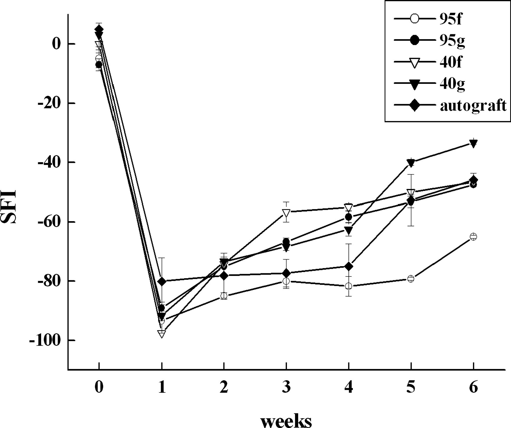
Assessment of functional recovery using SFI from the walking track analysis.
Discussion
Permeability and cell alignment are important factors that influence peripheral nerve regeneration. We have previously demonstrated that large outflow permeability caused by the asymmetric microporous structure of the poly(lactic-co-glycolic acid) (PLGA) conduits was beneficial for sciatic nerve reconstruction. 6 We have also optimized the microgroove dimensions for aligning Schwann cells in vitro.24,25 A combination of the two structural features, microporosity and microgrooves, could be achieved in one step by controlling the fabrication parameters of the conduit. To the best of our knowledge, this is the first study to combine the two design concepts, the high outflow permeability and the ability to align cells, in one conduit and to compare their effects on peripheral nerve regeneration in vivo.
The distinct microporous structures in various PLA substrates were resulted from the different solubility in the liquid–liquid demixing process. The skin layer was generated first and formed different liquid compositions (polymer rich and polymer poor) to reduce the free energy. 27 The polymer-poor area further becomes the pore, with the surrounding polymer-rich area becoming the wall. In general, a faster liquid–liquid demixing process leads to the formation of larger pores. In this study, the process was faster and more violent between dioxane and lower alcohol concentration (20% or 40%) because of the larger difference in solubility and polarity. Thus, the asymmetric distribution of pores in the cross section and the open pores on the microgrooves were more clearly observed in 20% and 40% groups. The asymmetric substrates comprised dense dual skins that covered the inner (lumen) and outer surfaces, 28 and a layer of macrovoids (40–80 μm), and a sublayer of micropores (3–20 μm) were generated between these two skin layers.29–31 Papenburg et al. 32 reported that the mass transfer was better in a scaffold without a skin layer. On the other hand, it is often assumed that the total membrane resistance is determined only by the thin skin layer of the membrane in membrane separation processes.31,33–35 Although the thickness of 20f was larger than that of 40f, we believed that a proper skin layer could control the permeability of the substrate.
The difference in permeability among various PLA substrates was ascribed to the distinct porous structure, agreeing with previous results on asymmetric PLGA substrates. 6 The additional microgroove feature did not influence the permeability. Differences, however, existed between PLA and PLGA. The asymmetric permeability previously reported for PLGA (20% alcohol) was not observed in PLA (20% alcohol). Some of the pores in the latter were too large to generate the asymmetric permeability successfully.
C6 cells were generally aligned by groove topography in this study. Nevertheless, the effectiveness of alignment depended strongly on the integrity of the microgrooves on the PLA substrates. It was possible that the depth of grooves on the substrates could change slightly during the phase transition process. For example, the phenomenon of shrinkage in the width/spacing/depth of microgrooves about 20–50% reduction was mentioned. 32 In our study, no significant change in the width/spacing/depth scale of the microgrooves was observed. This was probably due to the smaller disturbance caused by the nonsolvent used in our study. On the other hand, the integrity of the grooves on the asymmetric substrates was hampered by the presence of large open pores on the surface, which reduced the cell-guiding ability.
The histological results indicated the formation of new myelin sheaths in the symmetric groups and autograft group from 4 to 6 weeks. From our results, there was no correlation between blood vessel formation and number of new myelinated axons. The Wallerian degeneration process is known to be different in autograft and in conduits. 36 In autograft, the proximal nerve terminals send sprouts randomly toward the residual Schwann cell tubes of autograft transection, and endothelial cells will connect the original blood vessels. In conduits, the nerves are newly regenerated by Schwann cells, endothelial cells, and other cells in Wallerian degeneration process. When the new blood vessels are more, the nutrient supplied is more. Therefore, blood vessels were more abundant in conduits than in autograft. Besides, the newly generated myelin sheaths by Schwann cells are known to get thicker after the nerve successfully connects the muscle at the distal side (e.g., as 40g), and the myelin sheaths without nerve fiber passing through will be degraded in latter Wallerian degeneration process (e.g., as 40f). Therefore, 40f conduits had less blood vessels and more immature myelinated axons than 40g. The evolution of histology in the asymmetric conduits with implantation time also supported that regeneration was faster for asymmetric conduits. The better in vivo results for the asymmetric PLA conduits (without grooves) were parallel to our earlier findings with asymmetric PLGA conduits. 6 Unlike PLGA that was degraded during the implantation period, the asymmetric structure in PLA could be better maintained for a longer period in rats due to the lower degradation rate of PLA. This might benefit nerve regeneration and explain why the asymmetric PLA conduits were superior to the asymmetric PLGA conduits.
In the long term, the PLA conduits that continued to degrade among all and become fragment after 5 months were degraded into small pieces in our pilot study (data not shown). The surrounding regenerated tissue entangled into the conduit fragments. To compare with the available Food and Drug Administration/Conformit Europe (FDA/CE)-approved absorbable nerve conduits, Neurolac, we suggested that the microstructure of PLA-based conduits might increase in vivo degradation of conduits. 37
In our study, the asymmetric structure of PLA conduits may have drained out the wastage products at the initial stage of Wallerian degeneration in the conduit model, as hypothesized in our previous work. 6 At the same time, the ECM molecules in fluid may have adhered on the surface of the conduits. Surface microgeometry or smoothness of the conduits has been reported to influence the outcome of peripheral nerve regeneration, potentially by affecting the early arrangement of the fibrin matrix and/or inducing different cellular responses. 23 In our study, the significant increase in the number of capillaries in grooved conduits during nerve regeneration suggests an interaction between ECM and endothelial cells. The ECM plays an important role in the development of new blood vessels38,39 and in axonal regeneration.40,41 The interaction may be associated with the orientation of fibrin/fibronectin clot with neurotrophic factors to promote growth and migration of endothelial cells, Schwann cells, and axons during the early phase of Wallerian degeneration.42,43 On the other hand, the organized ECM may provide a pathway with structural and molecular cues that support the directed outgrowth of regenerating neurons.17,44,45 Therefore, it is hypothesized that the groove topography may have influenced the ECM remodeling process during Wallerian degeneration. The ECM bridge formed in grooved conduits may have been guided and developed into a more regular structure to further benefit the migration of endothelial cells and Schwann cells. After that, the axons regenerate from the proximal stump through the Schwann cell bridge in the conduit to their final contact. The endothelial cells start forming capillaries for nutrition supply. In brief, it seemed that the asymmetric porous structure transported waste drainage in the early stage of Wallerian degeneration, and the microgroove topography influenced ECM remodeling at the later stage based on our animal study.
Conclusion
In this study, two micrometric features, surface microgrooves and asymmetric microporous structure, were successfully combined on PLA substrates. The asymmetric microporosity equipped the substrate with asymmetric permeability, and the surface microgrooves induced cell alignment in vitro. The in vivo results obtained using the rat sciatic nerve transection model showed a higher degree of myelination at 4 weeks and at 6 weeks in the asymmetric conduits with surface microgrooves. Based on the study, the concepts of combining the waste drainage and extracellular matrix remodeling abilities could be adequately employed in the design of peripheral nerve conduits.
Footnotes
Acknowledgments
This work was supported by grants from National Science Council, National Health Research Institutes, and Veterans General Hospital Taichung-National Chung Hsing University Grant (VGH-NCHU Grant). The study was conducted in the Center of Tissue Engineering and Stem Cells Research of the university.
Disclosure Statement
No competing financial interests exist.