
Abstract
Vascular tissue engineering represents a promising approach for the development of living small-diameter vascular grafts that can be used for replacement therapy. The culture of strong human tissue-engineered (TE) vascular grafts has required long culture times, up to several months, whether or not combined with gene therapy. This article describes the culture of strong, genetically unmodified, human TE vascular grafts in 4 weeks Small-diameter vascular grafts were engineered using a fast-degrading polyglycolic acid scaffold coated with poly-4-hydroxybutyrate combined with fibrin gel and seeded with myofibroblasts isolated from discarded saphenous veins from patients undergoing coronary bypass surgery. The TE grafts were subjected to dynamic strain conditions. After 28 d of in vitro culture, the grafts demonstrated burst pressures of 903 ± 123 mmHg. Comparison with native vessels (intact human left internal mammary arteries (LIMAs) and saphenous veins) showed no significant differences in the amount of DNA, whereas the TE vessels contained approximately 50% of the native collagen content. In the physiological pressure range, up to 300 mmHg, the mechanical properties of the TE vessels were comparable to the LIMA. In this study, we showed that dynamic conditioning combined with fibrin gel cell seeding enhances the mechanical properties of small-diameter TE grafts. These grafts might provide a promising alternative to currently used vascular replacements.
Introduction
The goal of our study was to culture human vascular constructs with a burst pressure of at least 500 mmHg in a short culture period with genetically unmodified vascular cells. In our laboratory, Mol and co-workers recently succeeded in culturing strong tissue-engineered (TE) human heart valves using vascular myofibroblasts in a short culture period of 28 days. 10 The basis of such tissues was a PGA scaffold coated with poly-4-hydroxybutyrate (P4HB) in combination with fibrin gel. Hoerstrup et al. have already used the combination of PGA and P4HB in vascular tissue engineering. 11 These investigators did not use fibrin gel, and the constructs were seeded with ovine cells. Applying the approach of Mol and co-workers, 10 the present study describes the culture of strong human TE vascular grafts. Because the focus of the study was mechanical strength, the current grafts do not include endothelial cells. The engineered grafts were compared with human vessels routinely used for coronary bypass grafting (i.e., the left internal mammary artery (LIMA), which is the most successful bypass graft, and the saphenous vein (SV)). In the physiological pressure range, the mechanical behavior of the engineered grafts appeared to resemble that of the the LIMA more than the SV.
Methods
Construct preparation and tissue culture
Vascular graft constructs were based on a non-woven PGA fiber mesh (69 mg/cm3; Cellon, Luxemburg) with fiber diameters of 10 to 15 μm and pore sizes ranging from 50 to 200 μm. The tubular constructs were fabricated by wrapping a sheet of approximately 80 × 7 × 1 mm around a stainless steel rod with a diameter of 3 mm (Fig. 1). Afterwards, the constructs were dip-coated in 1 w/v% P4HB (Symetis Inc., Lausanne, Switzerland) in tetrahydrofuran (THF). The P4HB coating provides structural integrity to the PGA mesh by fixation of the randomly orientated fibers at crossing points (Fig. 1F). The scaffolds were dried under vacuum overnight to remove solvent remnants. The resulting tubular constructs were 3 mm in inner diameter, approximately 4 cm in length, and 1 mm in wall thickness.
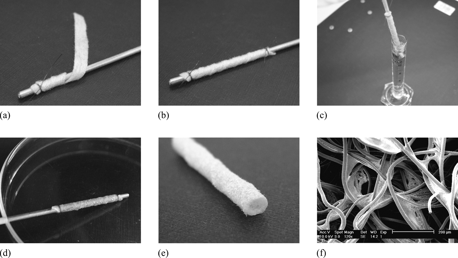
Fabrication of tubular construct of polyglycolic acid (PGA) scaffold coated with poly-4-hydroxybutyrate (P4HB). A rectangular PGA sheet (
Human myofibroblasts, harvested by plating 12 from the saphenous vein of two patients undergoing CABG (aged 47 (female) and 65 (male)) were used. Because the vein segments are discarded in the normal clinical setting, informed consent was not required. The cells were expanded using regular culture methods 12 up to passage 7 and characterized as a mixture of vimentin (V) and vimentin/actin (VA) type myofibroblasts. 10
Culture medium consisted of Dulbecco's modified Eagle medium (DMEM) Advanced (Gibco, Invitrogen, Breda, The Netherlands), supplemented with 10% FBS (Biochrom, Berlin, Germany), 1% Glutamax (Gibco) and 0.1% gentamycin (Biochrom). The medium for seeding and subsequent tissue culture, referred to as TE medium, contained 0.3% gentamicin and additional L-ascorbic acid 2-phosphate (0.5 mg/mL, Sigma, Zwijndrecht, The Netherlands).
The tubular constructs (n = 8) were slid over silicone tubing and mounted in a sterile bioreactor (see next section) and sterilized by exposing to ultraviolet radiation (germicidal lamp in LAF cabinet, 30 W, at a distance of approximately 30 cm) for 45 min and subsequent soaking in 70% alcohol for 3 h. The constructs were then washed in phosphate buffered saline (PBS) and placed in culture medium overnight. Afterwards, they were seeded using fibrin as a cell carrier. For this, human myofibroblasts were suspended in a sterile bovine thrombin solution with a concentration of 10 IU thrombin/mL medium (Sigma). The cell–thrombin mixture was subsequently added to a sterile fibrinogen solution (10 mg actual protein/mL medium) in a 1:1 mixture. The cell-containing fibrin gel (approximately 700 μL/tube) was mixed until onset of the polymerization process of the gel and subsequently dripped onto the tubular scaffolds (10,000–20,000 cells/mm3). Using this method, no cell solution is lost because the scaffold absorbs the solution while the fibrin rapidly polymerizes. It further ensures homogeneous cell distribution throughout the scaffold. 13
Bioreactor
In the bioreactor set-up (Fig. 2), tubular constructs were subjected to a circumferential strain of 1% while attached in the axial direction. Silicone tubing inside the constructs was used to apply strain. 7 A pressure device, consisting of a PBS-containing silicone tube that was compressed and decompressed using air, generated a pulsatile fluid flow through the silicone tubing, inducing straining of the constructs. To enable measurements of the pressure inside the silicone tubing, a pressure sensor (Beckton Dickinson, Erembodegem, Belgium) that was located in front of the construct was connected. During culture, the bioreactors could be placed on top of an inverted microscope such that the dynamic strain of the construct could be visualized.

Schematic drawing (
After seeding of the tubular constructs (see above), the bioreactors were placed in an incubator at 37°C and 5% carbon dioxide (CO2) to allow the fibrin gel to further polymerize for 30 min. Afterwards, the bioreactor was filled with 20 mL of TE medium, and the constructs were statically cultured for 1 week. The tubular constructs were then exposed to a circumferential strain of approximately 1% at 1 Hz for the remaining 3 weeks. To investigate the effect of the conditioning protocol, tubular control constructs (n = 4) were cultured statically, under no-strain conditions, in culture flasks (referred to as TE in flask). TE medium was changed twice a week.
Native human arteries and veins
To allow comparison between the TE vascular grafts and native equivalents, segments of discarded human SVs (n = 8, patient age 61 ± 10) and LIMAs (n = 6, patient age 51 ± 10) were obtained after completion of CABG from a local hospital. Because these segments are discarded in the normal clinical setting, informed consent was not required. The specimens were transported in DMEM Advanced (Gibco) at room temperature to the laboratory. As much loose adventitial tissue as possible was removed from the segments. Measurements of the mechanical properties were performed within 2 h. All vessels were investigated under maximal vasodilation with papaverine.
Histology
Ring segments of the TE constructs and native vessels were fixed in 4% phosphate-buffered formalin and embedded in paraffin. Ten-μm-thick sections were stained with Masson's Trichrome stains to visualize smooth muscle and the collagen. The viability was investigated by staining a representative portion of the constructs with viable CellTracker Green /propidium iodine staining.
Quantitative biochemical assays
Quantitative biochemical assays were performed on the TE (n = 8) and native (n = 6 for LIMA, n = 8 for SV) vessels. To determine the amount of DNA and sulfated glycosaminoglycans (GAGs) in the grafts, lyophilized tissue samples were digested in papain solution (100 mM phosphate buffer, 5 mM L-cystein, 5 mM ethylenediaminetetraacetic acid and 125-140 μg papain per mL) at 60°C overnight. The samples were centrifuged after digestion. The amount of DNA was determined using the Hoechst dye method. 14 GAG content was measured using a dimethylmethylene blue spectrophotometric assay. 15
Collagen content was assessed using hydroxyproline measurements, following a modified version of the protocol provided by Huszar. 16 In short, samples were hydrolyzed in 4 M sodium hydroxide for 10 min at 120°C followed by a reaction with chloramine-T and aldehyde/perchloric acid solution.
Mechanical testing
PGA degrades by hydrolysis and loses its mechanical properties after approximately 2 weeks because of bulk degradation, 17 so we first evaluated the mechanical properties of the bare PGA/P4HB/fibrin scaffold as a function of time. Tensile tests, as described below, were performed on rectangular scaffold samples that had been in culture conditions (i.e., in culture medium at 37º in 5% CO2) for up to 14 days.
The mechanical properties of the TE grafts were assessed using burst pressure measurements (n = 4) and uniaxial tensile testing (n = 4), as described below. The mechanical properties of the native vessels were analyzed using uniaxial tensile testing.
Burst pressure experiments were performed by inflating the TE vascular grafts to failure. To this end, the cultured vessels were taken out of the bioreactor, and both vessel ends were secured with a suture to a cannula (Medtronic, Heerlen, The Netherlands). The constructs were filled with PBS at a constant 7.5 mL/min flow rate. One cannula was connected to a pressure measurement dome (Beckton Dickinson, Erembodegem, Belgium) so that the luminal pressures could be measured using a pressure sensor (BD). The change in the diameter of the constructs was simultaneously captured using a high-speed camera (Phantom, V9.0, Photo-Sonics International Ltd, Oxfordshire, United Kingdom). Burst pressures were defined as the maximal attained pressures before rupture.
The tensile tests were performed in the circumferential and axial directions. Depending on the available size of the vessel segments, one to three rings of each construct were tested circumferentially and one to three rectangular strips axially. Stress–strain curves were obtained at room temperature using a uniaxial tensile tester (custom-built, equipped with a 20 N load cell) at a constant strain rate equal to the initial sample length/min. The stress (σ) was defined as the engineering stress, equaling the measured force divided by the initial cross-sectional area. From the engineering stress–strain curves, the ultimate tensile strength (UTS) was determined, as well as the elongation at break as a percentage of the initial length. The Young's modulus was calculated, defined as the slope of the linear portion of the stress–strain curve. The UTS in the circumferential direction can be used to estimate the burst pressure, as done previously, 18 using thin wall assumptions and Laplace's relationship: P × r = σ × h, where P is the pressure; r, the radius; σ, the Cauchy stress; and h, the wall thickness.
Statistics
All quantitative data were averaged per vessel, subsequently averaged per group, and represented as the average ± standard deviation. Comparison between the control group and the engineered grafts was performed using a Student t-test, assuming equal variances. Comparisons between the groups (TE, LIMA, and SV) were performed using one-way analysis of variance, assuming equal variances, using Bonferroni post hoc tests (STATGRAPHICS Plus 15.0.04, Statistical Graphics Corp., Herndon, VA) to determine significant differences (p < 0.05). For the uniaxial tensile tests, Student t-tests were used to elucidate differences between the properties in the axial and circumferential direction within each group.
Results
Macroscopic appearance
Macroscopically, the TE vascular grafts had a homogenous, shiny, tissue-like appearance (Fig. 3). After removal of the vessels from the silicone tubing, the vessels appeared as firm structures with a uniform inner diameter of 3 mm and an average wall thickness of 0.5 mm.

Photographs of a tissue-engineered vascular graft after 4 weeks of in vitro culture showing the graft attached in the bioreactor (
Histology
Visualization experiments using CellTracker Green and propidium iodine showed that, throughout the whole construct, most cells were alive (data not shown). Histological slides of a TE graft, LIMA, and SV are presented in Figure 4. Masson's trichrome staining clearly distinguished between muscle tissue (red) and collagen (blue). The TE constructs contained significant amounts of collagen, although less and not as organized as in the native vessels. The wall of the human artery (LIMA) demonstrated an abundant presence of smooth muscle (Fig 4B). The loose adventitial tissue mainly contained collagen. The human vein (SV) had a distinct different appearance, demonstrating more collagen in the vessel wall (Fig 4C). In addition, the lumen is much more irregular, which is a general characteristic of veins. The ‘folded’ structure illustrates the highly compliant behavior of veins at low pressures, because under these circumstances, the wall unfolds rather than resisting the pressure.
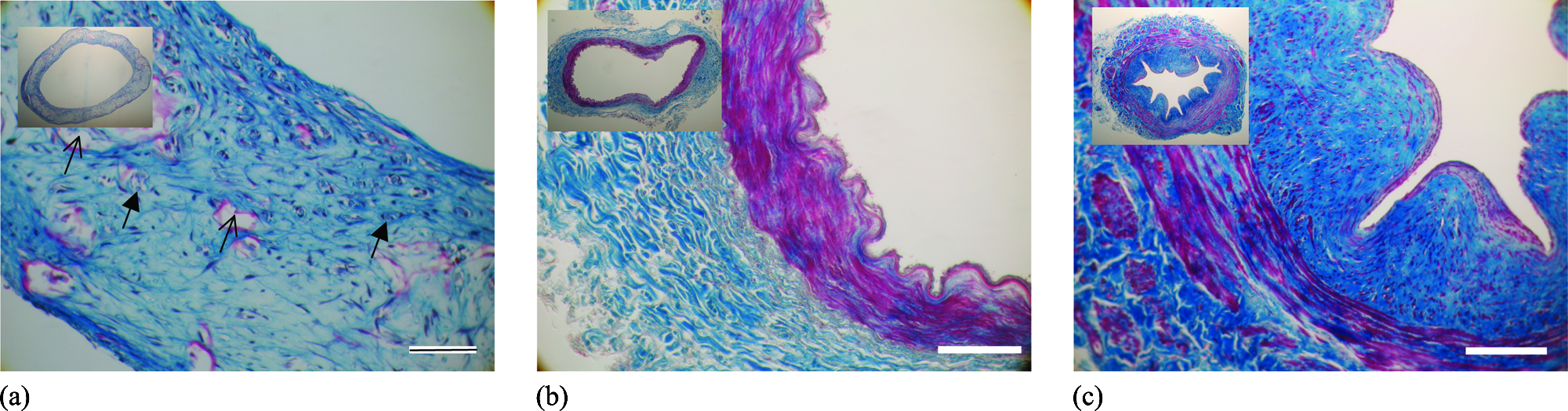
Masson's trichrome staining of the tissue-engineered graft (
Quantitative evaluation of tissue formation
The tissue content of the engineered and native vascular grafts was quantified according to the amounts of DNA, sulfated GAGs, and hydroxyproline (Table 1). No significant differences were found between the two groups of TE grafts resulting from the two different donors (aged 47 and 65). The conditioned TE grafts contained significantly more sulfated GAGs and hydroxyproline than the control constructs cultured in culture flasks (p < 0.001). Comparison of the TE grafts and the native vessels showed no significant differences between the amount of DNA in the TE, LIMA, and SV. The amount of GAG in the TE grafts was significantly higher than in the native vessels (p < 0.001). Conversely, the amount of hydroxyproline in the TE constructs was significantly lower than in the LIMA (p < 0.01) and SV (p < 0.001). On average, the hydroxyproline content of the TE constructs was approximately 50% of that of the analyzed native vessels. When normalized for the DNA content, the amount of GAG in the TE constructs was significantly higher than in the native equivalents (p < 0.001). The amounts of hydroxyproline per DNA were higher for the native vessels than for the TE constructs, although only significantly so for the SV (p < 0.01).
Values obtained from literature.6,35,36
Value different compared to LIMA (*: p < 0.05, **: p < 0.01, ***: p < 0.001).
Value different compared to SV (#: p < 0.05, ##: p < 0.01, ###: p < 0.001).
GAG, glycosaminoglycan.
Mechanical testing
Mechanical testing of the unseeded bare scaffold showed that the mechanical integrity of the scaffold, including the fibrin gel, started to decline after 1 week in culture conditions and was negligible after 2 weeks (data not shown). These results are in agreement with a study by Klouda. 19
The control constructs that were cultured in culture flasks were much weaker than the conditioned TE grafts, demonstrated by significantly lower Young's moduli and UTS (p < 0.001) (Table 1). The burst pressure experiments of the TE grafts revealed a burst pressure of 906 ± 123 mmHg (Table 1). During the burst pressure tests, changes in the vessel diameter were captured using a high-speed camera and analyzed at different axial positions (Fig. 5). Combination with the measured pressure signal resulted in pressure versus circumferential strain (Fig. 5A), showing, among other things, that the circumferential strain of the TE graft at 100 mmHg (i.e. in the physiological working range) was approximately 10%. In addition, these pressure-strain curves allow an estimation of the compliance of 0.03%/mmHg at 100 mmHg (dashed lines Fig. 5A).
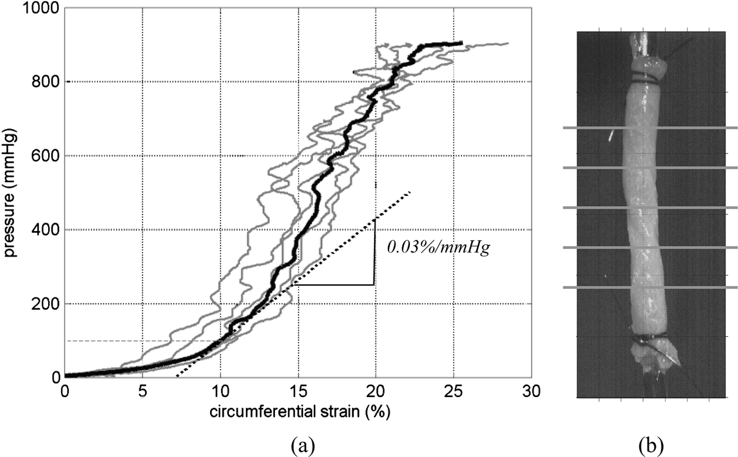
Pressure-strain curves (
Figure 6 shows the results of the tensile tests in the circumferential direction for the vascular grafts (TE vessels, the LIMA, and the SV). For the sake of clarity, one representative curve is shown for each type at stress values up to 1 MPa. To illustrate the variation in behavior, the stress–strain relations are presented, including envelopes within which all individual stress–strain relations are situated for each type of vascular graft. The dashed box illustrates the physiologically relevant stress–strain range. Quantification of the tensile tests, using the Young's modulus and UTS, in the circumferential and axial direction, is given in Table 1, showing, among other things, that the UTS of the native vessels is significantly higher than in the TE grafts. In the axial direction, all vascular grafts demonstrated stronger and stiffer behavior than in the circumferential direction, as quantified using the Young's modulus and UTS. The UTScirc of the TE vessels can be converted to a burst pressure of 886 mmHg using Laplace's equation, as described in the Methods section. This correlates well with the measured burst pressure of 906 ± 123 mmHg of the other group of TE vessels. Comparison between the LIMA and SV demonstrates that the vein was far more compliant than the artery. Comparing the TE vessel to the LIMA (i.e. the most successful graft for CABG) shows that, although the TE grafts are not as strong as the LIMAs, the mechanical behavior in the physiologically relevant range (up to pressures of 300 mmHg) does not deviate much from that of the LIMA.
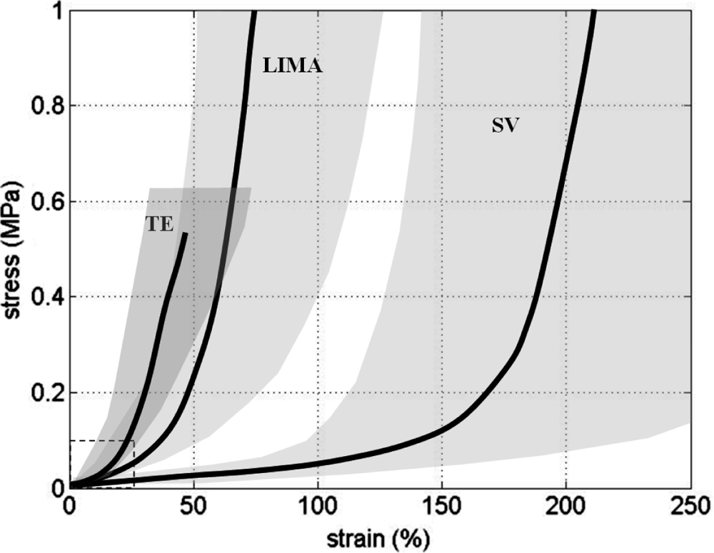
Representative engineering stress–strain curves in circumferential direction for the engineered vessels (TE) and native equivalents, i.e., the LIMA and SV. To visualize the variation in behavior, the figure includes envelopes within which all individual stress-strain relations are situated for each type of blood vessel. The dashed box indicates the physiologically relevant stress-strain range.
Discussion
Tissue engineering of small-diameter vascular grafts is the subject of ongoing research. Several approaches have already reached clinical trials.20–22 Optimizing the culture conditions of TE vascular grafts and reducing culture time will increase the potential for future use. Matching the (mechanical) properties of TE vascular grafts to those of a successful native graft may enhance the post-implantation performance of TE grafts. On the other hand, at implantation, the (mechanical) properties may not need to be identical to those of a native graft because a TE vessel is composed of viable tissue with the potential to remodel and grow. Nevertheless, some critical minimum demands remain; the construct should be strong enough to withstand surgical handling, it should withstand physiological pressures, and it should consist of viable tissue that is able to remodel. Until now, the culture of strong human TE vascular grafts required long culture times, up to several months, whether or not in combination with telomerase gene therapy.6,8 The present study describes the culture of strong human vascular grafts, based on a biodegradable scaffold, using genetically unmodified cells, over a 4-week period. The constructs contained significant amounts of collagen and possessed a burst pressure on the order of 900 mmHg. Tensile tests on the bare scaffold material indicated that the mechanical properties of the vascular TE grafts were the result of newly formed tissue and were not due to remaining scaffold material. The estimated compliance of the TE grafts was 0.03%/mmHg, whereas the strain at 100 mmHG was approximately 10%; these values correspond well with reported measures for human coronary arteries ( 0.06%/mmHg and 4–10%, respectively). 23 The presented TE grafts are based on biodegradable PGA. In studies by Niklason, 7 PGA tubular constructs were fabricated by sewing a PGA sheet using sutures into cylindrical constructs. This method creates an axially aligned suture line with a high concentration of PGA. It was observed that engineered vessels routinely ruptured at that suture line. 24 By wrapping a PGA sheet around a rod and including a P4HB coating to maintain dimensions, we avoided a suture line, which resulted in a more homogeneous construct.
The present TE grafts were cultured using myofibroblasts isolated from human SVs. These cells show good in vitro tissue generation and are easily obtainable and therefore suitable for tissue-engineering applications, 12 For application in, for example, second or third reoperative CABG, these cells could be harvested at the first operation, when the saphenous vein is harvested for use as bypass graft. A small piece of the vein could be used to isolate cells, after which they could be expanded in culture and stored cryogenically for future use. One month before reoperation, a vascular graft could be engineered from these cells.
The SV has already been used as cell source for the culture of human TE vascular grafts in studies by Niklason.8,9 Similar to our study, PGA was used as a scaffold material although not augmented with a P4HB coating or fibrin gel. After a 7- to 8-week culture period under comparable culture conditions, except for the frequency used (165 instead of 60 beats per min), the resulting constructs displayed a significantly lower burst pressure than in our experiments. In addition, these results could only be obtained when the cells were genetically modified (i.e., via human telomerase reverse transcriptase gene transfection). This transfection increases the replicative capacity of the fibroblasts and thereby possibly also the risk of tumorigenicity. Constructs cultured with non-modified cells resulted in weak vascular grafts. 9 Because one of the main differences between this study and our findings was the use of fibrin gel, it might be concluded that the use of fibrin strongly promotes tissue formation and the mechanical properties. This is in agreement with several other studies that have investigated the use of fibrin gels in regard to tissue formation.25–28 The effect of thrombin on the myofibroblasts is expected to be minimal in the three-dimensional fibrin gel. 29
To investigate the true potential of TE grafts for bypass grafting, it is important to use cells from elderly patients, because this age group is most likely to require such surgery. It is therefore worrisome that Niklason et al. found a strong dependence of the strength of TE vascular grafts on donor age. 8 Our study, which included cells from only two donors, did not show significant differences between the constructs from donors aged 47 and 65. However, additional experiments, using cells of more patients, should confirm whether our method is relatively donor-age independent.
Comparison of the presented TE vascular grafts with human native grafts (the LIMA and SV) showed no significant differences in the amount of DNA. This seems, at first sight, surprising, but other investigators have been reported comparable data. 30 Our findings were also confirmed using 4',6-diamidino-2-phenylindole staining, which showed comparable amounts of nuclei in TE and native constructs (data not shown). Rasmussen and colleagues, who found DNA contents in human aorta tissue ranging between 1.5 and 3.0 μg/mg dry weight, which is comparable with what we found in our native vascular samples, provides further support. 31
The amount of GAGs was higher in the TE grafts, whereas the amount of collagen was approximately 50% of native vessels. Histology revealed that, although significant amounts of collagen were present in the engineered grafts, it was not as organized as in native vessels. Comparison of the tensile tests of the TE vascular grafts and the native LIMA showed a strong resemblance in mechanical behavior in the physiological pressure range.
An aspect that is missing in the current TE vascular grafts is elastin. Although no measurements of elastin were performed in the present study, it is a general finding that human TE constructs do not contain significant amounts of elastin after in vitro culture. 32 Post-implant animal studies have revealed in vivo elastin development after a certain amount of time, 33 although it remains unknown whether this will happen in (older) patients.
In vivo testing also has to show how vulnerable our grafts erre to the formation of intimal hyperplasia (IH), one of the main causes of graft failure. IH is thought to be due to a variety of injuries that always involve some endothelial damage. 34 Therefore, by adding a confluent layer of endothelial cells to the graft and by carefully implanting the graft, we hope to minimize the risk of IH.
In conclusion, the presented TE vessels cultured from human SV myofibroblasts possessed good mechanical properties. The main difference from other TE grafts, based on a biodegradable scaffold, is the use of fibrin gel, which demonstrates the important contribution of this gel. The mechanical properties, assessed using tensile testing, of the presented TE grafts and native arteries were remarkably similar in the physiologically relevant range. Adding a functional endothelium at the lumen of the presented TE grafts will allow further investigation in animal models. These studies should reveal how the TE grafts remodel in the in vivo environment and whether they will be suitable for future use in bypass grafting or as arteriovenous shunts for patients undergoing hemodialysis.