
Abstract
Numerous reconstructive procedures result in wounds that require skin grafting. Often, the amount of tissue available from donor sites is limited. In vivo tissue expanders have been used clinically to generate larger sections of skin, and other methods exist to cover large wounds, but all have significant limitations. We investigated whether these difficulties could be overcome by increasing the surface area of skin in vitro while maintaining tissue viability. Human foreskin was incrementally expanded in a computer-controlled bioreactor system over 6 days to increase its surface dimensions under culture conditions. Morphological, ultrastructural, and mechanical properties of the foreskin were evaluated before and after expansion using histology, scanning electron microscopy, mercury porosimetry, and tensile testing. The surface area of the tissue was 110.7% ± 12.2% greater, with maintenance of cell viability and proliferative potential. Histomorphological and ultrastructural analyses showed that dermal structural integrity was preserved. The pore diameter of the expanded skin was 64.49% ± 32.8% greater. The mechanical properties were not adversely affected. These findings show that expansion of living skin matrices can be achieved using a computer-controlled bioreactor system. This technique provides an opportunity to generate large amounts of skin for reconstructive procedures.
Introduction
In vivo subcutaneous tissue expanders or meshed split thickness skin grafts (STSGs) are being used clinically to generate larger segments of autologous skin when donor-site tissue is limited.4,5 Subcutaneous tissue expanders are balloon implants that are sequentially filled with increasing amounts of saline to increase the amount of overlying skin. The physicomechanical stress of the tissue expander results in biologic creep, greater mitotic activity, and greater vascularity, which ultimately leads to expanded skin. Subsequently, the expanded skin can be used as a tissue flap or harvested for use as a skin graft.3,18,19 However, the drawbacks of this method are the requirement of an additional surgical procedure, which increases morbidity, and lengthy wait times (on the order of months) to obtain sufficient tissue for intervention. Moreover, discomfort associated with the increasing expander volume and frequent tissue fibrosis remain as major limitations. 20 Alternatively, meshed STSGs use a graft mesher that cuts the skin into a mesh pattern, which results in greater surface dimensions before application on the wound bed.1,2,4 Meshed STSGs are not considered ideal for many applications because they leave large gaps of the wound open, which requires longer healing times and results in a criss-cross or cobblestone pattern of healed skin when scar tissue fills the gaps.1,4 Despite these limitations, in vivo tissue expanders and meshed STSGs remain the mainstays for the generation of skin to cover skin defects in reconstructive procedures.
The problems associated with using autologous skin for various procedures would be solved if the dimensions of donor skin tissue could be increased in vitro while tissue viability was maintained for subsequent delivery in vivo. In this study, we investigated the possibility of enlarging the surface dimensions of human dermal tissue using a computerized bioreactor system under tissue culture conditions. We evaluated the effects of in vitro expansion of living skin matrices to determine the feasibility of using this technology clinically.
Materials and Methods
Preparation of foreskin
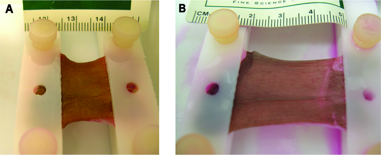
Foreskin samples were obtained from patients undergoing circumcision as approved by the Institutional Review Board at Wake Forest University Health Sciences. Excessive connective tissue was removed from the skin samples using sterile techniques in a tissue culture hood. The foreskins were rinsed twice for 5 min each in phosphate buffered saline (1 × PBS) solution containing 10% penicillin, streptomycin, and amphotericin (PSA) and a third time for 5 min in PBS with 3% PSA. The prepared foreskin was subsequently placed in the sterile skin bioreactor with the two opposite sides securely clamped (Fig. 1). The surface dimensions of each foreskin were measured and photographed (original lengths and areas ranged from 0.87-1.63 cm and 0.95-3.69 cm2, respectively). The bioreactor chamber was filled with 250 mL of high-glucose Dulbecco's modified Eagle medium containing 10% fetal bovine serum (FBS) and 2% PSA. All reagents for tissue culture were purchased from Invitrogen (Gibco Cell Culture, Carlsbad, CA).

Depiction of the expansion protocol curve (
Bioreactor setup
The bioreactor system consisted of an actuator mounted on a tissue culture container in which the skin samples were secured (Fig. 1). A linear motor-driven stimulator device (Linmot, Elkhorn, WI; E200-MT Controller, PS01-23x80 motor, Spreitenbach, Switzerland) controlled by a computer program was used for applying the mechanical stimulation, which consisted of uniaxial stretch. After loading the skin samples, the bioreactor was placed in an incubator at 37°C and 5% carbon dioxide. The bioreactor was programmed to uniaxially expand the skin to 20% of its initial length per day for 5 days to double the original length, and thus approximately double the surface area, of the skin. Each hour, the skin was stretched 4% per minute and then maintained at that length until the next hour, when the 4% stretching was repeated. This was performed five times, once per hour for 5 h, to achieve the 20% increase in length per day (Fig. 1). The average expansion time was 5.6 days. The medium was changed every other day. Digital imaging of each skin sample was obtained, and the length and surface area of each sample were measured using Image J software (1.37v, provided by the National Institutes of Health) before and after expansion (n = 5). The average change in length and area of all skin samples were compared using paired Student t-tests. Percentage increases in length and area were calculated as [(Dfinal-Dinitial)/Dinitial], where D was the dimension of interest (length or area; n = 5).
Porosimetric analyses
Pre- and post-expansion tissue specimens were fixed in 2.5% glutaraldehyde for 12 h and then were critically point dried before pore size analysis. The average pore size of the specimens was measured using a mercury intrusion porosimeter (AutoPore IV 9500, Micromeritics Co. Ltd., Norcross, GA). The porosimeter was set at a solid penometer volume of 6.7 to 7.3 mL. A mercury-filling pressure of 3.4 KPa was used and intruded to a maximum pressure of 414 MPa. Individual samples of 0.1 g were analyzed.
Scanning electron microscopy
Ultrastructure of the skin matrices before and after the bioreactor expansion was visualized using scanning electron microscopy (SEM, S-2600N, Hitachi, Tokyo, Japan). Briefly, a sample from each foreskin was cut and fixed with 2.5% glutaraldehyde in deionized water for 12 h, followed by rinsing in deionized water, freezing, and then lyophilizing. The samples were mounted on a SEM stand and sputter-coated with gold (Hummer 6.2, Anatech, Ltd., Denver, NC). The thickness of pre- and post-expansion samples was measured in five randomly selected areas, and the measurements were averaged. Paired t-tests were used to compare the changes in thickness between un-expanded and expanded groups (n = 4).
Histological evaluation
All histological samples (n = 5) were fixed in 10% formalin, embedded in paraffin, and sectioned using a microtome (6 μm, RM 2145, Leica Microsystems Inc., Bannockburn, IL). All skin samples were stained with hematoxylin and eosin (H&E), Masson's Trichrome, and immunohistochemistry for proliferating cell nuclear antigen. All histological samples were visualized and photographed using a Zeiss microscope (M1, Zeiss Axio Imager) and Zeiss AxioCam (Zeiss AxioCam MRc) using Axiovision software (Carl Zeiss Inc., Jena, Germany).
Tensile testing
Tensile testing was performed on all skin samples (2 cm × 0.5 cm) using an Instron 5544 (Instron, Norwood, MA). After the samples were loaded into the clamps, the gauge length was measured. The samples were kept moist with PBS during the entire period of mechanical testing.
Three independent mechanical tests were performed: evaluation of the mechanical properties of unexpanded skin; effects of rapidly straining the skin on the mechanical properties; and effects that slow, incremental, in vitro expansion had on the mechanical properties of the tissue. Two rectangular specimens were cut from the skin samples before expansion (pre-expansion specimens), and one rectangular specimen was cut from the skin after expansion (post-expansion specimens). One pre-expansion specimen from each skin sample was loaded to failure at an extension rate of 10.6 mm/min (Initial). 21 The other pre-expansion specimen was pre-strained to 100% of its starting gauge length at a rate of 100 mm/min and then loaded to failure at a rate of 10.6 mm/min (Control). Finally, the post-expansion specimens were loaded to failure at an extension rate of 10.6 mm/min (Expanded). Four foreskin samples underwent each mechanical testing. Young's modulus, ultimate tensile stress, and strain at failure from the initial and expanded samples were analyzed using a paired Student t-test. Control samples were not included in statistical analyses because two samples failed during testing.
Statistical analysis
All statistical analyses were performed using Microsoft Excel (Redmond, WA). Values are presented as means (standard errors of the mean) (SEM). Specific statistical tests performed are indicated in the above Methods sections. Statistical significance was determined as p- < 0.05.
Results
Planimetry
The length and surface area significantly increased after bioreactor expansion (p < 0.01) (Fig. 2A–C). The average percentage increase in length and surface area were 117.1 (7.1%) and 110.7% (12.2%), respectively. The dimensions of the skin samples were successfully increased in vitro using a bioreactor (Fig. 3).

Planimetry results showing dimensional changes of skin after expansion. (
(
Porosimetric analyses
Pore size measured from six skin samples initially, with only two successful measurements because of the inadequate tissue mass. Based on the two sets of samples analyzed, the mean pore diameter increased 64. 5% (32.8%) (Fig. 4A), with mean pore diameter of 94.0 (14.9) nm before expansion and 159.4 (55.2) nm (Fig. 4B) after expansion.

Porosimetric analyses. (
Scanning electron microscopy
The average thicknesses of the initial and expanded foreskins were 784.1 (95.9) μm and 889.0 (100.1) μm, respectively (Fig. 2D). The thickness changes between initial and expanded foreskins were not significantly different. The dermis was visualized, and the collagen fibers of the stretched group were found to be aligned in the direction of uniaxial extension (Fig. 5A, B). Furthermore, the dermal region of the expanded group appeared to have larger pores than the un-expanded group (Fig 5C, D). These data indicated that the collagen fibers of the skin aligned during stretching but that the thickness of skin matrices was not significantly affected at this level of dimensional increase.

Scanning electron microscopy images showing aligned collagen fibers and larger pores. (
Histological evaluation
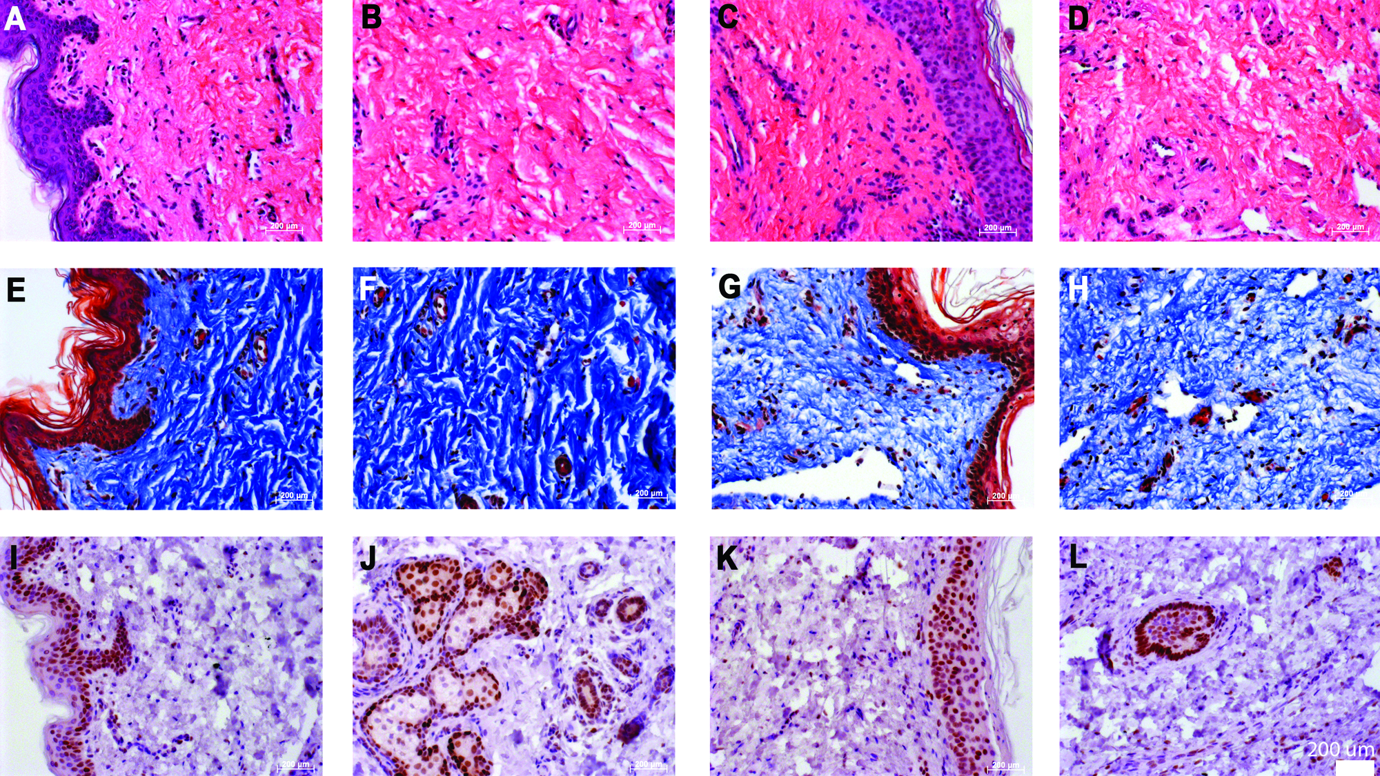
Tissue sections of expanded skin stained with H&E showed that the epidermis and dermis were intact and that the general structure of the skin was maintained (Fig. 6A–D). H&E demonstrated that cells retained normal morphology in the dermis, indicating that viable cells were maintained in the skin tissue during expansion. Masson's Trichrome staining further displayed the preservation of the tissue's structural integrity after uniaxial in vitro expansion (Fig. 6E–H). The expanded skin stained positively for proliferating cell nuclear antigen in the epidermal and dermal regions (Fig. 6I–L), indicating that the cells within the skin tissue were viable and maintained their ability to proliferate via extracellular signals produced by the mechanical stimulation in the bioreactor. These results show that the architecture of expanded skin is not disrupted, cell viability is preserved, and cells continue to proliferate throughout tissue expansion in the bioreactor.
(
Tensile testing
All specimens exhibited the typical viscoelastic properties of skin, including toe-in, linear, and failure regions (Fig. 7A). Young's modulus, a measure of stiffness, was lowest in the initial group (7.6 (2.7)) MPa, higher in the control group (9.0 (SM 6.5)) MPa, and highest in the expanded group (15.2 (4.1) Mpa; Fig 7B). The stiffness of the expanded group was significantly higher than that of the initial group (p < 0.05). Accordingly, the ultimate tensile stress was greater in the expanded group (4.9 (1.3 MPa)) than in the initial group (2.5 (0.6 MPa)), although this difference was not significant (Fig. 7C). The control group exhibited the lowest ultimate tensile stress (1.30 (0.87) MPa). In addition, there was no significant difference in strain at failure between the initial (109.1% (27.6%)) and expanded groups (72.7% (21.2%)). The initial group had the greatest strain at failure, whereas the control group (29.6% (10.9%)) showed the least strain at failure (Fig. 7D).

Tensile testing of skin. (
Discussion
Autologous skin grafts are considered the preferred method of skin defect treatment because the histocompatibility antigens and the pigmentation levels are identical. Although in vivo tissue expanders and graft meshers are a viable solution in instances in which availability of donor skin is limited, these approaches are associated with several limitations.4,5,20,22 This study aimed to increase surface dimensions of autologous donor skin tissue while maintaining tissue viability using a computerized bioreactor system. It showed that in vitro expansion of living skin matrices can be achieved and that expanded skin contains viable and proliferating cells. Thus, this method offers an advantage over currently available skin substitutes that do not contain cells, because the cells within the living skin matrices could contribute to wound healing in much the same way a skin graft would. Furthermore, the ability to expand skin in vitro while maintaining cell viability would eliminate the complications and disadvantages associated with in vivo tissue expanders, including pain, infection, erosion, hematoma, necrosis, rupture of skin expander, and long waiting times. 22
In addition to maintaining cell viability within the in vitro expanded skin, the residing cells show evidence of proliferation. Mechanical strain exerted on living tissues induces fibroblast proliferation, collagen production, and growth factor expression.23–26 The expansion system used in this study applies the same principle to skin tissue. Strain generated by the bioreactor is transmitted throughout the dermis and transduced to the fibroblasts via the components of the extracellular matrix, namely, collagen type I. Thus, bioreactor-facilitated expansion not only increases the surface dimensions of skin, but may also stimulate cellular proliferation. Furthermore, our results show that, although the surface area increased, the average thickness was not significantly changed at the current level of dimensional increase. In contrast, in vivo tissue expanders, which are implanted in the dermis, exert a tensile force primarily on the epidermis, resulting in a thickening of the epidermis and a decrease in dermal thickness.27–30 Cell proliferation might also result in better graft outcomes if the fibroblasts were proliferating when the expanded skin matrix was placed on the host wound bed. In essence, the in vitro expanded skin matrix could be primed for wound healing before use as a wound cover. Further investigations are currently being performed to evaluate these characteristics and determine whether they would result in better graft take in vivo.
Maintenance of the structural integrity of dermal tissues after expansion may be important for clinical outcome. In this study, we showed that the tissue structures remained intact with minimal disruption after the incremental strain. Partial disruption was observed between the dermis and epidermis, which may be due to the rapid expansion within a short period of time. This may be avoided by optimizing the rate and duration of expansion. The expansion process results in lower tissue density, which leads to larger pore size, as evidenced in this study. We showed that the expanded dermal tissue had a larger pore diameter than the non-expanded tissues. This response may be advantageous clinically because larger pore size may improve graft take by allowing better nutrient diffusion and angiogenesis.31,32 Another important factor that may influence the utility of the in vitro dermal expansion system is the retention of adequate mechanical properties. Currently, inadequate mechanical properties are a major drawback of current skin replacement products. The expanded skin matrix evaluated in this study showed greater values of the Young's modulus, whereas the strain at failure demonstrated a decreasing trend. Nonetheless, the skin matrices remained durable for use as skin grafts.
In this study, we used a computer-controlled bioreactor system that was designed to gradually expand dermal tissue to achieve a target size within a given time period. We used a uniaxial motor system to demonstrate the proof of principle that surface dimensions can be increased while viability of the skin tissue is maintained. Although the concept has been adequately demonstrated through the experiments performed in this study, improvements to the current prototype need to be made. One modification that could significantly enhance the outcome would be to develop a biaxial strain bioreactor. In the current study, using a uniaxial expansion system resulted in the alignment of the entire dermal fiber network, which includes elastic and collagen fibers. This leads to greater stiffness of the expanded tissue. A biaxial bioreactor design may minimize the unidirectional fiber alignment and reduce this stiffness.
To our knowledge, this is the first report in the literature that a computer-controlled bioreactor system has been applied to generate amplified living tissue in vitro for subsequent delivery in vivo. This novel technology allows for an accurate expansion rate for producing target tissue sizes over a defined time period. Although application of this system may be best suited for increasing surface dimensions of autologous dermal tissue, which avoids immunologic problems and retains the natural pigmentation of the recipient's skin, this technique could be applied to allogeneic skin with matching histocompatibility antigens. In this study, we used human foreskin, which is known to be more elastic than the skin tissues present in other regions of the body. Further studies are necessary to prepare this technology for full use in a clinical setting.
Conclusion
We showed that human skin can be incrementally expanded in vitro using a computer-controlled bioreactor system. Cell viability and proliferative potential of the tissue were maintained, dermal structural integrity was preserved, and pore size was increased. Thus, this technology provides an opportunity to generate large amounts of living skin matrix for reconstructive procedures. This may overcome the current limitations of other methods. Eventually, this technology could allow a surgeon to take a small skin biopsy and expand it in vitro, creating a large segment of skin for use in reconstructive procedures.
Footnotes
Acknowledgments
The authors thank Dr. Steve Hodges for acquisition of the skin samples, Dr. Jennifer Olson for editorial assistance, Cathy Mathis for technical assistance, and Kyle Binder for writing the computer software.