
Abstract
Currently, a tissue-engineered bone is usually constructed using a perfusion bioreactor in vitro. In the perfusion culture, fluid flow can exert shear stress on the cells seeded on scaffold, improving the mass transport of the cells. This experiment studied the effects of flow shear stress and mass transport, respectively, on the construction of a large-scale tissue-engineered bone using the critical-sized β-tricalcium phosphate scaffold seeded with human bone marrow–derived mesenchymal stem cells (hBMSCs). This was done by changing flow rate and adding dextran into the media, thus changing the media's viscosity. The cells were seeded onto the scaffolds and were cultured in a perfusion bioreactor for up to 28 days with different fluid flow shear stress or different mass transport. When the mass transport was 3 mL/min, the flow shear stress was, respectively, 0.005 Pa (0.004–0.007 Pa), 0.011 Pa (0.009–0.013 Pa), or 0.015 Pa (0.013–0.018 Pa) in different experiment group obtained by simulation and calculation using fluid dynamics. When the flow shear stress was 0.015 Pa (0.013–0.018 Pa), the mass transport was, respectively, 3, 6, or 9 mL/min. After 28 days of culture, the construction of the tissue-engineered bone was assessed by osteogenic differentiation of hBMSCs and histological assay of the constructs. Extracellular matrix (ECM) was distributed throughout the entire scaffold and was mineralized in the perfusion culture after 28 days. Increasing flow shear stress accelerated the osteogenic differentiation of hBMSCs and improved the mineralization of ECM. However, increasing mass transport inhibited the formation of mineralized ECM. So, both flow shear stress and transport affected the construction of the large-scale tissue-engineered bone. Moreover, the large-scale tissue-engineered bone could be better produced in the perfusion bioreactor with 0.015 Pa (0.013–0.018 Pa) of fluid flow shear stress and 3 mL/min of mass transport.
Introduction
Traditional tissue engineering is the static culture of cell-seeded three-dimensional (3D) scaffolds. However, this static culture pattern typically produces thin tissue growth localized to the construct periphery. 5 This phenomenon has resulted mainly from the lack of mass transport inside the construct. Hence, improving in vitro mass transport is a critical challenge in the production of thick cellular constructs. Many groups have developed bioreactors that perfuse cell-seeded constructs, and that have demonstrated the beneficial effects of perfusion on cell survival, proliferation, and tissue formation in the scaffold.6–9 However, most of these studies are about the construction of a small-size tissue-engineered bone. To simulate the long bone structure and to aid in the construction of the large-scale tissue-engineered bone, a perfusion bioreactor system and the critical-sized porous β-tricalcium phosphate (β-TCP) ceramic scaffold with a central tunnel have been successfully designed in our laboratory. 10 The perfusion bioreactor system can provide continuous and homogeneous nutrient supply inside the β-TCP scaffold, and permit cell proliferation during long-term incubation. 10
In addition to enhancing the exchange of nutrients and wastes, the perfusion bioreactor system simultaneously delivers mechanical stresses to cells seeded on the scaffold. Previous studies indicate that fluid flow shear stress is a potentially stronger stimulus for bone cell behavior than either hydrostatic compression or substrate deformation.6,11–13 In the perfusion bioreactor system, the two important factors influencing cell proliferation and differentiation are mass transport and flow shear stress. Therefore, the dynamic microenvironment including mass transport and flow shear stress could be considered as the fourth essential factor in tissue engineering. To date, the separate effects of mass transport and flow shear stress are not yet clearly understood, especially with regard to the construction of a large-scale tissue-engineered bone using the perfusion bioreactor system. Further investigation should therefore be made to elucidate the effects of different mechanical microenvironments on the construction of a large-scale tissue-engineered bone in vitro. Then, the mechanical microenvironment can be optimized to improve the construction of the large-scale tissue-engineered bone in vitro.
While separately studying flow shear stress' and mass transport's contributions to the construction of the tissue-engineered bone in a perfusion bioreactor system, a tackifier should be added to the media, enabling the media's viscosity change. As a result, media with different viscosities can be obtained by adding different quantities of tackifier. Additionally, based on the flow shear stress being proportional to the viscosity and flow rate of fluid, similar levels of mass transport can be maintained by giving the same flow rate while experiencing different flow shear stresses using media with different viscosities. This is also applicable vice versa, in which we can maintain similar levels of flow shear stress as well as experience different mass transport. This is by using media with different viscosities and giving different flow rates when the products of viscosities multiplied by the flow rates are equal. In this study, dextran was selected as the tackifier to change the media's viscosity.
In this study, our hypothesis is that both flow shear stress and mass transport affect the construction of large-scale tissue-engineered bone in vitro using perfusion bioreactor. So, the goal of this study is to verify the hypothesis and to find the appropriate conditions for construction of tissue-engineered bone using perfusion bioreactor.
Materials and Methods
Cell isolation and culture
Human bone marrow–derived mesenchymal stem cells (hBMSCs) were isolated and expanded using a modified method that was previously reported. 14 The donor was healthy and without metabolic diseases, inherited illnesses, or other diseases that may affect the current study. Bone marrow aspirates were obtained during routine orthopedic surgical procedures after obtaining written consent from the local ethics committee. Bone marrow aspirates (20 mL volumes) were harvested through a bone marrow biopsy needle inserted through the iliac crest. Each milliliter of the bone marrow aspirates was immediately inoculated into a 100 mm culture dish, and was cultured in a growth medium containing α minimal essential medium (α-MEM) supplemented with 10% fetal bovine serum (FBS) (Gibco, Carlsbad, CA), 100 IU/mL penicillin, and 100 mg/mL streptomycin (Hyclone, Logan, UT) in a humidified 37°C/5% CO2 incubator. On the basis of adhesion and proliferation on the tissue culture plastic substrate, the hBMSCs were selected. After 3 days, nonadherent cells were removed by two to three washes with PBS, and adherent cells were further cultured in α-MEM until 80–90% confluence. The attached cells that were left behind are mainly hBMSCs. The obtained hBMSCs were then enzymatically lifted from the culture dishes with trypsin–EDTA (0.25% [w/v] trypsin with 0.02% [w/v] EDTA; Sigma, St. Louis, MO), and subcultured at a density of 5 × 10³ cells/cm2. To inoculate the β-TCP scaffold, the third passage was used.
Preparation of the cells/β-TCP construct
The porous cylindrical β-TCP scaffold (Biocetis, Berck sur Mer, France) that was used in this study had a homogeneous porosity of 75%, spherical pores of 530 ± 100 μm in diameter, and interconnections of 150 ± 50 μm in diameter, and weighed about 3.35 g. The cylinder was 14 mm in diameter and 30 mm in length (Fig. 1), creating a tunnel with a blind end situated in the scaffold's center. The tunnel was 3.5 mm in diameter and 25 mm in length. According to the previous report, the bone defect greater than 20 mm in monkey man-dible was considered as critical-sized defect. 15 In this study, the scaffold was 30 mm in length, far greater than 20 mm, so the β-TCP scaffold can be considered as critical-sized scaffold. The cell/β-TCP construct was prepared using the previously reported method. 10 Briefly, the third passage of the hBMSCs was detached and resuspended at 2 × 10⁶ cells/mL. The β-TCP scaffold was immersed into the suspension. To make the cellular suspension penetrate into the scaffold, a vacuum (about 1.9 × 10⁴ Pa) was generated by a pump. Afterward, the cells/β-TCP construct was assembled to the perfusion bioreactor system. This perfusion bioreactor system was placed at 37°C in a humidified 5% CO2 incubator. After 2 h of cell attachment, 200 mL complete α-MEM was added to the perfusion bioreactor system. After another 2 h, the perfusion bioreactor system was switched on. The medium was completely changed every 3 days.
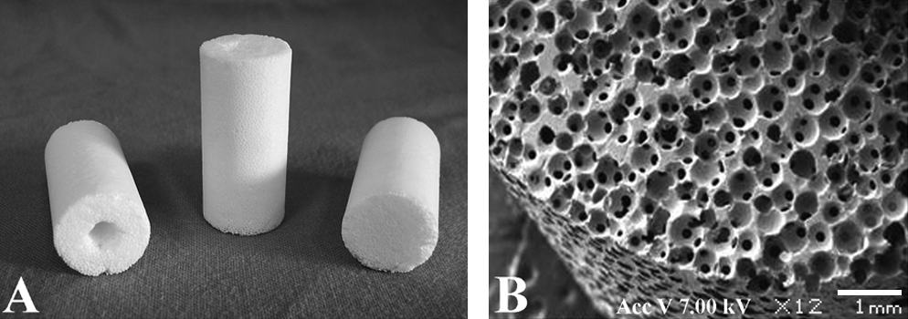
Porous β-TCP ceramic scaffold with a central tunnel. Holistic view (
Perfusion bioreactor system
The 3D perfusion bioreactor system has been previously described. 10 Briefly, the system consists mainly of three parts: the peristaltic pump, the reservoir, and the connective tubes (Fig. 2A). The reservoir is a 75 cm2 flask (Corning Incorporated, Corning, New York) that contains the perfusion medium, and the cells/β-TCP construct is placed into the flask and then suspended in the perfusion medium. The peristaltic pump drives the perfusion medium, enabling it to circulate in the connective tubes and the reservoir through the cells/β-TCP construct.

The perfusion bioreactor system used in this study (
In the perfusion bioreactor system, the cell culture medium contained α-MEM supplemented with 10% FBS, 100 U/mL penicillin, 100 mg/L streptomycin, and the osteogenic supplements ascorbic acid (50 μg/mL), β-glycerophosphate (10 mM), and dexamethasone (10 nM) (all from Sigma). The mass transport is positively proportional to the flow rate; hence, mass transport can be expressed by flow rate. When the mass transport was the same, that is, the flow rate being 3 mL/min, the media used were supplemented with 1% (w/v), 5%, or 7% dextran (average MW 70,000, lot OG0109A; Seebio Biotech, Shanghai, China). When the flow shear stress was the same, the flow rate was different: 3, 6, or 9 mL/min. Moreover, the media accordingly used were supplemented with 7%, 3%, or 1% dextran. The viscosity of the medium was measured with a viscometer (SNB-1; Shanghai Precision Instruments, Shanghai, China). The media containing 3% dextran had a viscosity that was one-half the viscosity of the media containing 1% dextran. The media containing 5% dextran had a viscosity twice the viscosity of the media containing 1% dextran. The media containing 7% dextran had a viscosity three times higher the viscosity of the media containing 1% dextran. When the flow rate is same, the flow shear stress is positively proportional to the viscosity of the fluid. So, when the flow rate was 3 mL/min, the flow shear stress in the three groups can be, respectively, represented by 1 ×, 2 ×, and 3 ×, and the three groups also can be, respectively, represented by 1 × group containing 1% dextran, 2 × group containing 5% dextran, and 3 × group containing 7% dextran.
Before this study, we have calculated the flow shear stress in the β-TCP scaffold when the flow rate was different or the media's viscosity was different. In brief, the model of the scaffold was first reconstructed in the computer, and then the flow field in the model of the scaffold was simulated and calculated using computed fluid dynamics method. After simulation and analysis of perfusion, we found that the flow shear stress in 75% region of the scaffold was similar. According to the above simulation and calculation using computed fluid dynamics method, the correlation between the flow rate, shear stress, and the dextran concentration in media was shown in Table 1. The flow shear stress was far lower than that reported (0.8–3.0 Pa) in vivo in previous study.16,17 The perfusion bioreactor system, during its normal operation, was placed in a cell culture incubator at 37°C with 5% CO2. The static culture was the control, and was represented by 0 × group or 0 mL/min. According to the previous study, 18 no significant difference was found for the effects of dextran on the proliferation and osteogenic differentiation when the concentration of dextran in media was 1–7%. So, the media used in static culture group contained 1% dextran. Additionally, in the static culture group, the cells/β-TCP construct was also connected to the perfusion bioreactor without perfusing.
Cell proliferation
Cell proliferation could be expressed in terms of cell viability. The MTT assay is one of the widely accepted cell viability assays. Cell viability was measured using a modified method that was previously reported. 10 Briefly, each cylindrical scaffold at day 28 was cut into two parts along the longitudinal axis. One part of the scaffold was fixed in 70% ethanol for histological study. The other part was then divided into three parts from top to bottom (Fig. 2B). The bottom part was washed with PBS and transferred to a six-well culture plate. Afterward, it was further cut into small fragments. Six milliliters of MTT (Sigma) solution (0.5 mg/mL in PBS) was added to each well followed by 4 h of incubation at 37°C. After the MTT solution was removed, 5 mL of the DMSO solution (Sigma) was added. The fragments were ground and extensively washed by repeatedly pipetting up and down to allow total color release. Moreover, the solution was centrifuged at 12,000 g for 10 min. The supernatant was collected and diluted whenever necessary. The absorbance was read via a spectrophotometer at 490 nm. The fragments were then weighed and dried overnight at 50°C. Cell viability was expressed as the absorbance per gram of the scaffold.
Glucose consumption
Glucose consumption can also indirectly reflect the cell proliferation and viability in the scaffold. The media were collected at days 4, 7, 14, and 28. The D-glucose in the media was determined via spectrometry according to the protocol provided by the D-glucose-hexokinase kit (Megazyme International Ireland Ltd., Co. Wicklow, Ireland). Briefly, the samples were appropriately diluted. First, 100 μL of nicotinamide-adenine dinucleotide phosphate/adenosine-5′-triphosphate and 100 μL of imidazole buffer were mixed with 100 μL of the sample and 2.0 mL of distilled water in the cuvette. After 3 min, the absorbance was read at 340 nm. Afterward, 20 μL of hexokinase/glucose-6-phosphate dehydrogenase solution was added. After 5 min, the absorbance was again read at 340 nm. If the reaction has not stopped after 5 min, the absorbance continued to be read during 2 min intervals until the absorbance remains the same over 2 min. The fresh medium and the D-glucose control solution were also processed and served as the reference. Finally, the absorbance difference and the glucose concentration were calculated. Glucose consumption was expressed as the daily reduction of glucose in the media for all the cells in the scaffold.
Alkaline phosphatase quantitative assay
The cylindrical scaffold was divided as described above. The middle part was used to assay alkaline phosphatase (AP) activity. AP activity was determined by using a modified method previously reported. 14 Briefly, the construct was rinsed with cold PBS, cut into small fragments, and stored in distilled deionized water at −70°C until assay. The samples were lysed using three cycles of thawing in a water bath at 37°C and were frozen in liquid nitrogen. Afterward, they were sonicated for 10 min and vortexed for 10 s to allow the enzyme and deoxyribonucleic acid (DNA) into the solution. The lysates obtained were centrifuged at 10,000 g for 10 min, and the DNA of the supernatant was quantified by assaying the fluorescence of Hoechst33258 (Sigma). This was then compared with DNA extracted from a known number of hBMSCs. AP activity in the same cell lysates was quantified using the conversion of paranitrophenylphosphate (Sigma) to p-nitrophenol. The results were normalized, with the cell number determined according to DNA assay.
Osteopontin assay
Osteopontin is a secreted protein that is a middle-stage marker of osteogenic differentiation. The osteopontin content was measured using an ELISA kit (Catalog No. DOST00) against human osteopontin available from R&D Systems (Minneapolis, MN). Medium samples were collected at days 4, 7, 14, and 28 during the culture period and stored at −70°C. These samples were thawed and vortexed for 10 s. The sample, or standard, was added at 50 μL/well to the plate, mixed with 100 μL/well assay diluent that was provided with the kit, and incubated at room temperature for 2 h. Each well was rinsed four times with a wash buffer, and 200 μL of secondary antibody conjugated with horseradish peroxidase was added to each well and allowed to incubate at room temperature for 2 h. Each well was thoroughly rinsed with a wash buffer for four times, and 200 μL of the substrate solution was added to each well. The plate was incubated at room temperature in the dark for 30 min before 50 μL of stop solution was added to each well. Absorbance was read at 450 nm with correction 570 nm. The samples were then run in duplicate and compared against the human osteopontin standard.
Osteocalcin assay
The amount of osteocalcin released into the media during the culture period was measured using an ELISA kit (Catalog No. BMS2020INST) available from Bender MedSystems (Vienna, Austria). Medium samples were collected at days 4, 7, 14, and 28 during the culture period and were stored at −70°C. These samples were thawed and vortexed for 10 s. Each sample well was added 100 μL of distilled water, and each standard and blank well was added 125 μL of distilled water. The sample was added at 25 μL/well to the sample well. The plate was incubated at room temperature in the dark for 2 h on a microplate shaker at 100 rpm. Each well was rinsed three times with a wash buffer, and 100 μL of the substrate solution was added to each well. The plate was incubated at room temperature in the dark for 15 min before 100 μL of the stop solution was added to each well. Absorbance was read at 450 nm with a correction of 620 nm. Afterward, the samples were run in duplicate and compared against human osteocalcin standard.
Scanning electron microscopy
Scanning electron microscopy (SEM) was performed to evaluate the morphological appearance of hBMSCs and the deposited matrix within the β-TCP scaffold. After 28 days of culture, the top part of the scaffold was washed with cold PBS. Fixation was carried out for 30 min in 2% glutaraldehyde at 4°C. Through a series of increasing graded ethanol, the samples were dehydrated and dried with a critical-point drier (HCP-2; Hitachi, Tokyo, Japan). The specimens were sputter-coated with gold, examined, and photographed with a scanning electron microscope (JSM-6360LV; Jeol, Tokyo, Japan) at an acceleration voltage of 7.0 kV. Then energy dispersive X-ray analysis was performed in another scanning electron microscope (XL30-FEG; Philips, Amsterdam, Netherlands) to identify the chemical composition of the structures present on the scaffolds.
Histological observation and histomorphometry
For histological analysis, the samples were fixed in 70% ethanol for 10 days. Afterward, they were dehydrated in a series of increasing graded ethanol, cleared with toluene, and embedded in methylmethacrylate. After polymerization, sec-tions about 150 μm thick were achieved via Leica SP 1600 (Leitz, Wetzlar, Germany) through the longitudinal axis of the cylindrical scaffold. They were then grounded and polished to about a 50 μm thickness with Exakt Grinder (Norderstedt, Germany). All sections were stained with Steven blue and van Gieson picro-fuchsin. The β-TCP scaffold could not be stained, but the mineralized extracellular matrix (ECM) was stained red. Three sections from each sample were examined with the Leica Microsystems (DM4000B; Wetzlar GmbH, Wetzlar, Germany). For samples after culture, the tissue area (all ECM forming in the section, including mineralized and un-mineralized ECM) and the bone area (all mineralized ECM in the section) was quantitatively measured with the software BIOQUANT OSTEO II (Nashville, TN). The percentage of tissue area/pore area and bone area/pore area was determined, respectively.
Statistical analysis
The SAS6.12 statistical software was used in this study. Multiple samples were collected in each measurement (n =4–6 per group), and the results were expressed in means ± standard deviation. Data for these measurements were analyzed using the analysis of variance (ANOVA) test. p-Values less than 0.05 were considered statistically significant.
Results
Proliferation of hBMSCs
Cell proliferation could be expressed as cell viability. The results revealed that cell viability in the 2 × group was higher than that in the other groups (p < 0.05) (Fig. 3A) when the flow rate was 3 mL/min. The flow shear stress in the 3 × group with 3 mL/min of flow rate inhibited the proliferation of hBMSCs. When the flow shear stress was the same, the 3–9 mL/min of flow rate did not result in a cell viability change (p > 0.05) (Fig. 3B). However, the cell viabilities in the perfusion culture were all higher (p < 0.05) than that in the static culture.
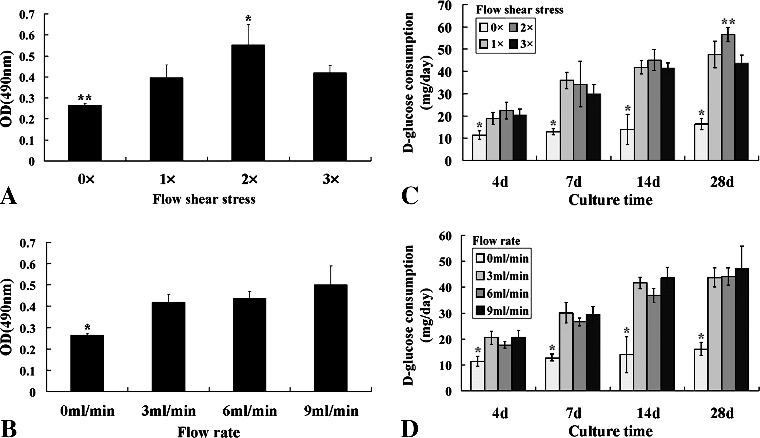
Cell viability measured after 28 days of culture (
Glucose consumption
The results showed that in all groups, the daily D-glucose consumption increased with time. When the flow rate was 3 mL/min, the daily D-glucose consumption in the 2 × group was higher (p < 0.05) than that in other groups at day 28 (Fig. 3C). When the flow shear stress was the same, no significant difference was found between all perfusion culture groups during the culture period (Fig. 3D). The daily D-glucose consumption in the static culture was lower (p < 0.05) than that in the perfusion culture. These results were similar to those of the cell viability investigation.
AP activity quantification
AP is a marker of early osteogenic differentiation and a commitment of mesenchymal stem cells toward the osteoblastic phenotype. AP activity was determined at day 28, and it was normalized according to cell number as determined by the DNA assay. When the flow rate was 3 mL/min, the AP activity in both 2 × and 3 × groups was higher (p < 0.05) than that in the 1 × group (Fig. 4A). When the flow shear stress was the same, the AP activity in the 6 mL/min group was higher (p < 0.05) than that of the other groups (Fig. 4B).
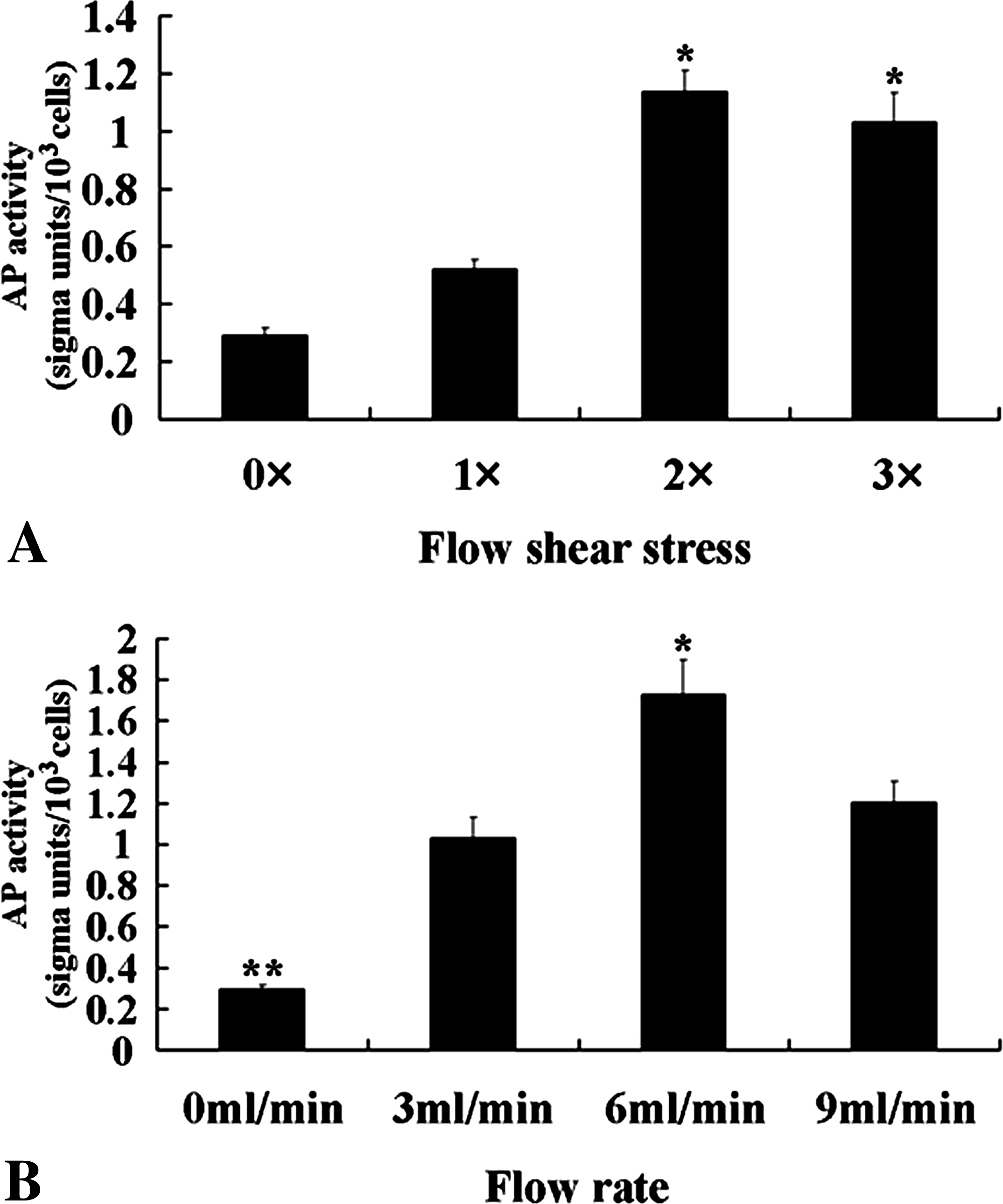
AP activity of hMSCs seeded on the β-TCP scaffold after 28 days of culture. When the flow rate was 3 mL/min, the AP activity was higher in the 2 × and 3 × groups than in other groups (
Osteopontin measurement
Osteopontin is an important middle-stage marker of osteogenic differentiation. It directly contributes to the regulation of mineral crystal formation and growth. When the flow rate was 3 mL/min, the secretion peak of osteopontin was reached at day 4 in the 3 × group, at day 7 in the 2 × group, and at day 14 in the 1 × group (Fig. 5A). When the flow shear stress was the same, increasing the flow rate resulted in an increase in osteopontin's secretion (Fig. 5B). In the static culture, osteopontin secretion increased during the culture period.
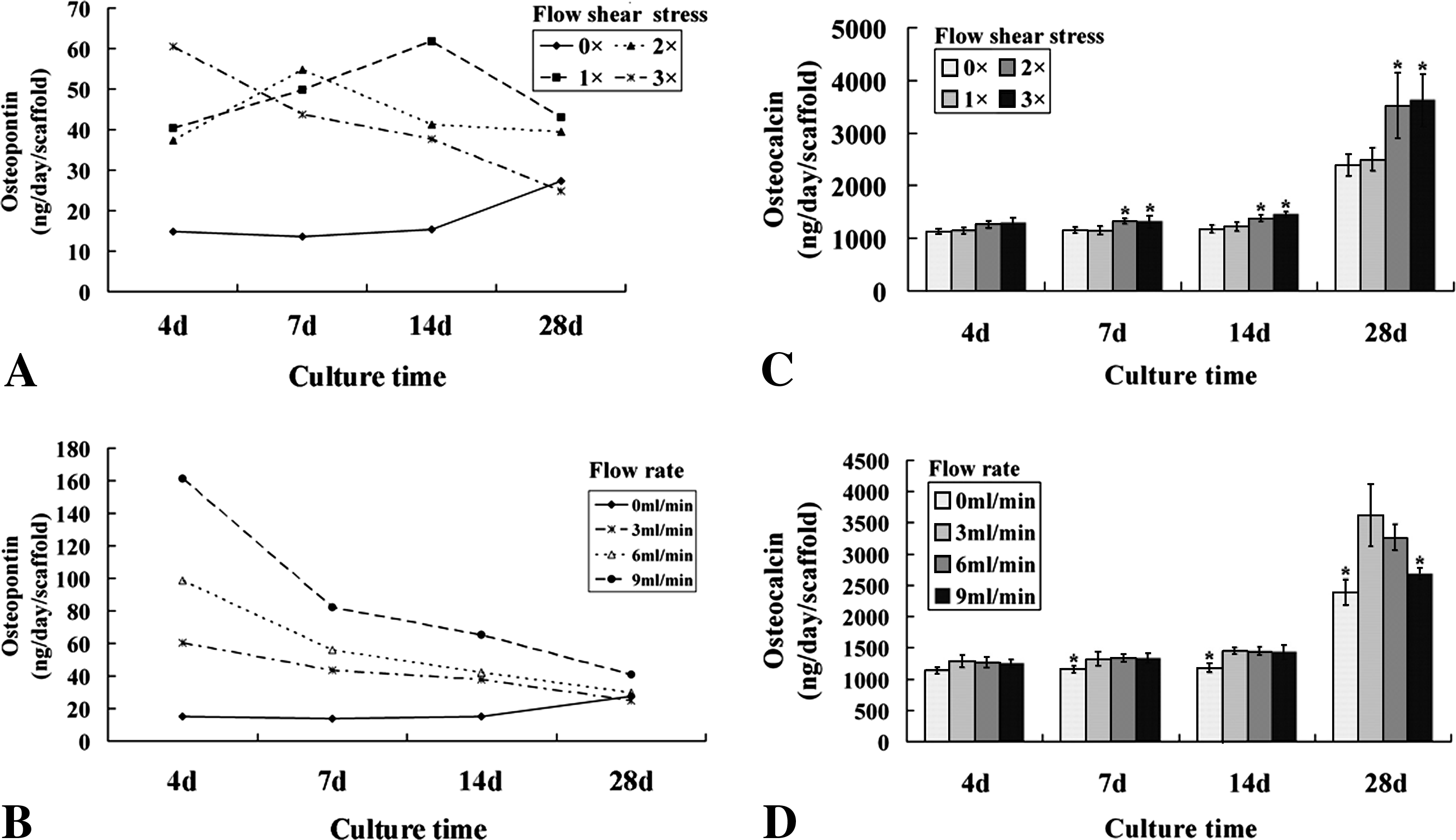
The daily secretion of osteopontin (
Osteocalcin measurement
Osteocalcin is the major noncollagenous protein of the bone matrix. It is synthesized in the bone by osteoblasts. Hence, osteocalcin is the late-stage marker of osteogenic differentiation. The free osteocalcin level can reflect the rate of bone formation. When the flow rate was 3 mL/min, the free osteocalcin in both 2 × and 3 × groups was higher (p < 0.05) than that in the other groups at days 7, 14, and 28 (Fig. 5C). When the flow shear stress was the same, the free osteocalcin in the 3 mL/min and 6 mL/min groups was higher (p < 0.05) than that in the other groups at day 28 (Fig. 5D). The secretion of osteocalcin in all groups dramatically increased after 2 weeks.
Scanning electron microscopy
SEM showed that hBMSCs adhered to the macroporous internal surface throughout the entire β-TCP scaffold after 4 h of incubation (Fig. 6A–C). After 28 days of culture, the ECM was only distributed in two to three pores near the exterior scaffold surface in the static culture (Fig. 6D). However, the ECM was distributed throughout the entire β-TCP scaffold after 28 days of perfusion culture. When the flow rate was 3 mL/min, the increasing flow shear stress resulted in an increased formation of ECM (Fig. 6E–G). When the flow shear stress was the same, the increasing flow rate resulted in a slight decreasing volume of ECM that was more compact (Fig. 6G–I). When ECM was further magnified, SEM showed that a few porous structures formed in the static culture (Fig. 6J), and many porous structures formed in the perfusion culture with 3 and 6 mL/min of flow rate (Fig. 6K–N). However, no porous structure was found when the flow rate was 9 mL/min, and the ECM was compact (Fig. 6O). In all constructs, sphere-like crystal structures were observed that are indicated in Figure 6P with a white circle. Energy dispersive X-ray analysis showed that these sphere-like structures consisted of calcium phosphate, which is depicted in Figure 6Q.
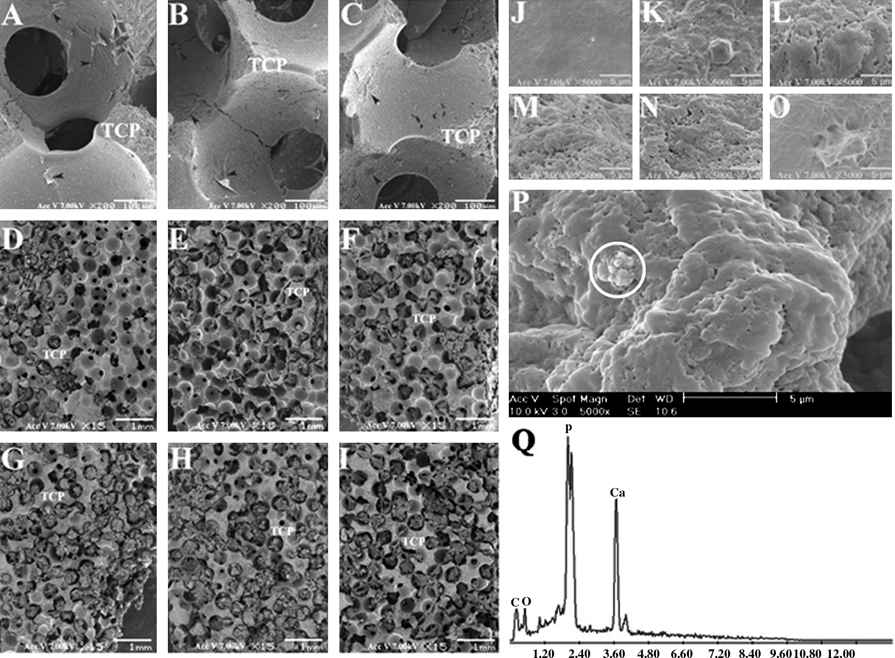
Representative scanning electron microscopy images of the cells/β-TCP construct. After 6 h inoculation, hMSC (black arrowheads) uniformly adhered to the inside (
Histological observation and histomorphometry
Histological sections stained with Steven blue and van Gieson picro-fuchsin demonstrated that the hBMSCs were sparsely distributed on most macropore's surface in the entire scaffold after 4 h of incubation (Fig. 7A–C). After 28 days of culture, the ECM was found in all groups. The distribution of ECM was only located in two to three pores near the exterior scaffold surface in the static culture (Fig. 7D). The ECM was distributed throughout the entire β-TCP scaffold in the perfusion culture. When the flow rate was 3 mL/min, the increasing flow shear stress resulted in an improvement of ECM distribution from the inside to the outside of the β-TCP (Fig. 7E–G). This is in agreement with previous studies that used titanium fiber meshes or collagen sponges.6,8,19,20 After 28 days of culture, a small amount of mineralized ECM formed in the static culture. The mineralized ECM or new bone was more obvious in the perfusion culture groups with a flow rate of 3 mL/min. However, the increasing flow rate depressed the mineralization of ECM when the flow shear stress was the same (Fig. 7G–I). Histomorphometrical study indicated that there was more tissue in 2 × and 3 × groups than in 1 × group (p < 0.05) (Fig. 8A), and that there was more new bone in 2 × group than in 1 × group, and more in 3 × group than in 2 × group (p < 0.05) (Fig. 8B) when the flow rate was 3 mL/min. When the flow shear stress was the same, the tissue volume was similar in the three perfusion culture groups (p > 0.05) (Fig. 8C). However, the new bone was most in the group with the 3 mL/min of flow rate when the flow shear stress was the same (p < 0.05) (Fig. 8D).
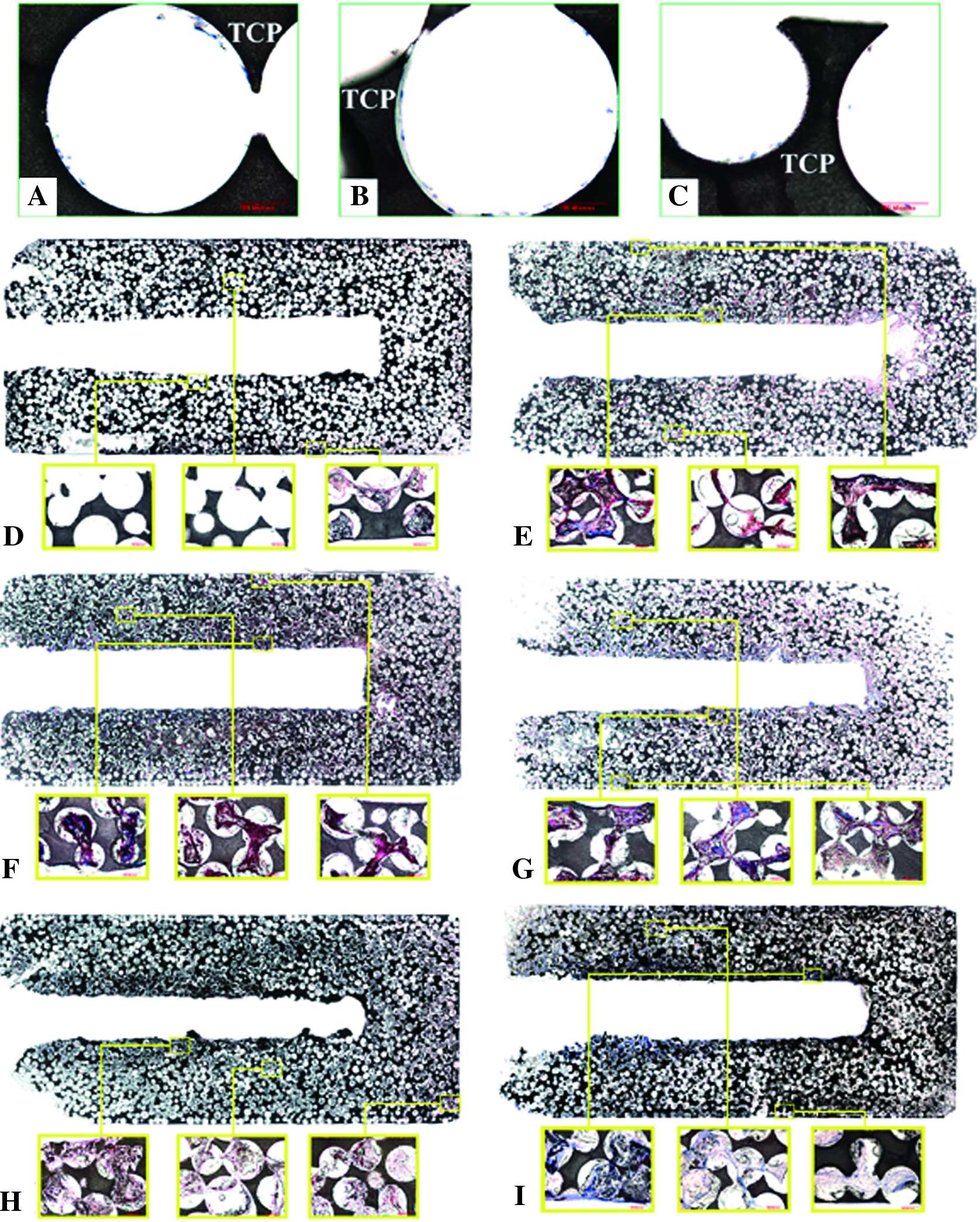
Histological sections of the cells/β-TCP construct stained with Steven blue and van Gieson picro-fuchsin. After 6 h inoculation, human mesenchymal stem cells (hMSCs) (blue) uniformly attached to the inside (
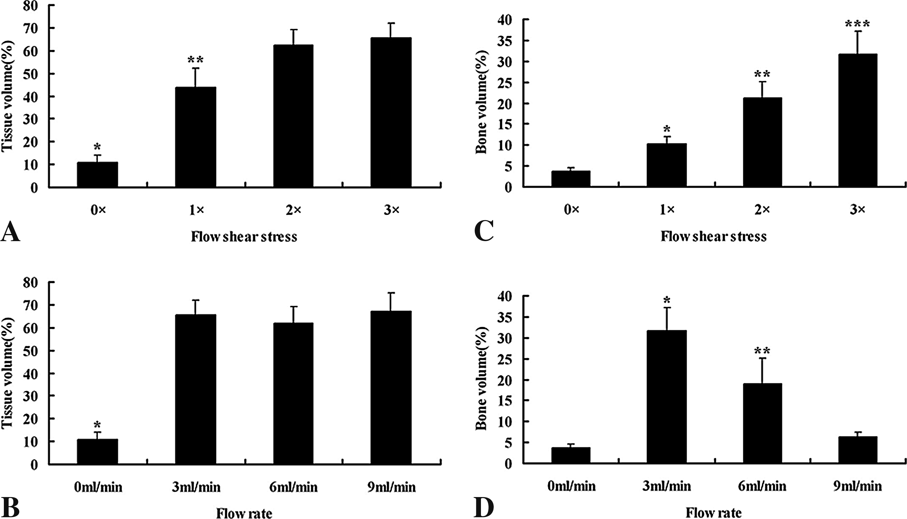
The percentage of tissue area/pore area (
Discussion
This study investigated the effects of flow shear stress and mass transport, respectively, on the construction of a large-scale tissue-engineered bone using a perfusion bioreactor. The results demonstrated that hBMSCs seeded on the critical-sized β-TCP scaffold and cultured in the perfusion bioreactor for a long period of time responded to fluid flow shear stress and mass transport. In this study, the dextran was added into the media to change the media's viscosity. According to the relationship among flow shear stress, viscosity, and flow rate in the Navier-Stokes equations, different flow shear stresses can be obtained with the same flow rate by changing the viscosity of the media. Moreover, different mass transport can be obtained with the same flow shear stress by changing the viscosity of the media. Hence, the effects of flow shear stress and mass transport on the construction of a tissue-engineered bone in the perfusion bioreactor can be investigated, respectively, through this approach.
Cell proliferation can be estimated by cell viability and glucose consumption. In previous studies, cell proliferation is measured through DNA assay. Although the quantity of DNA can directly reflect the cell number, DNA of the cells embedded in a mineralized matrix cannot be completely released into the solution. Hence, the cell proliferation in a tissue-engineered bone can be better estimated by cell viability and glucose consumption. Previous studies showed different findings on the effects of flow shear stress on cell proliferation.20–23 In this study, the flow shear stress in the 2 × group with 3 mL/min of flow rate improved the proliferation of hBMSCs. However, when the flow shear stress further increased, the proliferation of hBMSCs decreased. The result was in accordance with previous reports. 22 Thus, when the mass transport was already adequate for the proliferation of hBMSCs, the cells' biological behavior mainly responded to the flow shear stress. Moreover, the flow shear stress benefiting the proliferation of hBMSCs had an appropriate range. In this study, the appropriate flow shear stress was found in the 2 × group with 3 mL/min of flow rate. In addition, 3–9 mL/min of mass transport did not result in a change in the proliferation of hBMSCs when the flow shear stress was the same. In this study, the results suggested that the 3 mL/min flow rate was already adequate for the proliferation of hBMSCs seeded on the critical-sized β-TCP scaffold and cultured in the perfusion bioreactor. Moreover, the cell viability and the glucose consumption were lower in the static culture than in the flow perfusion culture. The main reason is that the cells only survived and proliferated around the scaffold periphery, and that the cells inside the scaffold died because of the poor supply of nutrients and the accumulation of metabolic products.
In this study, the osteogenic differentiation of hBMSCs seeded in the β-TCP scaffold responded to flow shear stress and mass transport in a dose-dependent manner. AP activity is an important marker of early osteogenic differentiation and the commitment of mesenchymal stem cells toward the osteogenic phenotype. 24 Increasing the flow shear stress resulted in an increased AP activity. The result was not in complete agreement with that of previous studies. 20 The different genus of MSC, the different flow shear stress caused by the different microarchitecture of the scaffolds, and the different scaffold materials may be the reasons for the incongruent results. When the flow shear stress was the same, the AP activity first increased and then decreased, while the mass transport increased. The decreasing AP activity was perhaps related with the high hydrostatic pressure caused by the increasing mass transport. Previous studies have demonstrated that hydrostatic pressure is an important inductor benefiting chondrogenic differentiation.25–27 However, the hydrostatic pressure did not significantly influence the osteogenic differentiation of hBMSCs.25,26 Moreover, increasing mass transport decreased the local concentration of cytokines around the cells, which perhaps also affected cell osteogenic differentiation.
With the exception of AP activity as the early marker of osteogenic differentiation, the expression of osteopontin and osteocalcin can be measured as the mid-stage and late-stage markers. Osteopontin is an extracellular protein that is produced by matrix-producing osteoblasts and bone-resorbing osteoclasts. 28 With hBMSCs cultured under flow conditions, there was a temporal pattern in the rise-and-fall expression of osteopontin 24 with peak levels occurring at day 4 for the higher flow shear stress, at day 7 for the middle flow shear stress, and at day 14 for the lower flow shear stress. This suggested an acceleration in the osteogenic differentiation of the perfusion-cultured hBMSCs seeded on the β-TCP scaffold that depended on the increasing flow shear stress. At day 28, the quantity of osteopontin in the perfusion culture has decreased and was even similar with the static culture. We attribute this phenomenon to the fact that osteopontin is a calcium-binding protein and will therefore be sequestered within the mineralized matrix of the construct. 29 Additionally, the mineralized ECM prevents the physical release of osteopontin. When the flow shear stress was maintained constant, the patterns of secretion of osteopontin in the perfusion culture with different mass transport were similar. Moreover, increasing the mass transport improved the expression of osteopontin. This result was not in agreement with that of AP activity.
As a late-stage marker of osteoblast maturation,24,30 osteocalcin dramatically increased after 14 days of culture in both static culture and perfusion culture. This result suggested that hBMSCs had differentiated into mature osteoblasts after 14 days of culture in accordance with previous studies. 6 Moreover, the expression pattern of osteocalcin was in accordance with that of AP. These data demonstrated that the increasing flow shear stress benefited osteogenic differentiation and the maturation of hBMSCs. When the flow shear stress was the same, the level of osteocalcin was lower in the higher mass transport than that in the lower mass transport at day 28. This result was in agreement with that of AP activity. The reason may be that the higher hydrostatic pressure derived from the faster mass transport influenced the expression of osteocalcin.
The SEM and histological sections showed the characteristics of the holistic view and microscopic view of the large-scale tissue-engineered bone constructed in vitro. When the mass transport was the same in the perfusion culture with higher flow shear stress, the mineralized ECM was distributed well from the inside to the outside of the scaffold. One reason was that the increasing flow shear stress improved and accelerated the osteogenic differentiation of the hBMSCs seeded on the scaffold, as discussed above. Another reason may be that the higher flow shear stress pushes the inside ECM outside of the scaffold. At a flow rate of 3 mL/min, the mineralization of ECM was obvious, and thus a new bone can be observed. However, when the flow shear stress was the same, increasing the mass transport depressed the mineralization of the ECM. It is in agreement with the expression of osteocalcin and AP activity. We presumed that this phenomenon was mainly caused by the faster mass transport and high hydrostatic pressure. As we know, a high hydrostatic pressure can improve the chondrogenic differentiation of cells.25–27 Moreover, the hydrostatic pressure is in positive proportion to mass transport according to the Navier-Stokes equations. Additionally, a faster mass transport does not benefit the local assemble of all kinds of cytokines and ions that are essential for the mineralization of ECM.
According to the above discussion, the cell proliferation was lower in 3 × group than in 2 × group with 3 mL/min of flow rate. However, the bone volume was highest in 3 × group with 3 mL/min of flow rate. These data agree that there appears to be a benefit in using a higher shear stress even though proliferation and glucose consumption are decreased. This would match with the fact that during culture, cells slow proliferation as they begin to differentiate. This study primarily investigated the effects of flow shear stress and mass transport, respectively, on the construction of a large-scale large tissue-engineered bone. To further optimize the fluid flow microenvironment and predict the appropriate perfusion parameters for different specification of scaffolds, we will establish a 3D mathematical geometrical model, simulate the distribution of flow field in the critical-sized β-TCP scaffold.
In conclusion, this study demonstrated that a large-scale tissue-engineered bone can be constructed in vitro by seeding and culturing hBMSCs on the critical-sized β-TCP ceramic scaffold in a perfusion bioreactor. The two fluid flow parameters, flow shear stress and mass transport, were important biological factors in the construction of the large-scale tissue-engineered bone. There was an individual appropriate range for flow shear stress and mass transport in the construction of a large-scale tissue-engineered bone. In this study, the large-scale tissue-engineered bone could be better produced in the perfusion bioreactor with about 0.015 Pa (0.013–0.018 Pa) of fluid flow shear stress and 3 mL/min of mass transport. Hence, when the tissue-engineered bone was constructed using scaffolds with different microarchitecture and shape, the flow shear stress and mass transport should be adjusted to benefit the construction of the large-scale tissue-engineered bone by changing the perfusion rate and viscosity of the media.
Footnotes
Acknowledgments
This research was accomplished in Shanghai Key Laboratory of Orthopaedic Implant (08DZ2230300). This research was supported by the Key Project of International Science and Technology Cooperation (2005DFA30120), the National Basic Science Research Program of China (973 Program) (2005CB522700), the National Natural Science Foundation of China (30600629), Specialized Research Fund for the Doctoral Program of Higher Education (200802480070), and the Ph.D. Programs Foundation of Shanghai Jiaotong University, School of Medicine (BXJ0821).
Disclosure Statement
No competing financial interests exist.