
Abstract
The aim of this study was to investigate the effect of implant location on bone formation in goats using autologous bone marrow–derived stromal cells in porous calcium phosphate scaffolds. Intramuscular locations were compared to posterolateral spine fusion locations in eight goats. As scaffolds, we used biphasic calcium phosphate porous blocks of 5 × 5 × 5 mm. Cell-seeded implants were compared to empty controls. Bone marrow–derived stromal cells were seeded at 8 million cells per cm3 scaffold and cultured for 1 week. The follow-up time was 12 weeks. Fluorochromes were administered intravenously at 4, 6, and 8 weeks. Ectopic implants showed 21 ± 3.6% bone formation for the cell seeded and 2.0 ± 3.0% for the controls (p < 0.001). Paraspinal implants, however, showed 0.10 ± 0.13% in the cell seeded compared to 0.023 ± 0.027% in the control group (p = 0.09). A benefit of the cells was only found in the area closest to the paraspinal muscles (p < 0.01). Bone formation in the control samples was of later onset compared to the cell-seeded implants. In conclusion, cell-based bone tissue engineering in an ectopic environment was clearly effective. Similar constructs implanted in a posterolateral spine fusion location hardly showed any effect.
Introduction
Sheep and goat segmental defect studies represent the majority of potentially clinically relevant cell-based bone tissue engineering publications. These involve defects in the cranium, 5 mandibula,6,7 femur, 8 tibia,9,10 and metatarsals.11–13 Successful TE posterolateral spine fusions (PLF), however, appear to be more challenging.14,15 PLF relies on the formation of a paraspinal bone mass, with the formation of a bone bridge in an area that is normally occupied by soft tissues. This model may therefore be considered an ectopic as well as an orthotopic model. Part of its vascularization is derived from the surrounding paraspinal muscles in addition to the decorticated host bone.
Histological studies in rabbits, undergoing noninstrumented PLF surgery, show that a process of delayed vascularization takes place in the central zone of the healing fusion mass, in between the transverse processes. 16 A delayed influx of bone marrow–derived osteoprogenitor cells into the center of the fusion mass may contribute to a nonunion rate that is similar to the 44% of nonunions that is seen in the noninstrumented PLF in the clinical situation. 17 Whether this phenomenon is similar in goats is unknown; however, the rate of neovascularization in these large animals is not expected to be faster than in rabbits. What is known from literature is that goat bone has a very similar composition and a similar bone remodeling compared to human bone. 18 With a repeated success in ectopic (intramuscular) bone formation19–21 and with a benefit in the speed of osteointegration 22 for cell-seeded implants compared to empty controls in true orthotopic locations (os ilium), the PLF location in these animals may give more insight in the exact working mechanisms of cell-based bone tissue engineering. The PLF model can be considered both an orthotopic as well as an ectopic model 17 since this model has a host osteoconduction effect on one side (decorticated host bone) and cell- or scaffold-based osteoinduction potential on the other side (overlying paraspinal muscles). This would, theoretically, limit the overwhelming osteoconduction effect on the potential (early) benefits of these implanted constructs compared to the (circumferentially present) os ilium model as we observed in our group previously.21,22 The (limited?) role of the seeded cells may therefore be more clearly observed in this model compared to the critical-sized os ilium model.
The main purpose of this study was to observe the effect of implant location using cell-seeded and control scaffolds, not to achieve a mere solid spine fusion. The latter would necessitate a stiff spinal instrumentation construct 23 using, for example, pedicle screw instrumentation. 24 This instrumentation has a bulky nature that does not, in our opinion, allow as much of a direct contact between the paraspinal muscles to the scaffold compared to a noninstrumented PLF model. Since Hurley et al. already showed in 1959 the importance of these muscles for successful fusion, 44 we considered this to be an essential potential contributor to successful bone formation in the PLF location and thus making a noninstrumented PLF model most optimal for our study purpose.
In summary, the aim of this study was to investigate the influence of implant location on cell-based osteogenesis. The well-established intramuscular ectopic implantation model in the goat was used for comparison to the potential of this technique in a noninstrumented PLF location.
Materials and Methods
Experimental design
A group of eight adult Dutch milk goats was included in this study after approval from the local animal care committee. Each goat was subjected to the aspiration of 8 mL of peripheral blood per kg body weight (to produce autologous serum) and a 25–30 mL aspirate of iliac crest–derived bone marrow (for cell culture, cryopreservation, and subsequent seeding of the cells on the implants). In a second procedure, goats received autologous cell-seeded implants (TE group) and control implants without cells (no cells [NC] group) in ectopic intramuscular locations (paraspinal muscles) and in PLF sites at levels L1–L2 and L4–L5. TE and NC implants were positioned on alternating levels.
Scaffold material
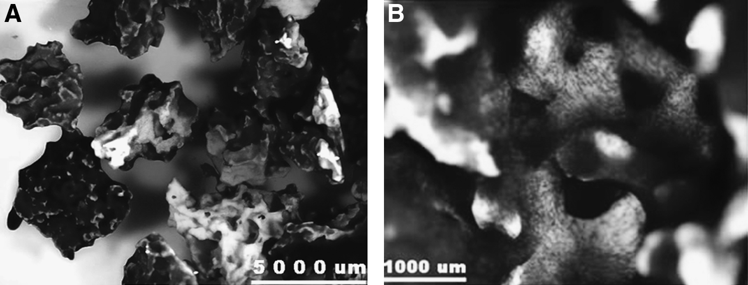
Porous ceramic cubes (5 × 5 × 5 mm) from a biphasic calcium phosphate (BCP; IsoTis SA, Bilthoven, The Netherlands) were used, containing 20 ± 5% tricalcium phosphate (TCP) and 80 ± 5% hydroxyapatite (HA). 25 X-ray diffraction and Fourier Transform Infrared spectroscopy confirmed the biphasic nature without impurities. The material had a porosity of 60–70% and a mean pore size of 350 μm. Sintering temperature of the material was 1200°C. Before use, the cubes were cleaned in an ultrasonic bath and steam-sterilized at 121°C for 30 min. This material was previously shown to be osteoinductive when implanted ectopically with varying success rates depending on the animal species used.20,25–27
Cell culture and scaffold seeding
Autologous serum was obtained after the blood was allowed to clot overnight in sterile bottles. Serum was collected and centrifuged at room temperature for 10 min at 2500 g. Heat inactivation was followed for 30 min at 56–58°C before a second centrifugation step. The heat-inactivated serum was then sterilized by passing it through 0.22 μm filters and subsequently stored at −20°C.
Goat bone marrow aspirates were cultured as described previously. 28 In short, cells were resuspended with the culture medium that was supplemented with 1 ng/mL basic fibroblast growth factor (Instruchemie, Delfzijl, The Netherlands) before plating at 5 × 10⁵ nucleated cells per cm2. The medium was refreshed twice weekly, and cells were trypsinized at confluency using 0.05% trypsin in ethylenediaminetetraacetic acid (Sigma, Zwÿndrecht, The Netherlands). Cells were replated at 5000 cells/cm2 in the next passages. After passages 1–2, all cells were cryopreserved for several weeks. Ten days before implantation, the cells were thawed and replated in the standard culture medium without fetal bovine serum but with the addition of autologous goat serum as described previously. 21 After 3 days of culture, cells were detached and Trypan Blue vitality staining was performed upon counting. 29 Subsequently, 100 μL of a 10 × 106 viable cells/mL suspension was statically seeded onto each scaffold using a cell droplet technique. This corresponded to approximately 8 × 10⁶ cells per cm3 of scaffold material. After 2 h, the differentiation medium was added containing 15% v/v autologous serum, and 10 nM dexamethasone and 10 mM β–glycerophosphate (both from Sigma). Control implants without cells were incubated under the same culture conditions and for the same culture time of 7 days. Additional implants were seeded for stereomicroscopy imaging of cell attachment to the constructs using methylene blue staining. Based on previous work, using the same culture conditions and seeding concentration in the same BCP scaffolds, a 49.1 ± 14.7% seeding efficiency and a cell count of 8.7 ± 3.7 × 10⁶ cells/cm3 were found before implantation after 1 week of static seeding conditions. 21
Surgical procedure
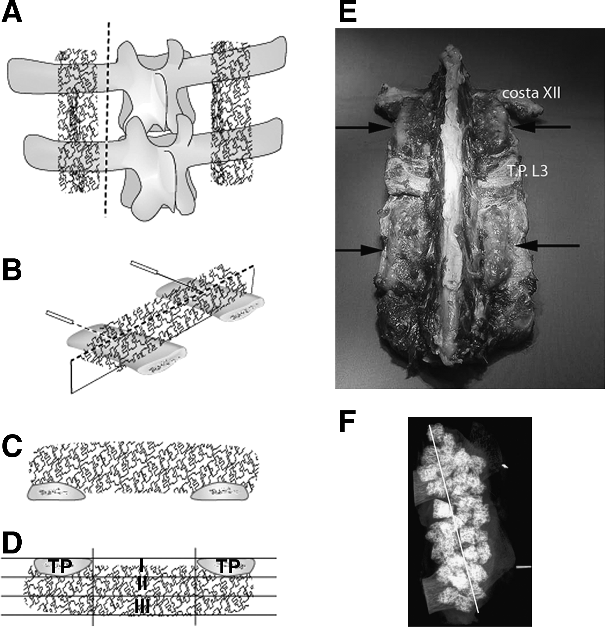
All surgeries were performed under standard conditions. 19 Antibiotic prophylaxis (Clamoxyl 1 g/70 kg body weight) was given perioperatively. A dorsal midline skin incision was made from T10 to the lumbosacral junction. Ectopic implants (one scaffold for each condition) with and without cells were inserted in intramuscular pouches as previously described. 19 The transverse processes of L1–L2 and L4–L5 were exposed through parallel (Wiltse type) incisions, leaving the facet joints and the laminae unaffected. 15 The middle 2 cm of the bilateral transverse processes were decorticated using a high-speed burr under continuous saline irrigation. Bone remnants were washed away before the addition of 10 cc of graft material (= 50 cubes, now referred to as the paraspinal implants) on each side of the spine for each level. The cubes were carefully positioned across the denuded areas of both transverse processes and in the area in between, on top of the intertransverse membranes (Fig. 1). The ectopic implants were introduced into the dorsal paraspinal muscles using several stab incisions and blunt spreading with scissors. The muscle fascias in all locations were closed using a nonresorbable suture to easily relocate the surgery site upon explantation. The level was alternated for each condition per goat. Postoperatively, buprenorfine 0.3–0.6 mg (Temgesic®; Shering-Plough, Oss, The Netherlands) was administered intramuscularly for pain relief. Fluorochrome labels (Sigma) were administered intravenously: at 4 weeks, 10 mg/kg Calcein Green; at 6 weeks, 35 mg/kg Alizarin Red; at 8 weeks, 75 mg/kg Xylenol Orange. After 12 weeks, the animals were killed by an overdose of pentobarbital (Euthesate®; Organon, Oss, The Netherlands) and potassium chloride.
Sequence of chronological events (
Postmortem sample acquisition and histology
Ectopic implants were excised and fixated in phosphate buffered 1.5% glutaraldehyde. The L1–L2 and L4–L5 spinal segments were excised entirely. The paraspinal implants were assessed by radiograms to study any potential bone or callus formation visible by X-rays. The transverse processes with the paraspinal implants were separated en bloc from the vertebral bodies in the sagittal plane (Fig. 1A). The samples were fixated in cold Karnovsky fixative containing 4% v/v paraformaldehyde and 5% v/v glutaraldehyde for at least 1 week.
All implants were dehydrated in alcohol series and embedded in polymethyl-methacrylate. Semithin sections of approximately 10 μm thicknesses were made on a Leica sawing microtome (Leica, Nussloch, Germany). Basic fuchsine and methylene blue were used for staining the sections for light microscopy; additional sections were left unstained for fluorescence microscopy (E600; Nikon, Tokyo, Japan).
After mounting the paraspinal implants on a sample holder, sections through the middle were made in the sagittal plane (parallel to the spine of the goat, Fig. 1B). Using size-calibrated radiograms with two metal clips, the center-implant line was determined and the polymethyl methacrylate (PMMA) blocks were grinded and sectioned parallel to this line in the sagittal plane, resulting in slides as shown in Figure 1C. For the ectopic implants the midsections were determined and used after sectioning the entire implants.
Histomorphometry
After 180° rotation for the paraspinal slides (Fig. 1D), to bring the transverse processes at the top level of each slide, all sections were digitized using a Nikon D100 digital camera (300 dpi), blinded, and loaded into Adobe Photoshop. For the ectopic implants, pores that were open at the exterior contour of the single implants were crossed with straight lines to obtain the area of interest. The paraspinal masses were fully captured, including some surrounding fibrous and muscular tissue and part of the transverse processes that was in contact with the individual implants. To increase the contrast, the images were pseudo colored before analysis with a customized KS400 histomorphometry program (Zeiss, Munich, Germany).
Slides, as displayed in Figure 1D, were divided into a central zone, in between the transverse processes, and the cranial and caudal outer zones that are in direct contact with the transverse processes. 30 The central zone was the area of interest. A custom-built macro was used to analyze bone formation in the central zone. Three areas of interest were determined within the central zone for subgroup analysis: the intertransverse zone (between the transverse processes), the intermediate zone (the most central area), and the submuscular zone. The intertransverse and intermediate zones were defined based on the individual thicknesses of the transverse processes of each goat; the submuscular zone comprised the remainder of the implants up to the fascia of the overlying paravertebral musculature.
All implants were analyzed for bone–scaffold contact length percentage (Cont. % = percentage of total available scaffold contact length occupied by the contact with the newly formed bone).19,31 In standard nomenclature 32 this could be formulated as
where Bd = boundary, B = bone, BCP = BCP scaffold, and Pm = perimeter.
Statistics
Ectopic bone formation was analyzed by comparing the TE to the NC condition using the Student's t-test for paired samples. For the paraspinal implants a nonparametric test was used since, in contrast to the ectopic implants, these data were not normally distributed. The Friedman paired-ranks test and post hoc Wilcoxon signed-rank test for comparisons between the three zones were used (level of significance at p < 0.05).
Results
In vitro results
For all goats, the adherent cell population proliferated well both before and after cryopreservation. After seeding on the BCP scaffolds, methylene blue cell staining revealed that confluent cell layers were present on the outer aspects and the inner contours of the open pores of the implants, as far as these were visible by stereomicroscopy. However, minimal amounts of cells were present in areas that had been in contact with the deep-well plates (bottom side) (Fig. 2A, B).
(
In vivo results
General
There were no surgical complications, and all animals remained in good health during the course of the experiment. Macroscopically, the intramuscular samples were well incorporated into the surrounding muscle bed. The paraspinal implants were solidly grouped but not as firmly integrated with the overlying muscles as the ectopic implants (Fig. 1E). Macroscopically and microscopically there were no apparent signs of resorption of the material used since the shape and size of the implants was well maintained upon explantation, similar to the results in other animals at 3 months after implantation. 27 Radiograms showed no signs of callus or bone formation between the implants in any of the goats in either treatment group (Fig. 1F).
Ectopic implants
Histology showed newly formed bone in 8/8 goats in the cell-seeded implants (TE group) and in 4/8 goats in the control implants (NC group). Although not quantified, vascularization was observed in sections from all of the implants regardless of the presence or absence of new bone formation.
In the TE group, an even distribution of the newly formed bone had developed throughout the implants, although no bone was found on the outer surface of the implants that was in direct contact with the surrounding muscles. In 2/8 goats, an impressive amount of fat-like tissue in large areas in the center of the implants was observed (Fig. 3A) in contrast to the other goats (Fig. 3B). Fluorescent microscopy showed the presence of the 4, 6, and 8 weeks labels in all of the TE implants in the goats, always present in a time-related order, indicating that bone formation initiated before 4 weeks of implantation and progressed away from the implant surface area toward the center of the pores (Fig. 3C).

An example of the histology obtained from intramuscular implants (stained with methylene blue and basic fuchsine) from the cell-seeded condition (TE) showing in 2/8 goats fat-like tissue in the center of the implants (
In the NC group containing bone (4/8 goats), only the 8 weeks' label was observed, indicating that bone formation on these implants was of later onset compared to the TE implants.
Histomorphometry showed a bone–scaffold contact of 21 ± 3.6% for the TE and 2.0 ± 3.0% for the NC group, respectively (p < 0.001) (Table 1).
The paraspinal implant data (bottom) are summarized for the central zone I (intertransverse), zone II (intermediate), and zone III (submuscular). Note the difference in magnitude between the ectopic and paraspinal data. Areas indicated in gray represent significant differences between the TE and NC samples in the ectopic group (p < 0.001) and the paraspinal implants (p < 0.01).
TE, tissue-engineered; NC, no cells.
Paraspinal implants
Microscopically, the consolidation (packing density 33 ) of the paraspinal implants was often poor. Interimplant distances could sometimes reach up to 2.5 mm. Histology of the implants showed very limited amounts of bone formation. No signs of bone bridging from one transverse process (a histological sign of fusion) to the next were observed in any of the goats. The overall histology showed bone formation in the contact zones closest to the transverse processes and varying degrees of bone formation in the areas closest to the overlying muscles in both groups. In between, areas of fibrous tissue were observed. Although not quantified, within these areas no signs of vascularization were noted. This was in contrast to the areas of newly formed bone where signs of vascularization were observed.
In the TE group, the outer zones (as well as adjacent areas in the central zone) showed an area of bone in contact with the decorticated host bone (Fig. 4A, vertical arrows). This bone, sometimes incorporating entire individual implants, had a trabecular-like structure similar in aspect to the transverse processes. This was clearly different from the woven-like bone structure that was formed in the intertransverse zone (within the central zone) not containing any central bone marrow depositions such as seen in the transverse processes (Fig. 4A, asterisk). Further away from the transverse processes toward the overlying paraspinal muscles, there was an area in all of the goats without bone where implants were surrounded by fibrous tissue. There was never bone in continuity from the transverse processes reaching into the submuscular zone (toward the paraspinal muscles). In the submuscular zone, a distinct cluster of particles containing newly formed bone was observed (8/8 goats) in contact with the overlying muscles, again with a woven-like bone structure (horizontal arrows in Fig. 4A). In comparison to the ectopic (intramuscular TE) group implants, no bone was found on the outer surfaces of these implants in contact with the overlying muscles or in between the implants. All three fluorescent labels were observed in both the intertransverse and intermediate zones. The submuscular zone showed also six out of eight goats with the earliest (4 weeks) and subsequent labels in the areas with woven-like bone (Fig. 4C).

Histological slide of a midsection through the paraspinal implants of a spinal segment treated with cell-seeded implants (TE group) in (
In the NC group a similar trabecular-like bone bridge between the implants and the transverse processes was seen as observed in the TE group (outer zones) (Fig. 4B, vertical arrows). Again, in most cases complete individual particles were incorporated into the transverse processes by trabecular-like bone formation. Further into the intermediate zone, also fibrous tissue was found between several of the implants. Similar to the TE group, bone was also present in the submuscular zone (5/8 goats) but clearly smaller in amount and in fewer particles compared to the TE group (outer and central zones). All three fluorescent labels were observed in both the intertransverse and intermediate zones where bone was present (always as a continuous bone bridge reaching from the transverse processes into these zones, similar to that observed in the TE group). However, none of the early labels (4 and 6 weeks) but only the 8 weeks' label was found in the implants in 5/8 goats (Fig. 4C). This indicates a later onset in osteogenesis in this area compared to the TE group.
Histomorphometry data on both the ectopic and paraspinal implants are shown in Table 1.
Discussion
A striking difference was observed in the extent of bone formation in the posterolateral spine location compared to the intramuscular location, especially for the cell-seeded group. Scaffolds (with or without seeded cells) in contact with the host transverse processes were abundantly covered with bone that appears to be derived from osteoconduction from the decorticated host bone. This concurs with results that were obtained with a transverse process cassette model using the same material.31,34 Scaffolds in between the transverse processes, where a potential fusion should take place, 35 were surrounded by fibrous tissue in both groups. Only the most ectopic location in the posterolateral spine, the area covered by the paraspinal muscles, showed signs of limited bone formation that may be derived from the seeded cells. The onset of the bone formation in this area is similar to that found in the intramuscular location based on the presence of the early and late fluorochromes, both for the cell-seeded and control scaffolds. This same phenomenon in this submuscular location was previously shown for both cell-seeded BCP scaffolds as vital autograft bone. 34
Studies on factors affecting bone graft incorporation
36
show that even for the most optimal bone graft, a fresh cancellous autograft, several important aspects are crucial for its success:
A mechanically stable environment. Early lifting of rabbits out of their cage (to promote motion at the arthrodesis site) after autogenous PLF surgery leads to significantly more nonunions than nonlifted animals.
37
The presence of adequate vascularization. Local irradiation may have significant limitations on the incorporation of a fresh autologous cancellous graft due to limited revascularization of the graft.
38
An intimate contact with the host bone. Spine fusion does not occur without decortication of the host bone.
39
It is the sum of these interactions that determines the success rate of a bone graft. Considering the current experiment, these items can be discussed in relation to the poor amount of bone observed in the posterolateral spine location:
Mechanical stability. Based on clinical results, alternatives to the use of autografts such as morcellized frozen or freeze-dried allografts are less successful in achieving PLF in humans,
40
whereas their success in acetabular or femoral reconstructions, where the graft is impaction grafted, is quite satisfying.41,42 Mechanical stability therefore could be an increasing prerequisite when bone grafts are less biologically active compared to cancellous autografts.
36
In our study, the mechanical stability was suboptimal since no instrumentation was used and the posterior elements, such as the facet joints and laminae, were left intact during surgery. Another mechanical factor could have been the poor consolidation (or packing density
33
) of the current 5 mm cubic particles as a graft. Compared to successful ovine posterolateral fusion studies, where 1–3 mm particles where used,
15
our implants had a much smaller intrinsic stability. The lower packing density, in large particle porous media,
43
could also have had consequence on the process of osteoconduction, which is challenged with larger interparticle distances. In addition, high packing density leads to a better coherence of the particle mass compared to large-sized particles where mechanical forces lead to increased individual particle movements.
43
The situation for the ectopic implants as investigated in this study is essentially different, as these were enclosed in an envelope of muscles. In addition, particles in between a network of parallel muscle fibers will cocontract with this network, whereas loosely packed particles in the posterolateral spine location will experience more shear and bending forces between the (relatively rigid) lumbar spine on one side and contracting muscles on the other side. Despite all this, successful fusions were observed after 6 months in a similar noninstrumented PLF study in sheep using 2–3 mm TCP implants exposed to (cell enriched) bone marrow seeding.
15
Vascularization. Vascular supply in fusion mass formation in the PLF site is mainly derived from the decorticated transverse processes, the pars interarticularis (mainly superior pars), the lamina,
35
and the overlying muscles.
44
Based on the paraspinal histology that showed clear trabecular-like bone outgrowth incorporating the implants that were in direct contact with the transverse processes both in the TE and NC conditions (Fig. 4A, B), a deficit in decortication can be considered unlikely. Vascularization from the area of the facet joint and lamina was not expected since these were intentionally not included in the implantation site. On the other hand, little intimate contact was observed with the overlying musculature and the graft. This was in contrast to the well-integrated ectopic implants. Although the fundamental question still remains as to why this was the case, it is likely that vascularization from the muscle side was not fully developed at this stage, possibly as a result of relatively increased motion in this area based on the previously mentioned arguments. Host bone contact. Decortication alone without grafting does not achieve successful fusion in rabbits.
39
Void space, that is, the physical absence of paravertebral musculature in the intended fusion site and the available graft porosity is deemed necessary to achieve a successful fusion.
44
In support of this, a flexible sponge-like, collagen scaffold in combination with recombinant human bone morphogenetic protein-2 is less successful in achieving spine fusion when compressed by the paravertebral muscles compared to a more solid (noncompressible) ceramic HA/TCP graft in a large animal (monkey) spine fusion model.
45
In the current study, this void space was more than sufficient based on our histology. However, the low packing density and subsequent little host–scaffold contact may have reduced the potential for new bone formation.
Overall, the mobile setting of the posterolateral spine in comparison to the solidly enveloped area intramuscularly in addition to the relatively loose packing of cell-seeded constructs could be part of the explanation for the approximately 100-fold difference in bone scaffold contact length between the two locations. In addition, for more optimal comparisons, the intramuscular location should ideally be implanted with several particles with interparticle dynamics that may negatively influence the osteogenic potential more similar to the posterolateral implant location in the spine in this study.
The follow-up time of 3 months for our goat model may be short. Gupta et al. found an increase in fusion rate in the cell-enriched TCP implants between the 3 and 6 months follow-up. 15 Others showed that HA/calcium carbonate constructs without cells in the ectopic location in a nonhuman primate model failed to show any osteoinduction at 60 and 90 days but a striking 25% bone fill in the 13% HA/calcium carbonate group after 365 days of follow-up. Similarly, in the orthotopic calvarial location, a sixfold increase was seen between the 60 and 365 days of follow-up for the same type of implants with an average of 49% bone fill at 365 days in the same study. 46
Finally, cell concentration on the implanted scaffolds may be of crucial importance to the fusion rate in the posterolateral implant location. In rabbits this was shown to be a relevant difference for the concentrations of 1 × 10⁶ bone marrow cells/mL (all seven rabbits has nonunions) compared to 1 × 10⁸ bone marrow cells/mL (five out of seven rabbits achieved a solid fusion) on a type I collagen sheet for 6 weeks. 47
Conclusion
Ectopic implants showed a clear effect of the seeded cells with 21 ± 3.6% bone–scaffold contact for the TE and 2.0 ± 3.0% for the NC group (p < 0.001). Paraspinal implants, however, showed 0.10 ± 0.13% in the cell seeded compared to a 0.023 ± 0.027% in the control group with p = 0.09. A benefit of the cell seeding was observed only in the area closest to the overlying paraspinal muscles (p < 0.01). The fluorochrome labels revealed a BCP surface-derived process of bone formation. Bone formation in the control samples was of later onset compared to the cell-seeded implants. In conclusion, cell-based bone tissue engineering in an ectopic environment was clearly effective. Similar constructs implanted in a PLF location hardly showed any effect at all.
Footnotes
Acknowledgment
The authors acknowledge The Netherlands Technology Foundation (grant number UGN.4966) for financial support of this study.
Disclosure Statement
No competing financial interests exist.