
Abstract
Bioprinting by the codeposition of cells and biomaterials is constrained by the availability of printable materials. Herein we describe a novel macromonomer, a new two-step photocrosslinking strategy, and the use of a simple rapid prototyping system to print a proof-of-concept tubular construct. First, we synthesized the methacrylated ethanolamide derivative of gelatin (GE-MA). Second, partial photochemical cocrosslinking of GE-MA with methacrylated hyaluronic acid (HA-MA) gave an extrudable gel-like fluid. Third, the new HA-MA:GE-MA hydrogels were biocompatible, supporting cell attachment and proliferation of HepG2 C3A, Int-407, and NIH 3T3 cells in vitro. Moreover, hydrogels injected subcutaneously in nude mice produced no inflammatory response. Fourth, using the Fab@Home printing system, we printed a tubular tissue construct. The partially crosslinked hydrogels were extruded from a syringe into a designed base layer, and irradiated again to create a firmer structure. The computer-driven protocol was iterated to complete a cellularized tubular construct with a cell-free core and a cell-free structural halo. Cells encapsulated within this printed construct were viable in culture, and gradually remodeled the synthetic extracellular matrix environment to a naturally secreted extracellular matrix. This two-step photocrosslinkable biomaterial addresses an unmet need for printable hydrogels useful in tissue engineering.
Introduction
Bioprinting and the development of extrudable biomaterials are rapidly expanding research areas that may provide solutions to these challenges. However, many technological hurdles exist to print a functional organ. Paramount among these challenges are the need to recreate the complex cellular organization within the neotissues of an engineered organ, and the need to create a vascular network within the construct that can be functionally connected to the recipient. 4
The technique of bioprinting consists of two printable components. First, cell aggregates, cellularized synthetic extracellular matrix (sECM) hydrogels, or cell-seeded microspheres comprise the bioink. Second, the cell-free polymers that provide a scaffolding or substratum for the bioink are often referred to as the biopaper. Bioprinting allows the stepwise assembly of bioink and biopaper components into an organ-appropriate three-dimensional (3D) arrangements using a three-axis printer.5,6 Vascular networks can be printed by a scaffold-free process involving automated deposition of sausage-like cell aggregates and agarose tubes. 7 In each case, a computer-assisted design program can be used to guide deposition of precise geometries that mimic the structure of an actual tissue or organ. 8 After printing, the engineered construct is allowed to mature and gain functionality in a bioreactor or in vivo environment.7,9
To date, a complete organ has not been printed; the basic technologies are still in the proof-of-concept stage. Nonetheless, the field is advancing. Cell aggregates and cellular macrofilaments have been printed layer by layer into tubular formations within agarose,10,11 and then fused into singular seamless structures,12,13 showing the feasibility of printing vessels and other tubular structures. A printed vessel network or duct structure would constitute a significant milestone in bioprinting. Once a tubular structure can be built, most other biological structures are attainable, as they are comprised of a combination of tubular or hollow structures and simpler cell arrangements.14,15 A major hurdle has been that few biomaterials have been developed with design criteria specific to bioprinting.
For a biomaterial to function successfully in a bioprinting setting, it should at least meet three criteria. First, it must be mechanically suitable for printing, whether it be by drop deposition, extrusion from a syringe, or some other method. Second, the material should maintain its structural integrity after the deposition process. Third, the material must provide a cytocompatible environment before, during, and after deposition. Many materials fail to meet one or more of these criteria. Materials that are extrudable and maintain structural integrity often do so by using high cure temperatures or solvents for polymerization, and thus cannot be printed together with cells. Other more cell-friendly hydrogels lack the appropriate mechanical properties for printing.
A biocompatible sECM composed of the thiol-modified hyaluronic acid (HA) and gelatin derivatives, thiol-modified carboxymethyl hyaluronic acid (CMHA-S) and gelatin-3,3′-dithiobis(propanoic hydrazide) (DTPH), was developed to provide a microenvironment suitable for cell growth.16–18 These sECMs have proven to be versatile tools for wound healing and reparative medicine, including controlled release of growth factors for increasing angiogenesis, neovascularization, and vessel maturation.19–26 The ease with which 3D tissue culture can be performed in vitro and in vivo has made this biomaterial appropriate for new tissue engineering research applications such as development of bladder tissues, centrifugally cast vessel-like tubes, and tumor xenograft models for drug and discovery.14,18,27–32
Despite these many applications, however, the polyethylene glycol diacrylate (PEGDA)-crosslinked thiolated HA-based sECMs were found to be unsuitable for bioprinting. Because they could not maintain structural integrity during printing and would frequently clog the print heads, a new crosslinking chemistry was needed. Thus, we investigated the use of methacrylated HA (HA-MA), since the rate and degree of crosslinking could be easily controlled during photopolymerization. HA-MA has been used successfully in research on cutaneous and corneal wound healing, 33 embryonic stem cell expansion, 34 and drug and growth factor delivery.35–37 In addition, photo-crosslinked HA-MA provided a 3D microenvironment suitable for mesenchymal stem cells to differentiate into a chondrogenic phenotype. 38 However, most cells are unable to attach to HA-MA alone, thus limiting its utility as a biopaper for bioprinting applications.
Herein we describe the preparation of a new hydrogel composition and the use of a two-step crosslinking strategy to address the need for bioprintable materials. In addition to the HA-MA component, we synthesized a novel photocrosslinkable gelatin derivative, gelatin ethanolamide methacrylate (GE-MA). GE-MA was prepared in two steps. First, the abundant carboxylic acid groups of gelatin were converted to ethanolamide derivatives, analogous to the modification of gelatin carboxylates to thiol functionalities in gelatin-DTPH. 39 The primary alcohol functionalities of GE were then methacrylated, affording a higher degree of substitution by crosslinkable chemical groups than could be achieved by direct methacrylation of the lysine groups in gelatin. By combining HA-MA and GE-MA, we obtained a photocrosslinkable sECM that is easy to work with, biocompatible, and supports cell attachment.
To demonstrate the utility of the new hydrogel components for bioprinting, we have chosen to work with a simple, modular device that was developed as part of the open source Fab@Home project. This economical and versatile printing machine can print a variety of materials. Surprisingly, relatively few biologically relevant applications of this technology have been described. Several examples are the building of clear silicone heart and aorta models for educational purposes, and the printing of an ovine meniscus-like construct using alginate.40–42 We now show that the Fab@Home printing system can be used to print HA-MA:GE-MA hydrogels in the form of tubular constructs.
Materials and Methods
Synthesis of HA methacrylate
HA-MA was synthesized by modification of a literature preparation. 43 Thus, HA (1.0 g, Novozymes, 950 kDa) was dissolved in 100 mL of water, and a 20-fold excess of methacrylic anhydride (7.5 mL) was added. The reagent excess was calculated relative to a total of four hydroxyl groups per HA disaccharide unit. The solution was stirred overnight at room temperature, and the pH of the reaction mixture was maintained at 8.5 by adding 5 N NaOH. The resulting clear solution was adjusted to pH 7.0 and dialyzed against water for 24 h, with four changes of water during dialysis. The solution was frozen (−80°C) and lyophilized to obtain 0.85 g dry HA-MA (Fig. 1A). The structure of the product was confirmed by proton nuclear magnetic resonance 1 (H-NMR) (D2O) and a substitution degree of 5% was determined by integration of one of the two vinyl protons relative to methinyl (CHOH and CH2OH) proton resonances.

(
Synthesis of GE-MA
Gelatin (5.0 g; Sigma-Aldrich) was dissolved in 500 mL water at 37°C, and 3 mL neat ethanolamine was added with stirring. The pH was adjusted to 4.75, and then 1-ethyl-3-(3-dimethylaminopropyl) carbodiimide (EDCI, 1.0 g) was added; the pH of the stirred mixture was maintained at 4.75 for 4 h. The pH was then increased to 7.0 to stop the reaction, and the solution was dialyzed against water for 24 h, with four changes of water during dialysis. The resulting solution was frozen and lyophilized to obtain 4.1 g of GE.
Next, GE (1.0 g) was dissolved in 100 mL of water, and a 20-fold excess of methacrylic anhydride relative to GE hydroxyl groups (7.5 mL) was added. The pH of the reaction of mixture was adjusted to 8.5 by adding 5 N NaOH, and the solution was stirred overnight at room temperature while adding 5 N NaOH to maintain pH 8.5. The resulting clear solution was adjusted to pH 7.0 and dialyzed against water for 24 h, with four changes of water. The dialyzed solution was frozen and lyophilized to obtain 0.80 g dry GE-MA (Fig. 1B). The structure of the product was confirmed by 1H-NMR (D2O), and the molecular size of GE-MA was determined by gel permeation chromatography: Mw = 15.5 kDa, Mn = 11.2 kDa, and polydispersity = 1.4. The substitution degree was estimated to be 2.6% based on integration of the one of the vinyl protons to the combined aromatic protons, assuming complete conversion of the gelatin carboxyl groups to ethanolamide groups, and using relative amino acid compositions of 2.9% aromatic residues and 16% carboxylic acid residues in gelatin. 44 An essentially identical methacrylation protocol was used to convert gelatin to methacrylated gelatin (G-MA) as a comparison compound.
To synthesize G-MA, unmodified gelatin was methacrylated using the same protocol as that employed for the methacrylation of GE. Thus, 500 mg of gelatin was dissolved in 50 mL of water, and 3.75 mL of methacrylic anhydride was added. The remainder of the above protocol was followed to give 370 mg of dry G-MA. It is known that the (Z)- and (E)-vinyl protons of butyl methacrylate show downfield chemical shifts at δ 6.15 and 5.58, while those of butyl methacrylamide appear at δ 5.89 and 5.49. The 1H-NMR spectrum of GE-MA showed the distinctive presence of the vinyl protons of the GE-MA methacrylate esters at lower field than the vinyl protons of the G-MA methacrylamide functionalities modifying lysine residues. In the gelatin derivatives, the vinyl protons of G-MA were observed as complex multiplets centered at approximately δ 5.70 and 5.40, whereas those of GE-MA were observed at δ 5.95 and 5.50.
Preparation of hydrogels
Several formulations were initially tested to obtain hydrogels with suitable printing properties and hydrogel stability. In general, HA-MA was dissolved in 1× phosphate-buffered saline to form a 1.5% w/v solution. A second 1.5% w/v solution was prepared for cell attachment in which HA-MA was supplemented with GE-MA to give a final HA-MA:GE-MA ratio of 4:1 (w/w). Greater percentages of gelatin resulted in hydrogels with shear storage modulus values below 50 Pa. These gels were structurally weak and could not hold their shape after printing. An aliquot of the photoinitiator stock solution (2,2-dimethoxy-2-phenyl-acetophenone in N-vinylpyrrolidone, 300 mg/mL) was added at the rate of 10 μL per mL of hydrogel solution. Solutions were irradiated with ultraviolet (UV) light (365 nm, 180 mW/cm2) for at least 3 min to crosslink. For cell culture use, all solutions were sterile filtered through 0.45 μm filters (Millipore) before gelation. Standard 1% w/v Extracel™ hydrogels (Glycosan BioSystems) were prepared according to the manufacturer's directions for use as the hydrogel control. Thus, Glycosil and Gelin-S, the thiolated HA and gelatin components of Extracel, were dissolved in sterile degassed water to make 1% w/v solutions. Extralink, a PEGDA crosslinker, was dissolved in sterile degassed water to make a 2% w/v solution. Glycosil, Gelin-S, and Extralink solutions were then combined in a 2:2:1 volume ratio and mixed thoroughly. Gels typically formed within 30 min.
Rheology
HA-MA hydrogels with a concentration of 1.5% w/v were cast in 60 mm Petri dishes and tested as previously described, 45 with the difference that the hydrogels were irradiated at 365 nm for different time periods immediately before testing. Samples (n = 3) were tested after each subsequent irradiation period, resulting in cumulative exposure times of 30, 45, 60, 120, 180, 240, 300, 360, and 420 s. A 40-mm steel disc was lowered until it contacted the gel surface, and then G′ and G″, the shear storage and loss modulus, respectively, were measured using a shear stress sweep test ranging from 0.6 to 20 Pa at an oscillation frequency of 1 Hz applied by the rheometer (TA Instruments).
In vitro biocompatibility
The HA-MA:GE-MA hydrogels were used to encapsulate 25,000 cells per 100 μL hydrogel in tissue culture inserts (Corning) in 24-well plates. Human hepatoma cells (HepG2 C3A), human intestinal epithelial cells (Int-407), and murine fibroblasts (NIH 3T3) were evaluated. The cells were cultured with the minimum essential medium Eagle, basal medium Eagle, and Dulbecco's minimum essential medium (all Sigma), respectively, each containing 10% fetal bovine serum (HyClone). The medium was changed on day 3. Viability was then determined by quantifying mitochondrial activity using an 3-(4,5-dimethylthiazol-2-yl)-5-(3-carboxymethoxyphenyl)-2-(4-sulfophenyl)-2H-tetrazolium (MTS) assay 46 (Promega) immediately after encapsulation (day 0) and on days 3 and 7 (n = 4). Aliquots (100 μL) were removed and absorbance readings determined at 490 nm using an Optimax Tunable Microplate Reader (Molecular Devices). Absorbance levels are directly proportional to the number of live cells.
Three pilot studies were performed to assess the effects of UV irradiation and extrusion. HepG2 C3A cells were encapsulated in HA-MA:GE-MA hydrogels at an increased density of 25 million cells/mL to mirror bioprinting conditions. First, 100 μL aliquots of the cell suspension were subjected to irradiation for 0, 120, 120 + 60 (180), or 120 + 120 (240 s) (n = 3). Viability was determined using the MTS assay as described above. Second, 100 μL aliquots of the cell suspension (n = 3) were transferred to 48-well plates and irradiated for 120 s to form 100 μL soft gels. Third, 100 μL aliquots of the cell suspension were prepared for printing by irradiation for 120 s and then printed into a 48-well plates using the Fab@Home Model 1 printer (NextFab). Viability was assessed as described above.
In vivo biocompatibility
The HA-MA:GE-MA hydrogel (1.5% w/v) and the control Extracel hydrogel (1% w/v) were prepared as described above and injected (without cells) subcutaneously into the flanks of nude mice, with four injection sites per mouse. Two 100-μL hydrogels were injected into the anterior region, and two 400-μL hydrogels were injected into the posterior region following a protocol approved by the University of Utah Institutional Animal Care and Use Committee. A total of 12 mice were employed, n = 3 per time point. Mice were sacrificed at 2 and 4 weeks. The hydrogels and surrounding tissue were excised and fixed in 4% formaldehyde for 4 h. Samples were then dehydrated with graded ethanol washes, followed by Citrisolv (Fisher Scientific). Samples were paraffin embedded and sectioned at 4 μm. Sections were then stained with hematoxylin and eosin for histology, and slides were imaged under light microscopy to determine the presence or absence of unhealthy or inflamed tissue.
Bioprinting
The two hydrogel formulations, HA-MA alone and the 4:1 HA-MA:GE-MA blend, were prepared for printing as follows. First, each solution was adjusted to a physiological pH of 7.4 (1 M NaOH) and were sterile filtered. HepG2 C3A cells were cultured to confluency on tissue culture plastic in minimum essential medium Eagle with 10% fetal bovine serum, treated with accutase (Innovative Cell Technologies) to detach them from the substrate and counted. Then, an aliquot of the cell suspension was centrifuged to provide a cell pellet that was resuspended in the hydrogel solution. The cell density in the HA-MA:GE-MA solution was 25 million cells/mL. Solutions were irradiated at 365 nm for 120 s to partially crosslink the hydrogels. Subsequently, the soft hydrogels were taken up into 10-mL syringes in preparation for printing.
To print a cellular structure, a vertical ring-stacking protocol was used. Hydrogel-containing syringes were placed into the Fab@Home printing device. Using a 3D. STL file to represent the desired structure, the ring stacking protocol was implemented by the computer-controlled printing device, thereby building the construct layer by layer. First, a central cell-free HA-MA hydrogel was printed with a diameter of 1–2 mm. Second, a 2-mm-thick ring of cell-containing HA-MA:GE-MA hydrogel was deposited around the central disc. Third, an additional ring of cell-free HA-MA hydrogel was deposited around the first ring. Fourth, the printed rings were irradiated at 365 nm for an addition 60 s to further photocrosslink the gels, thereby both increasing the rigidity of each hydrogel as well as creating hydrogel-to-hydrogel crosslinks. Fifth, steps 1–4 were repeated for several iterations to build up a tube of cellularized hydrogel that was contained within a cell-free hydrogel. Each successive UV irradiation further glued the layers together. The medium was then added to each dish, and the constructs were placed in culture (37°C, 5% CO2) for 3 weeks to allow the constructs to mature. In several constructs, a fluorescent HA-BODIPY tracer (Invitrogen) was encapsulated in the cell-containing hydrogel to label the HA-MA:GE-MA portion of the construct for improved observation and imaging immediately after printing.
Histology and immunohistochemistry
After 3 weeks of culture, the medium was aspirated and constructs were fixed, sectioned, and embedded as described above. Masson Trichrome staining was accomplished utilizing a standard kit (Sigma), and slides were imaged under light microscopy to determine the presence of collagen. A cell-free HA-MA:GE-MA hydrogel was used as a negative control.
For immunohistochemistry (IHC), incubations were carried out at room temperature unless otherwise stated. Slides were deparaffinized and hydrated through Citrisolv and graded ethanol washes. Endogenous peroxidase activity was blocked with 1% hydrogen peroxide solution in 1× phosphate-buffered saline containing 0.1% Tween-20 (PBT) for 20 min. Antigen retrieval was performed on each slide by microwaving in 1% antigen unmasking solution (Vector Laboratories) for 20 min, and then left at room temperature for 30 min. IHC was performed using the Vectastain Elite ABC peroxidase kit (Vector Laboratories) according to the manufacturer's protocol. Briefly, nonspecific antibody binding was minimized by incubating sections in diluted normal blocking serum for 90 min. Sections were incubated overnight at 4°C in a humidified chamber with primary antiprocollagen antibodies (Lot #: LV1541013; Millipore) at a 1:500 dilution. After overnight incubation, slides were washed in PBT for 9 min (3 × 3 min). Sections were then incubated for 90 min with biotinylated secondary antibody solution diluted to 5 μg/mL in PBT, followed by Vectastain Elite ABC Reagent (Vector) diluted in PBT for 30 min. Between incubations, sections were washed for 9 min (3 × 3 min) in PBT. Immunoreactive regions were observed by incubating sections in the DAB peroxidase substrate kit (Vector) for 1–2 min. The sections were washed in nanopure H2O, counterstained with hematoxylin, dehydrated, and cover slipped. Positive control slides of previously sectioned epidermal and dermal tissue were used for comparison. Negative controls were performed in parallel with the primary antibody incubations and included incubation with blocking serum in place of the primary antibody. No immunoreactivity was observed in the negative control sections.
Statistical analysis
The data are presented as the means of the number of replicates, unless there is no accompanying graph, in which case the data presented as the means ± standard deviation. Values were compared using Student's t-test (two-tailed) with two sample unequal variance, and p < 0.05 or less was considered statistically significant. Additionally, in some data sets, p is specified as <0.01.
Results
Synthesis of GE-MA
To develop a novel printable, photocrosslinkable hydrogel that would support cell attachment and proliferation, we prepared a new derivative of gelatin (Fig. 1B). GE-MA was prepared by first converting all gelatin carboxylic acid groups to ethanolamide moieties using a 3–5-fold excess of ethanolamine. Together, aspartic acid and glutamic acid comprise 16% of the amino acids of gelatin. Essentially, the carboxyl groups became hydroxyl functionalities. This conversion was necessary because of the low abundance of hydroxyl groups (serine or threonine) in gelatin. These newly installed hydroxyl groups were methacrylated to introduce the photoreactive component. The gelatin ethanolamide derivative (GE) was allowed to react overnight with a 20-fold excess of methacrylic anhydride in an aqueous solution at pH 8.5. The resulting solution was dialyzed and lyophilized, and the structure of the product was confirmed by 1H-NMR. From the NMR, we estimated that approximately 20% of the newly installed hydroxyl groups had become methacrylated. Unmodified gelatin was also derivatized by methacrylation to give G-MA, which was expected to be modified primarily on the relatively rare (4%) lysine residues. The 1H-NMR spectrum of GE-MA showed downfield shifted vinyl protons of the methacrylate ester relative to the vinyl protons of the methacrylamide groups of the G-MA. Moreover, the downfield-shifted vinyl resonances were significantly more abundant in GE-MA than the methacrylamide vinyl resonances of G-MA.
In vitro biocompatibility
Extracel was selected as the control hydrogel for several reasons. First, compared to other common matrices for 3D cell culture, Extracel has a more reproducible and simpler composition. Second, all manipulations could be carried out under ambient conditions and physiological pH. Third, cell viability in Extracel equals or exceeds that of cells encapsulated in other common commercial matrices. Fourth, the composition of Extracel, modified HA and modified gelatin, was most similar to the novel hydrogel being evaluated.
Cell number increased significantly (p < 0.01) from day 0 to 3 and day 3 to 7 within each sample set (Fig. 2). For each cell type and at both 3- and 7-day time points, cells cultured within HA-MA:GE-MA hydrogels appeared to have lower average absorbance values than those in Extracel, but this difference was only statistically significant with NIH 3T3 cells at days 3 and 7 and Int-407 cells at day 7. This might be explained by the fact that Extracel is a 50:50 ratio of modified HA and gelatin by weight, whereas the new HA-MA:GE-MA hydrogel has an 80:20 ratio of the nonadherent to adherent macromolecular components. As discussed in more detail below, the 20% GE-MA composition allowed retention of the desired mechanical properties, while still providing an adequate titer of adherent sites for cell attachment and growth.
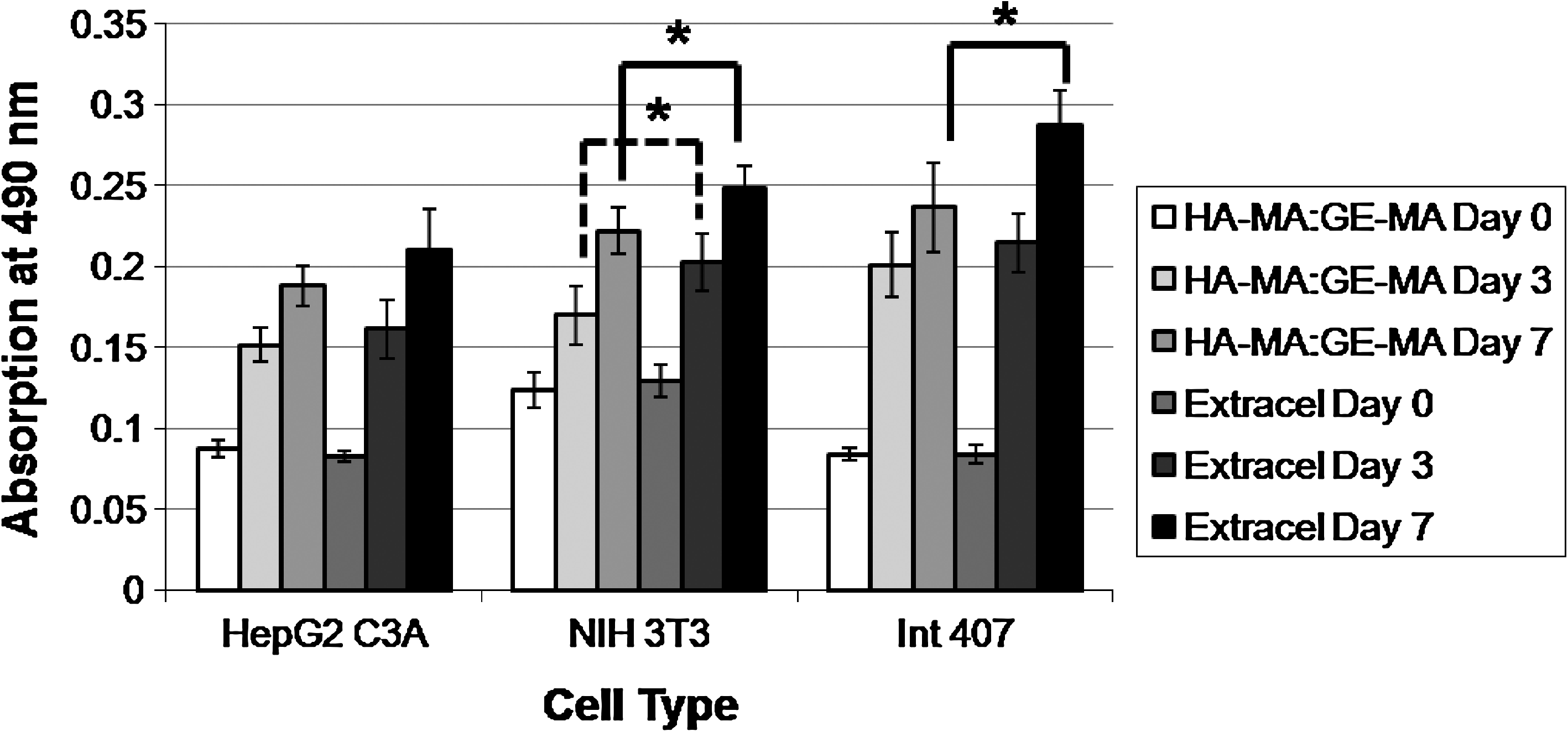
New hydrogels (HA-MA:GE-MA [4:1]) support cell growth and proliferation. HepG2 C3A, NIH 3T3, and Int-407 cells were encapsulated in either Extracel™ or the new hydrogels. Biocompatibility was determined using an 3-(4,5-dimethylthiazol-2-yl)-5-(3-carboxymethoxyphenyl)-2-(4-sulfophenyl)-2H-tetrazolium (MTS) assay (see text for details); increased absorbance from day 0 to 3 and day 3 to 7 indicate proliferation of cells on both materials. *p < 0.05.
UV irradiation at 365 nm for 0, 120, 180, and 240 s resulted in MTS absorbance values of 0.95 ± 0.08, 0.92 ± .04, 0.94 ± 0.09, and 0.93 ± 0.04 respectively. The 120-, 180-, and 240-s irradiated groups were not statistically different from each other or from the nonirradiated group. Thus, the range of UV exposures relevant to our bioprinting protocol does not adversely affect the viability of the encapsulated cells. Further, reported irradiation times for cell encapsulation in hydrogels can vary from 20 s 47 to 5 min with no observable decrease in cell viability.48,49
Moreover, unprinted and printed cell-containing hydrogels had MTS absorbance values of 0.99 ± 0.09 and 0.95 ± 0.03. These were not significantly different, suggesting that the extrusion conditions were not detrimental to cell viability. This is expected, since only a small percentage of cells were actually exposed to the walls of the printing tip and subjected to the shear forces during printing. Cells have also been shown to maintain viability in high stress situations such as centrifugal casting, in which they are subjected to 5–20 g of rotational force. 50 Further, extrusion techniques such as injections of cell suspensions through syringes are performed regularly in research and clinical practice.
In vivo biocompatibility
Extracel was chosen as the control based on extensive validation in in vivo models for reparative and regenerative medicine. 29 No differences were observed between Extracel hydrogels and the HA-MA:GE-MA hydrogels, nor were any qualitative differences observed between the 100 and 400 μL injections. Hematoxylin and eosin–stained sections showed the injected hydrogels to be intact underneath the dermis (Fig. 3). Adjacent tissue appeared healthy, showing no signs of inflammation or necrosis.

Cell-free hydrogels are biocompatible in vivo. Hydrogels were injected subcutaneously in nude mice and evaluated at 2 or 4 weeks. (
Rheology
For the first three UV exposure time points (30, 45, and 60 s), G′, the storage modulus, was ∼10 Pascals (Pa), whereas G″, the loss modulus, was ∼20 Pa. During these periods, G″ was significantly greater than G′ (p < 0.01), resulting in a material with fluid behavior. At 120 s, G″ was still significantly greater than G′ (p < 0.01), but the values were both close to ∼30 Pa, resulting in a material that was either a viscous fluid or loose hydrogel. At 180 s, G′ had increased to ∼50 Pa and was significantly higher than G″ at ∼45 Pa (p < 0.01), the point at which the material would be considered a true hydrogel. Over the next 4 min, G′ continued to increase until reaching a plateau at 80–90 Pa, while G″ decreased and leveled off at ∼40 Pa (Fig. 4). At these time points, G′ was again significantly greater that G″ (p < 0.01). These results led to the selection of 120 s for the initial photocrosslinking to provide a printable gel. The hydrogel was sufficiently fluid to be taken up into a syringe and extruded through a needle tip without visibly damaging the hydrogel structure. In addition, the 120-s crosslinked hydrogel was solid enough to temporarily retain its shape during printing and before the secondary photocrosslinking step.

Stiffness increases with time of irradiation. The moduli G′ and G″ were determined as a function of the time of irradiation at 365 nm. On the basis of these data, 120 s was selected for the first irradiation period to obtain a gel-like fluid.
Bioprinting
Following the ring-stacking protocol (Fig. 5A), several sets of constructs were printed with the Fab@Home device (Fig. 5B) to provide adequate tissue for imaging and histology. Printed constructs containing the HA-BODIPY tracer in the cellularized hydrogels fluoresced brightly when irradiated at 365 nm, clearly distinguishing the cell-containing tubular portion of the construct from the cell-free lumen and outer ring (Fig. 5C). Over the 3-week culture period, the neotissue appeared to remain within the HA-MA:GE-MA hydrogel, as cells did not invade the nonadherent HA-MA hydrogel. Indeed, this was an intentional design feature, in which the HA-MA only hydrogel served as a migratory barrier to maintain the tubular shape and the central lumen of the cellularized construct. Adherent cells can only attach to, and proliferate in, hydrogels containing the gelatin component. During culture, the cellular portion became noticeably more opaque, as the cells proliferated and secreted their own ECM (Fig. 6A). At the end of the maturation period, each of the constructs had retained mechanical properties that permitted easy handling during the histology protocols.
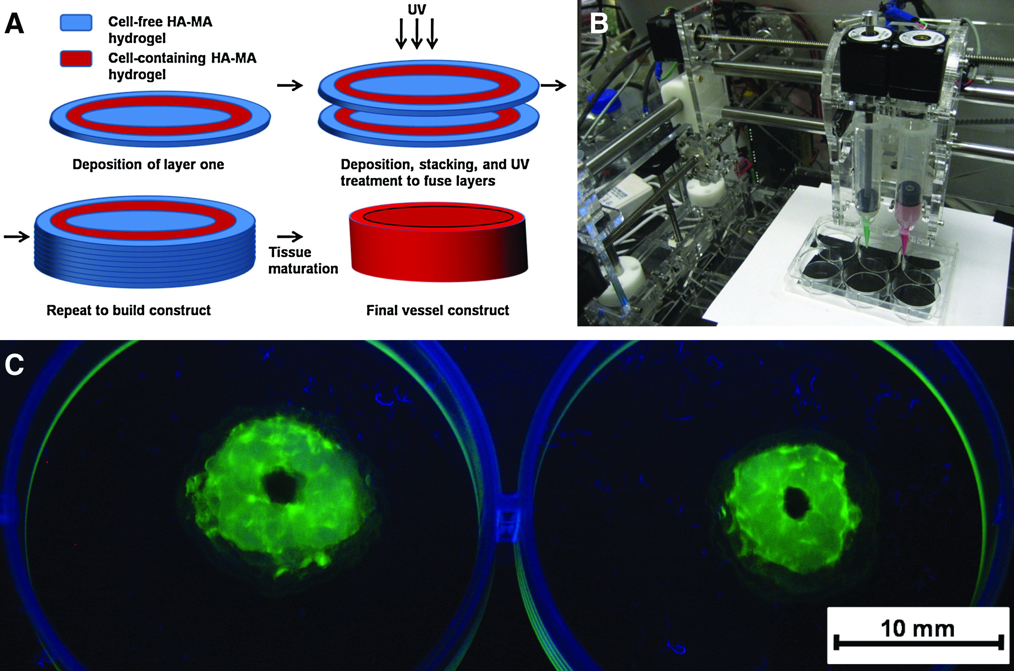
Bioprinting of the new hydrogel. (
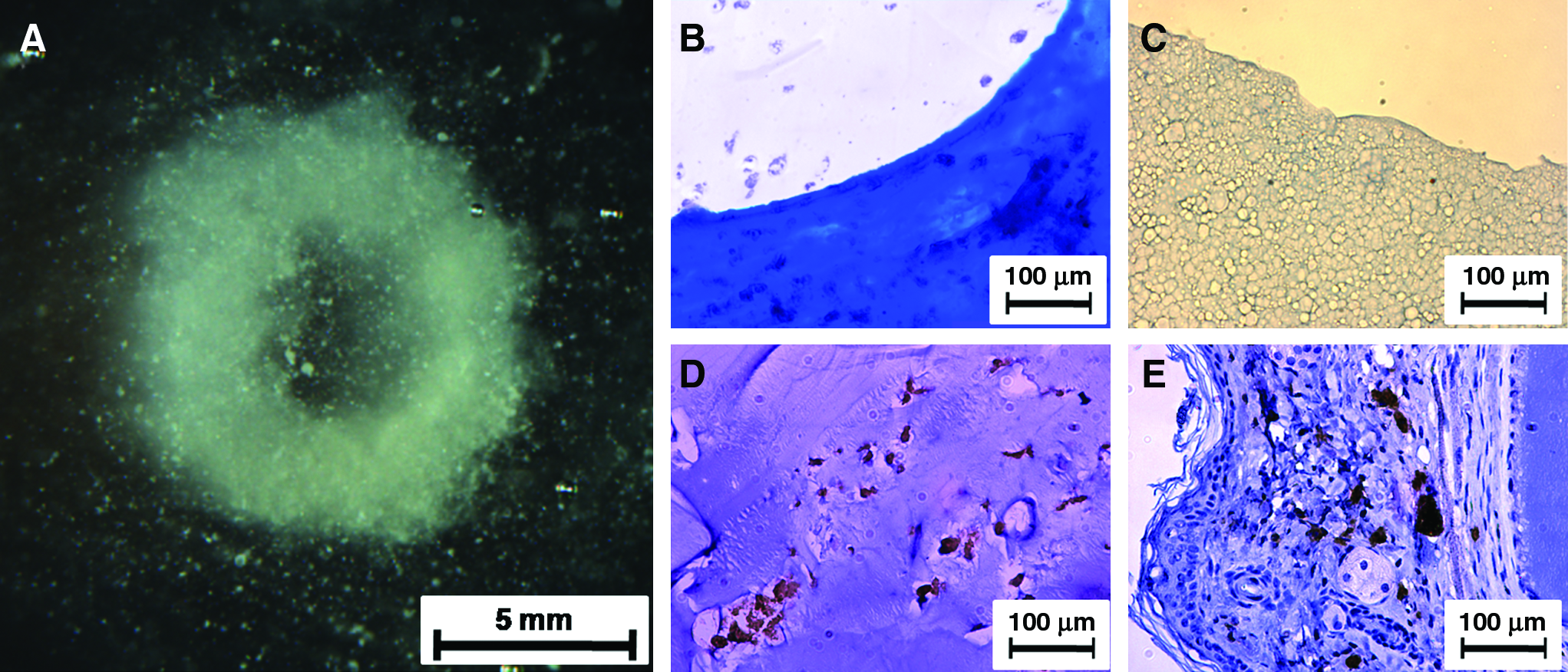
Printed tissue construct after 3 weeks of culture. (
Histology and immunohistochemistry
Masson Trichrome staining of sectioned tissue constructs showed cells encased in sheets of collagen fibrils (Fig. 6B). In sectioned areas that were noncellularized, printed hydrogel material was still evident and lacked collagen, as evidenced by the contrast between the unstained lumen and collagen-containing construct walls. The negative control (cell-free HA-MA:GE-MA hydrogel) failed to stain positive for collagen, showing that histology can distinguish between native collagen and gelatin, a denatured and hydrolyzed collagen product (Fig. 6C). As a second demonstration of collagen production in vitro, IHC showed that procollagen, the intracellular precursor of collagen, was present, verifying that the cells in the constructs were indeed actively producing collagen (Fig. 6D). Similarly, the positive controls showed antibody specificity and areas of procollagen that were surrounded by denser and more varied tissue (Fig. 6E). The formation of procollagen indicates that the cells had remodeled the synthetic ECM environment, secreting a cell-produced ECM as they matured into viable tissue.
Discussion
Developing biomaterials compatible with bioprinting devices is inherently difficult. For a syringe extrusion approach, a material is required that can support cells and yet still retain sufficient fluidity to be extruded intact through a narrow tube. The 80% HA-MA–20% GE-MA gel accomplished this goal; it possessed the desired mechanical properties while still providing an adequate number of sites for cell attachment and growth. Higher levels of gelatin have been shown to result in better cell attachment. For example, thiolated HA:thiolated gelatin mixtures with ratios of 100:0, 75:25, 50:50, 25:75, and 0:100 were compared with respect to cell attachment and proliferation. Increasing the gelatin component from 0% to 75% gradually increased attachment, but 75% and 100% were not significantly different. 39 However, in rheological work with similar hydrogels, addition of the crosslinkable gelatin component decreased G′ of the polymer network. 45 Chemically modified gelatins, for example, Gelin-S and GE-MA, have smaller molecular sizes (ca. 10–15 kDa) relative to the linear, chemically modified HA derivatives (typically 120–150 kDa). The crosslinkable gelatin components therefore do not contribute to structural component of the hydrogel, but rather serve to dilute the modified HA and thereby decrease the hydrogel modulus. Thus, on one hand, we were limited by the need for cell adherence, but, on the other hand, we were constrained by the need to maintain mechanical properties. High HA-MA:GE-MA ratios would result in stiffer gels with poorer cell adherence, whereas low ratios would result in softer gels with better cell adherence. An 80:20 ratio resulted in a material that best balanced the two properties desired.
Since both macromonomers shared a common photocrosslinking moiety, it was possible to develop extrudable hydrogels by an initial 120-s light-induced cocrosslinking period. These gels—either loaded with cells or cell-free—were then printed into the desired form, and crosslinked with an additional period of irradiation to fix their shapes and crosslink the separately extruded hydrogels to each other. These hydrogel materials were demonstrated to be biocompatible both in vitro and in vivo. Further, we investigated the effects of extrusion and repeated irradiation on the viability of cells, and observed no adverse effects.
One concern with building structures by sequential vertical stacking is that layers of tissue that were deposited early in the printing process might be damaged by additional irradiation required to crosslink subsequent layers. However, the design of the experiment mitigates this concern. First, as one builds consecutive layers with this translucent hydrogel, the UV light intensity is attenuated by absorption and scattering within the construct. Thus, deeper layers are exposed to increasingly lower light intensities. Second, there is an endpoint to the photopolymerization reaction. In our rheology data, G′ plateaus after 5 min; thus, whether a particular layer is irradiated for 5 or 10 min, the material properties should not differ significantly.
A factor that remains untested in many biomaterials is the effect of added cells on material properties. In appropriate environments, cells will build and stabilize their own matrix, as observed in the printed constructs described herein. How does cell density impact a material upon encapsulation? At the cell densities of 25 million cells/mL employed herein, the cells only comprise approximately one thousandth of the total hydrogel and cell volume. Only at cell densities of an order of magnitude or more higher would cells make up a substantially greater portion of the total volume. It is likely that at sufficiently high cell densities, the crosslinking of the hydrogel and its mechanical properties would likely be compromised.
In scaling up size and complexity of tissue constructs there may be limitations on printing height. These printable gels are relatively soft, so at some point, gravity will compromise the ability of the structure to support itself. However, several techniques can mitigate this problem. First, biologically inert or degradable structural supports could be added to the construct. Second, small organ modules could be printed with vertical or horizontal printing approaches, with subsequent assembly of the modules. Ideally, the constructs can be sized such that the temporary stabilization provided by the hydrogel will allow cells to remodel the synthetic sECM into a cell-derived ECM with the mechanical properties of a mature tissue. Nonetheless, the two-step photocrosslinking process demonstrated herein offers a proof of concept for the printing of tissue constructs in which cells can remodel the printed sECM during growth and proliferation.
Currently, bioprinting devices vary from simple inkjet printer designs to complex, environmentally contained 3D computer-directed extrusion systems. Printing from syringes is attractive from the point of view of greatest ease of preparation and maximum flexibility in generating constructs of different sizes and geometric complexity at a reasonable cost. If we compare extrusion from a syringe to extrusion from glass microcapillary tubes or to droplet deposition, syringe extrusion allows for larger printing volumes, and less time is required to print an entire construct. Importantly, the syringes allow modularity; different syringes in a multisyringe system can be charged with suspensions of cells of different types or at different cell densities. The cartridges can be replaced less often, allowing printing to occur uninterrupted.
We chose to use the Fab@Home printing system for several reasons. First, as an open source product, it is economical. Second, the Fab@Home printing system is versatile and can be easily customized. Third, this printing system has been used to print a wide variety of different materials, from epoxy resins and silicone rubbers, to cake frosting and cheese. In contrast, the Fab@Home system has seen limited use for cell culture and tissue engineering purposes. The results described herein demonstrate that this system has the capability to print soft tissue constructs.
We chose to print a tubular geometry as a demonstration that a viable hollow structure could be built to maintain patency during tissue culture and maturation. This proof-of-concept experiment makes it feasible to design printing strategies for tissues or organs with one or more lumens or cavities, for example, engineered blood vessel networks, bladders, or heart constructs. Further, because diffusion is limited in tissue constructs of any substantial size, a vascular network will be required to allow passage of oxygen, nutrients to the tissue and removal of metabolic by-products from the tissue. Incorporating lumens into printed tissues is one way to increase viability of sizeable constructs. Bioprinting is one of the only methods that explicitly addresses the complexity needed to build these kinds of structures.
These tissue constructs matured over time in culture and began secreting their own ECM, as evidenced by the presence of collagen and procollagen. In contrast, the cell-free gelatin-containing hydrogel HA-MA:GE-MA showed no evidence of collagen staining. Thus, the collagen present in the tissue constructs was produced by the encapsulated cells; a false-positive result due to the gelatin component in the hydrogel can be ruled out. The ability to secrete a native ECM is an important attribute of a viable tissue.
Several key challenges still remain before bioprinting can become a routine procedure, including (1) the dimensions of the construct, (2) the cell types used to create the construct, (3) the viability of the printed cells in the sECMs, (4) the ability to form a branched network of interconnected tubes of different diameters, (5) and the creation of lumens in the constructs. Both the HA-MA:GE-MA and the use of the Fab@Home printing system still require optimization needed to print tissue constructs that better simulate real tissues. First, the printed proof-of-concept tissues had walls that were thicker than would be desired in a tissue-engineered blood vessel. Future research will be directed toward printing of thinner-walled tubular constructs to more accurately simulate blood vessel constructs. Second, this goal will be accompanied by the use of smooth muscle and endothelial cells, at a minimum, in creating the construct.
Third, HA-derived sECM hydrogels have demonstrated that multimillimeter–sized tissues can be produced in vivo. In one case, fibroblasts were encapsulated in PEGDA-crosslinked thiol-HA/thiol-gelatin hydrogels and injected subcutaneously in mice. After 4 weeks in vivo, healthy fibrous tissue plugs with sizes over 1 cm in diameter were recovered at necropsy. 18 In a rabbit osteochondral defect model, autologous mesenchymal stem cells were encapsulated in the same crosslinked HA-gelatin hydrogel at a density of 50 million cells/mL and placed into 5-mm-diamter, 5-mm-deep defects. After 12 weeks, healthy hyaline cartilage and trabecular bone had been produced within the defect. 51 In a final example with this sECM, orthotopic human tumors were produced by injection of in situ–crossling cell suspensions in nude mice. The resulting tumors were well vascularized in the mammary fat pad, subserosally in colon, and in the ovarian capsule, and reached diameters of up to 7 mm. Tumors in sECMs were significantly larger and less necrotic that those formed from cell suspensions in buffer alone. 52 These sECMs employed different chemical modifications of the same macromolecular components of the bioprinted HA-MA:GE-MA gels, and were used at analogous concentrations and cell densities as those described herein. Additional evidence on viability of printed tissues will be produced in due course.
The final two points, the branched network and lumen generation, are interlinked and are among the most prominent challenges for bioprinting, as is an active research area in its own right. The proof-of-concept tissue constructs that were printed contained lumens that are actually comprised of cell-free hydrogels. Unfortunately, crosslinked HA-MA hydrogels are not readily removed material in such a setting, except by hydrolysis or hyaluronidase treatment, which would adversely affect the cellular portion of the constructs. In previous work, we reported a method for successfully recovering cells from 3D hydrogel culture by the degradation of disulfide-containing crosslinkers with mild cysteine solutions. 27 However, disulfides are incompatible with the photocrosslinking chemistry, and could not be used here. Separately, we are investigating additional hydrogels and crosslinking strategies to allow printing but incorporate the ability to remove the noncellularized lumen gels under mild conditions.
How might such a printing approach be used? Recently, the Hutmacher group explored several strategies for zonal cartilage repair. 53 In one approach, rapid prototyping was used to print cell-containing alginate rods using cells from different zones of articular cartilage. One can envision adapting this protocol with a more suitably chondrogenic 3D microenvironment provided by an HA-rich biopaper. In addition, a sequential crosslinking strategy to control cellular spreading in 3D was recently demonstrated by the Burdick group. Methacrylated HA was partially crosslinked with a bis-thiol containing peptide and subsequently formed into a hydrogel using a patterned photocrosslinking strategy. 54 Combining bioprinting advances with different cell types and novel biomaterials has potential for success in engineering tissues that are physiologically relevant, both geometrically and functionally.
Conclusions
We presented four components of a successful bioprinting system. First, we described the synthesis of a new G-MA derivative, GE-MA. Second, we combined GE-MA with HA-MA to produce a biocompatible hydrogel using a two-step photocrosslinking strategy. Third, we showed that the new HA-MA:GE-MA hydrogels were both cytocompatible in vitro and biocompatible in vivo. Fourth, we used an open source rapid prototyping system to print a tubular construct. Under computer control, a gel-like fluid was printed by extrusion from a syringe into a defined pattern of cell-free and fibroblast-loaded hydrogels. A second irradiation of the printed base layer made a more rigid structure, and the printing-irradiation sequence was iterated to build a tubular construct. Cells remodeled the printed ECM and secreted collagen. Taken together, these results suggest that the printable HA-MA:GE-MA hydrogels may have potential utility as the biopaper component in bioprinting protocols.
Footnotes
Acknowledgments
This research was supported by an NSF FIBR Grant (EF-0526854) and by the State of Utah Centers of Excellence Program. The authors thank Glycosan BioSystems Inc. for providing the Extracel kits and selected crosslinkable sECM components.
Disclosure Statement
G.D.P. holds equity in Glycosan Biosystems (Salt Lake City, UT).