
Abstract
Porous, tubular, flexible, and elastic poly(trimethylene carbonate) (PTMC) scaffolds (length 8 cm and inner diameter 3 mm) for vascular tissue engineering were prepared by means of a dip-coating and particulate leaching procedure. Using NaCl as porogen, scaffolds with an average pore size of 110 μm and a porosity of 85% were obtained. Before leaching the salt, the structures were made creep-resistant by means of crosslinking at 25 kGy gamma irradiation. To increase the efficiency of cell seeding, the scaffolds were provided with a microporous outer layer of 0.2 mm with an average pore size of 28 μm and a porosity of 65% (total wall thickness 1 mm). Human smooth muscle cells (SMCs) were seeded in these scaffolds with an efficiency of 43%, as determined after 24 h cell adhesion. SMCs were cultured in the scaffolds up to 14 days under stationary conditions or under pulsatile flow conditions in a bioreactor (pressure 70–130 mmHg, 69 pulsations/min, and average wall shear rate 320 s−1). Although SMCs proliferated under both conditions, cell numbers were three to five times higher in case of dynamic culturing. This was qualitatively confirmed by means of histology. Also, in terms of mechanical properties, the dynamically cultured constructs performed better than the statically cultured constructs. After culturing for 14 days, the maximum tensile strengths of the constructs, determined in the radial direction, had increased from 0.16 MPa (unseeded scaffold) to 0.48 MPa (dynamic culturing) and 0.38 MPa (static culturing). The results of this study indicate that a potentially useful medial layer for tissue-engineered vascular grafts can be prepared by dynamic culturing of human SMCs seeded in porous tubular poly(trimethylene carbonate) scaffolds.
Introduction
Tissue engineering is a promising technique to prepare functional small-diameter arterial replacements. To this end, autologous vascular cells are seeded in biodegradable (tubular) scaffolds and subsequently cultured in a bioreactor or immediately implanted. 2 Among others, Niklason et al. 3 implanted bioreactor-cultured blood vessel equivalents based on poly(glycolic acid), smooth muscle cells (SMCs), and endothelial cells in experimental animals. Although the constructs remained patent for several weeks, the mechanical properties of these grafts are considered to be insufficient for implantations in humans. 4 Shin'oka et al. 5 implanted porous scaffolds (patches or tubes), prepared from a copolymer of L-lactide and ε-caprolactone, directly after seeding with autologous mononuclear bone marrow cells in the pulmonary artery of 1–24-year-old patients with a congenital defect. Follow-up during 30 months showed no complications, and all arteries remained patent. However, the pulmonary artery is a relatively large-diameter low-pressure artery, indicating that additional research is necessary for replacement of arteries in the systemic circulation. L'Heureux et al. introduced the concept of cell sheet-based tissue engineering without the use of a polymer scaffold. 6 Several layers of fibroblast sheets are wound around a mandrel and subsequently matured. After removal of the mandrel, the luminal surface is seeded with endothelial cells. Although these grafts show good mechanical properties and perform well as arterio-venous fistulas in humans, the main draw-back of this approach is the long preparation time of 6–9 months. 4
Our vascular tissue engineering approach is based on cell seeding in flexible and elastic tubular poly(trimethylene carbonate) (PTMC) structures. 7 This material shows excellent biocompatibility and enzymatic degradation by surface erosion in vivo.8–10 Compliant and creep-resistant PTMC networks are obtained by means of gamma irradiation 11 and interconnected pores are formed by particulate leaching. 12 To prepare the medial layer of the vascular graft, human SMCs are seeded by perfusion of a cell suspension from the lumen (inner diameter 3 mm) through the wall of a tubular scaffold. 7
Mechanical stimulation of SMCs seeded in vascular tissue engineering scaffolds promotes the circumferential orientation of the cells as well as the deposition of extracellular matrix.13,14 The aim of the present study was to subject SMCs seeded in the tubular PTMC scaffolds to cyclic mechanical strain in a pulsatile flow bioreactor to evaluate the histology, cell proliferation, and mechanical properties of the constructs up to 14 days of culturing.
Materials and Methods
Preparation of porous tubular PTMC scaffolds
High-molecular-weight PTMC (Mw = 8.78 × 10⁵ g/mol) was synthesized from trimethylene carbonate (1,3-dioxane-2-one; Boehringer Ingelheim) as previously described. 7 Glass mandrels (φ = 3.0 mm) were dipped in a PTMC solution in chloroform (2.5% w/v) containing homogeneously dispersed NaCl particles (Acros Organics) sieved to a size range of 106–250 μm. The polymer to porogen weight ratio was 10:90. The dip-coating process was repeated several times until an outer diameter of 7–8 mm was reached. Subsequently, the coated mandrels were dried at room temperature for 2–3 days, vacuum sealed, and subjected to 60Co gamma irradiation (25 kGy; Isotron) to obtain crosslinked structures.
To increase the cell seeding efficiency, the gamma-irradiated salt-containing PTMC-coated mandrels were dipped two times in a PTMC solution in chloroform (1.5% w/v) containing homogeneously dispersed NaCl particles sieved to a size range of 0–60 μm. The polymer-to-porogen weight ratio was 30:70. The coated mandrels were dried at room temperature for 1 day. Finally, the scaffolds were removed from the mandrels and the salt particles were leached out with demineralized water. To prevent shrinkage and collapse of the pores, the scaffolds were stored in water at 4°C.
Analysis of porous tubular PTMC scaffolds
Cross sections of the porous tubular PTMC scaffolds were evaluated by means of scanning electron microscopy (SEM). The samples were cut in liquid nitrogen, rinsed with methanol, and dried. After coating of the specimens with a gold–platinum layer using a Polaron E5600 sputter-coater, images were taken with a Hitachi S800 field emission scanning electron microscope operating at 6 kV. Specimens cut from cell-seeded scaffolds cultured up to 14 days were dried using a series of incremental ethanol solutions (60–100%), sputter-coated, and evaluated in the same way.
The tensile properties of hydrated porous tubular scaffolds with a length of 5 mm were measured in the radial direction according to standards of the American National Standards Institute and the Association for the Advancement of Medical Instrumentation (ANSI/AAMI, VP20: 1994, paragraph 8.3). Tensile tests were carried out using a Zwick Z020 universal tensile testing machine at room temperature. The tensile testing machine was equipped with a 500 N load cell and was operated at a crosshead speed of 1 mm/min. The specimen deformation was derived from the grip to grip separation. The initial stiffness was determined from the slope of the tensile curve from 2.5% to 5% of strain. Tensile properties of porcine carotid artery, human arteria mesenterica inferior, and cell-seeded scaffolds cultured up to 14 days were determined in the same way.
Seeding and culturing of SMCs in porous tubular PTMC scaffolds
SMCs were isolated from human umbilical veins as previously described. 15 The cells were cultured in gelatin-coated (0.5% w/v) tissue culture polystyrene flasks using Dulbecco's modified Eagle's medium (Invitrogen) containing 20% (v/v) heat-inactivated (30 min, 56°C) fetal bovine serum (Lonza), 50 units/mL penicillin, and 50 μg/mL streptomycin. Cell culturing was carried out in a humidified atmosphere at 37°C and 5% CO2 in an incubator. The culture medium was refreshed every 2–3 days. When cultures were almost confluent, SMCs were detached with 0.125% (w/v) trypsin/0.05% (w/v) EDTA and subcultured with a split ratio of 1:3 up to 10 passages.
Porous tubular PTMC scaffolds with a length of 4 cm were disinfected with 70% (v/v) ethanol, rinsed with phosphate-buffered saline (PBS), and placed overnight in the culture medium. SMCs were seeded in a scaffold by perfusing 20 mL SMC suspension in the culture medium (0.5 × 10⁶ cells/mL) from the lumen through the wall with two syringes connected to both ends of the scaffold. During the first 2 h, the seeded scaffolds were rotated 90° every 10 min to ensure a homogeneous cell distribution. The cells were allowed to adhere to the scaffolds for 1 day. Subsequently, cell culturing was continued under stationary conditions in the incubator or under dynamic conditions in a bioreactor (see below) up to 7 or 14 days. Culture medium was refreshed every 3 days.
Pulsatile flow bioreactor
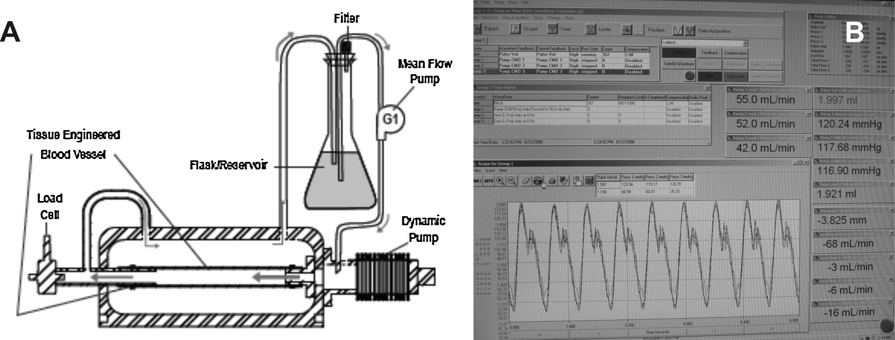
The SMC-seeded tubular PTMC scaffolds were cultured under dynamic conditions in a Bose-Electroforce pulsatile flow bioreactor (Fig. 1). Three culture chambers, each containing one construct, were operated in parallel. Culture media were independently perfused through the chambers using three gear pumps (G1 in Fig. 1A). Pulses were generated by a linear displacement pump (Dynamic Pump in Fig. 1A), which was shared between the culture chambers. The shape of the intraluminal pressure profile was programmed with WinTest software and mimicked that of the human carotid artery (Fig. 1B). 16 The bioreactor was placed in a 5% CO2 incubator operating at 37°C.
Analysis of cell-seeded scaffolds
PTMC scaffolds seeded with SMCs and cultured up to 14 days were rinsed with PBS. Transverse sections of 2 mm were cut, fixed overnight in a 4% (w/v) paraformaldehyde solution, rinsed with demineralized water, and incubated for 1 h in a 1% methylene blue solution. Subsequently, the samples were rinsed with demineralized water until the water was clear and evaluated using a stereomicroscope. Alternatively, samples were fixed as described above and embedded in glycol methacrylate. Transverse sections with a thickness of 5 μm were cut, stained with hematoxylin and eosin, and observed by light microscopy.
Numbers of SMCs present in the constructs were quantified by means of the CyQuant cell proliferation assay (Molecular Probes). 17 After culturing up to 14 days, constructs were rinsed with PBS, and tubular samples were cut with a length of 7 mm and digested with 200 μL proteinase K solution (1 mg/mL) for 16 h at 56°C. Subsequently, the solutions were diluted various times with cell-lysis buffer (Molecular Probes) containing 1.35 Kunitz units/mL RNAse and incubated for 1 h at room temperature to remove RNA and single-stranded DNA. Finally, the samples were mixed with CyQuant® dye and after 2 min, fluorescence was measured with a Victor fluorescence analyzer (Perkin-Elmer). Excitation and emission wavelengths were 480 and 520 nm, respectively. The measured fluorescence intensities were correlated to cell numbers by means of a calibration curve using suspensions with known concentrations of SMCs.
Results and Discussion
As recently reported, porous, tubular, flexible, and elastic PTMC scaffolds for vascular tissue engineering were prepared by means of a dip-coating and particulate leaching procedure. 7 Using NaCl as porogen, scaffolds with an average pore size of 110 μm and a porosity of 85% were obtained, which were made creep-resistant by means of crosslinking at 25 kGy gamma irradiation. The length of the tubular scaffolds was 8 cm, the inner diameter 3 mm, and the wall thickness 0.8 mm. To increase the efficiency of cell seeding, the scaffolds were provided with a microporous outer layer of 0.2 mm with an average pore size of 28 μm and a porosity of 65%. As shown in Figure 2, the porous inner and outer layers are firmly attached to each other.
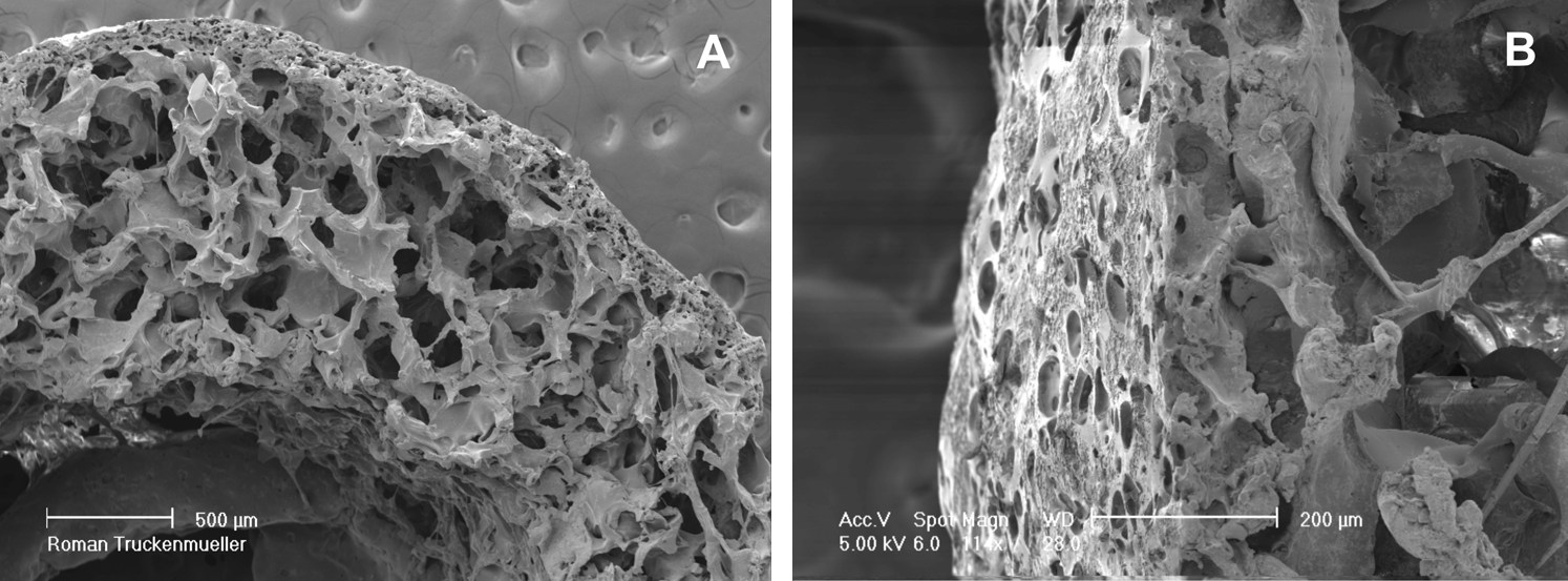
Scanning electron microscopy images of cross sections of tubular PTMC scaffolds provided with a microporous outer layer. Scale bars:
In this study, we used a Bose-Electroforce pulsatile flow bioreactor for dynamic culturing of human SMCs seeded in the tubular PTMC scaffolds. As programmed with WinTest software, the intraluminal pressure in the SMC-seeded scaffolds ranged from 70 to 130 mmHg, the number of pulsations was 69/min, and the flow rate was 50 mL/min, corresponding to an average wall shear rate of 320 s−1. From 70 to 130 mmHg, the outer diameter of the tubular PTMC scaffolds increased ∼7%, as determined with a laser micrometer (data not shown). These values fit within the range reported for human arteries of comparable size.18–20
During the first week of culturing, the seeded SMCs proliferated both under stationary and dynamic conditions (Fig. 3). Cell numbers were significantly higher in case of dynamic culturing, indicating that improved transport of nutrients and waste products and/or cyclic mechanical strain stimulates SMC proliferation. As previously shown by us, SMCs subjected to a comparable cyclic strain showed significantly increased expressions of cyclin E and collagen I at the mRNA level, as compared to cells cultured under static conditions. 17 Cell numbers after 1 day cell adhesion corresponded to a seeding efficiency of 43%. Without microporous outer layer, the efficiency of cell seeding in the tubular PTMC scaffolds would have been ∼15%. 7
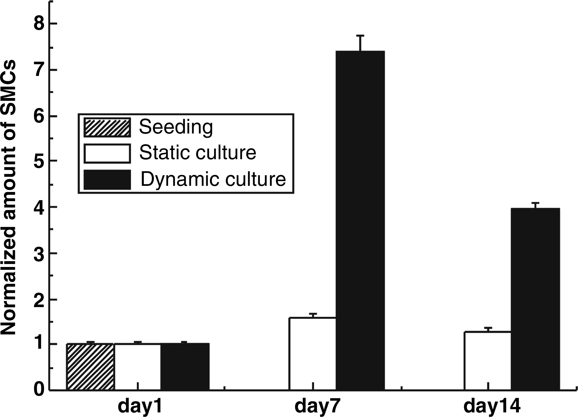
Numbers of SMCs present in the tubular PTMC scaffolds during culturing, normalized to the amount of cells present on day 1. Cell numbers were determined with the CyQuant cell proliferation assay (n = 3, ±SD).
During the second week of culturing, SMC numbers decreased both under stationary and dynamic conditions (Fig. 3). Cell numbers were still significantly higher in case of dynamic culturing. Possibly, increased cell mass and/or pore occlusion resulting from SMC proliferation adversely affected the diffusion of nutrients and waste products.21,22 These data indicate that this approach may benefit from an adventitial-like layer containing small channels or capillaries protruding into the medial layer, facilitating sufficient transport of nutrients and waste products. 23
Methylene blue staining of cross sections of SMC-seeded tubular PTMC scaffolds showed homogeneous cell adhesion in the scaffolds after 1 day of culturing under stationary conditions (Fig. 4B). Moreover, methylene blue staining confirmed significant SMC proliferation after 7 and 14 days of culturing under pulsatile flow conditions (Fig. 4C, D).

Stereomicroscopic images of cross sections of tubular PTMC scaffolds stained with methylene blue.
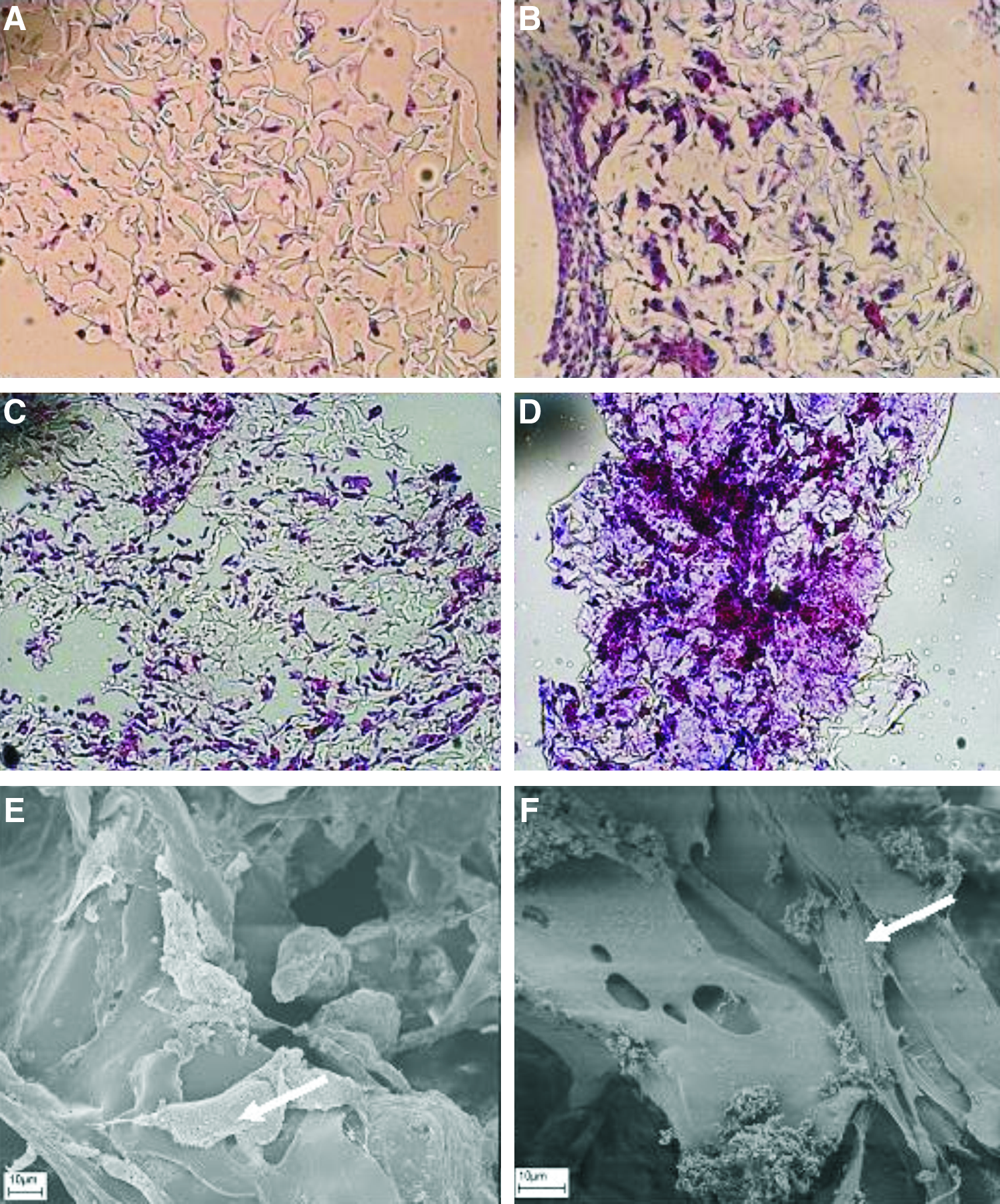
Homogeneous adhesion of SMCs in the tubular PTMC scaffolds 1 day after seeding was confirmed by means of histological staining with hematoxylin and eosin (Fig. 5A). Subsequent culturing under stationary conditions until day 7 showed SMC proliferation predominantly on the adventitial side (outside) of the scaffolds (Fig. 5B). In contrast, culturing under pulsatile flow conditions resulted in a more homogeneous cell distribution, although some parts of the scaffolds contained more cells than other parts (Fig. 5C, D). After 7 days of culturing under dynamic conditions, SMCs could be observed on the luminal side of the constructs by means of SEM (Fig. 5E, F).
The mechanical properties of SMC-seeded tubular PTMC scaffolds cultured up to 14 days were investigated by means of tensile testing of freshly recovered samples in the radial direction. The maximum tensile strengths of constructs cultured for 7–14 days under stationary conditions were higher than that of hydrated scaffolds without cells (Fig. 6). The highest tensile strengths were measured with constructs cultured for 7–14 days under dynamic conditions. With increasing tensile strength, the elongation at break decreased.
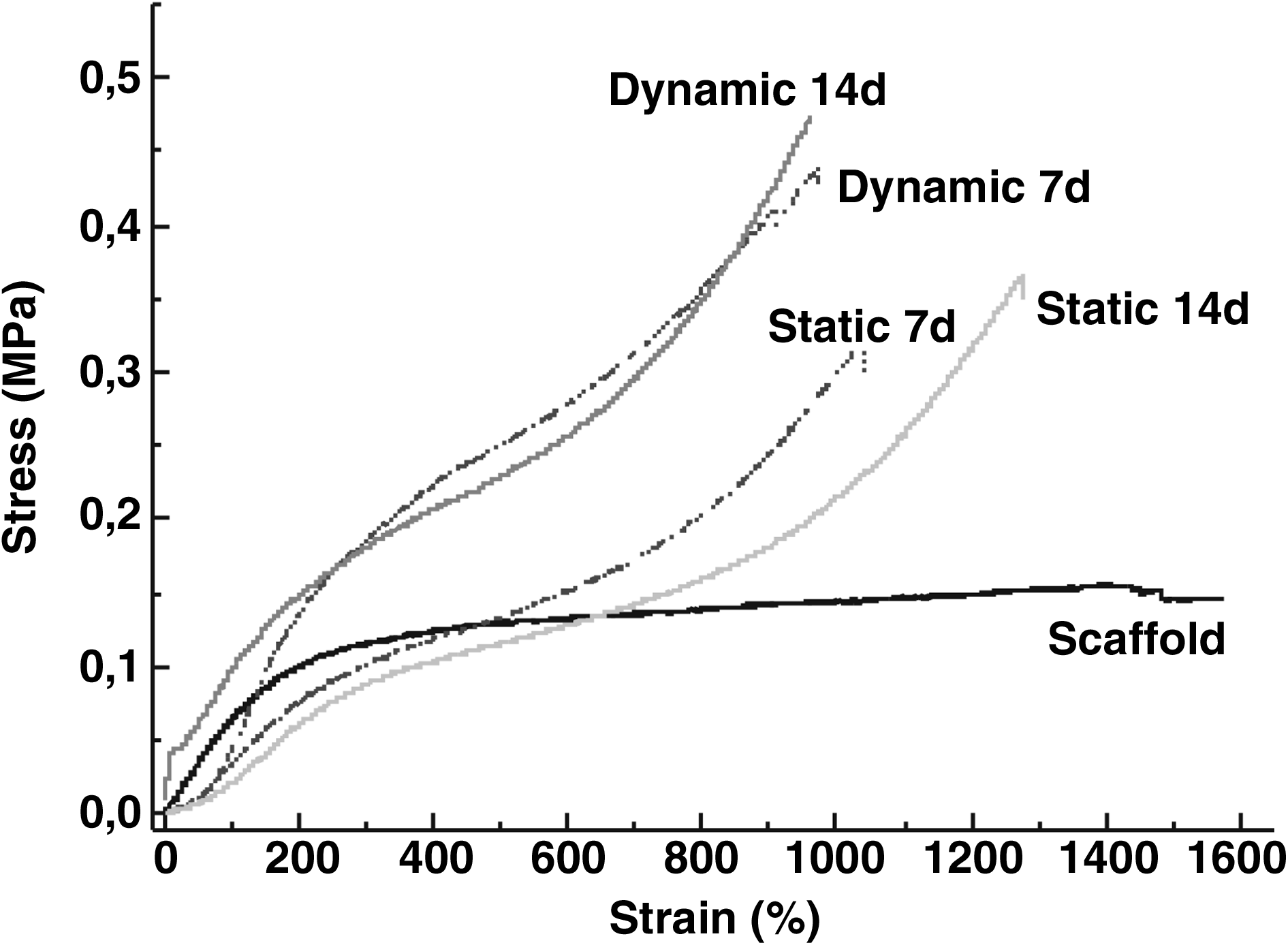
Tensile properties (determined in the radial direction) of SMC-seeded tubular PTMC scaffolds cultured up to 14 days under static or dynamic conditions. Scaffolds without cells were used as reference.
Combining these data with the histological data, it can be concluded that during the first week of culturing the tensile properties of the constructs increased with increasing numbers of SMCs present in the constructs. The decrease in cell numbers from day 7 to 14, as determined with the CyQuant assay, did not adversely affect the tensile properties of the constructs, indicating that the cells had deposited extracellular matrix proteins, which mainly determine the mechanical properties of the constructs.
In Table 1, the mechanical properties of the constructs cultured under pulsatile flow conditions are summarized and compared to those of porcine carotid artery and human arteria mesenterica inferior. In addition to the data discussed above, Table 1 shows that the initial stiffness of the SMC-seeded tubular PTMC scaffolds tended to decrease somewhat upon dynamic culturing. Probably, the PTMC was degraded to some extent. As shown in Figure 6, the stress–strain curves of the constructs display a toe-region at low strain values, which may also be an indication for the presence of extracellular matrix. The toe-region, which is also present in stress–strain curves of native blood vessels and tendons, is characteristic for the removal of macroscopic crimp in collagen fibres.24,25
n = 3, ±SD, except human arteria mesenterica inferior, n = 1.
On the basis of the above results, it can be concluded that the mechanical properties of the constructs do not significantly improve during the second week of dynamic culturing. Moreover, the values of the maximum tensile strength and initial stiffness of the constructs cultured for 1 week under dynamic conditions were lower than those of native arteries. In vivo studies will be initiated to investigate the performance of the vascular grafts with respect to mechanical properties as well as patency rates in small-diameter arterial reconstructions. Before in vivo experimentation, the luminal surface of the grafts needs to be endothelialized. Previously, we have investigated the endothelialization of synthetic vascular grafts. Because Dacron and Teflon do not support the adhesion and proliferation of endothelial cells, our strategy was to coat these materials with albumin–heparin or collagen–heparin conjugates. These coatings were loaded with the heparin-binding growth factor basic fibroblast growth factor, which significantly improved endothelialization of the surfaces.26,27 This approach could also be used for endothelialization of the constructs described in the present article. However, extracellular matrix will be deposited by the SMCs proliferating in the PTMC scaffolds. It has been reported that this is a good substrate for direct endothelial cell seeding, obviating the need to coat the luminal surface of the constructs to promote endothelialization.28,29
Conclusions
Porous tubular PTMC scaffolds were seeded with human SMCs and subsequently cultured up to 14 days under stationary conditions or pulsatile flow conditions in a bioreactor. Both determination of cell numbers and histology showed that dynamic culture conditions significantly improved SMC proliferation, as compared to stationary culture conditions. Also in terms of mechanical properties, the dynamically cultured constructs performed better. The results of this study indicate that a potentially useful medial layer for tissue-engineered vascular grafts can be prepared by dynamic culturing of human SMCs seeded in porous tubular PTMC scaffolds.
Footnotes
Acknowledgments
This work was financially supported by the Dutch Program for Tissue Engineering (DPTE). The authors wish to thank M. Smithers for the SEM work.
Disclosure Statement
No competing financial interests exist.