
Abstract
Macroporous networks of poly(ethylene glycol) (PEG) with interconnected pores can be created by cryogelation techniques. In this study, we describe the potential application of such PEG cryogels as scaffolds for cartilage tissue engineering. Three-dimensional macroporous cryogels were evaluated for chondrocyte growth and production of cartilage-specific extracellular matrix (ECM). Seeded primary bovine chondrocytes showed homogeneous distribution throughout the cryogels. DNA content suggests continuous cell proliferation over 4 weeks of in vitro culture. Analysis of the composition of cell-secreted ECM showed a culture-time-dependent increase in the amount of glycosaminoglycan and collagen. The production of ECM by chondrocytes was confirmed using scanning electron microscopy analysis. Further histological and immunohistological analysis of the cell-laden scaffold confirmed the presence of accumulated cartilage-specific ECM within the scaffold. The interconnected macroporous network promoted diffusion of cell-secreted matrix within the cryogels. Our results indicated that interconnected macroporous PEG cryogels successfully supported attachment, viability, proliferation, and biosynthetic activity of seeded chondrocytes.
Introduction
More recently, gelation technique at subzero temperatures, known as cryogelation has been employed to create hydrogels with macroporous interconnected microstructures. During cryogelation at subzero temperatures, the reactants restricted to the unfrozen/semi-frozen phases form the crosslinked network, while ice crystals nucleated from the aqueous phase (formed by the freezing of water at low temperatures) function as porogens. The melting of these ice crystals at temperature above the freezing temperature (e.g., room temperature) gives rise to interconnected macroporous networks. 21 The resultant cryogels show enhanced mechanical properties and swelling kinetics.22,23 Recent studies have shown the potential application of cryogels as scaffolds, owing to their hydrophilicity and macroporous interconnected microstructures. 24 For instance, Kathuria et al. reported that chitosan-gelatin cryogels promoted the attachment and proliferation of fibroblasts (Cos-7). 25 Bolgen et al. have demonstrated the ingrowth and biosynthetic activity of osteoblast-like cells (MG63) within biodegradable 2-hydroxyethyl methacrylate-lactate-dextran cryogels. 26
Recently, we have synthesized interconnected macroporous poly(ethylene glycol) (PEG) cryogels with varying microstructures by manipulating the rate of polymerization, freezing temperature, or degree of supercooling. 27 In this study, we evaluated the potential of these PEG cryogels as scaffold for engineering cartilage tissue using primary bovine chondrocytes.
Materials and Methods
Materials
Toluene and triethylamine were purchased from Fisher Scientific, and dichloromethane and diethyl ether were purchased from Sigma-Aldrich. PEG (Mn 3400) and acryloyl chloride were purchased from Aldrich and were used without further purification. Ammonium persulfate and N,N,N′,N′-tetramethylethylenediamine were obtained from Sigma. Live/Dead Viability/Cytotoxicity Kit (Molecular Probes; Cat. no. L-3224) for cell viability test and Quanti-iT™ PicoGreen® dsDNA Assay Kit (Molecular Probes; Cat. no. P7589) for DNA assay were purchased from Molecular Probes. Collagenase type II (Cat. no. 4177) and papain (Cat. no. LS003126) were purchased from Worthington Biochemical Corporation.
Synthesis of PEG diacrylate oligomer
PEG diacrylate (PEGDA) oligomers were prepared as described elsewhere. 28 Briefly, 18.0 g of PEG was dissolved in 300 mL of toluene in a 500 mL round-bottomed flask in an oil bath at 150°C. The solution was then refluxed for 4 h under vigorous stirring. Trace amounts of water in the reaction mixture were removed by azeotropic distillation. Upon cooling the solution to room temperature, 3.262 g (32.2 mmol) of triethylamine was added with vigorous stirring. The flask was then moved to an ice bath and stirred for 30 min. About 2.918 g (32.2 mmol) of acryloyl chloride in 15 mL of anhydrous dichloromethane was then added to the reaction mixture drop wise for 30 min. After keeping the reaction mixture in an ice bath for another 30 min, the flask was heated to 45°C overnight. The reaction mixture was then cooled to room temperature and the quaternary ammonium salt was removed from the reaction mixture by filtering through diatomaceous earth (2–3 cm) on a fritted glass funnel. The filtrate was condensed using a rotary evaporator and then precipitated in excess diethyl ether. The white precipitate was collected and vacuum dried at 40°C for 24 h. The resultant PEGDA oligomer was purified by precipitation followed by column chromatography and dialysis before its usage.
Preparation of PEG cryogels
PEGDA oligomers were dissolved in phosphate-buffered saline (PBS) to prepare a solution of 10%w/v. At 4°C, the initiator–accelerator mixture (0.5%w/v of ammonium persulfate and 0.05%w/v of N,N,N′,N′-tetramethylethylenediamine) was added to this solution, and the reaction mixture was polymerized at −20°C for 20 h. The gelled reaction mixture was thawed at room temperature, which resulted in the formation of interconnected macroporous network (cryogels). The cryogels were washed with excess water to remove the unreacted reactants. Cryogels were synthesized in cylindrical forms having diameter and height of 10 and 5 mm, respectively.
Scanning electron microscopy of cryogels
The microstructures of PEG cryogels were examined using a scanning electron microscope (SEM; Philips XL30 ESEM). Briefly, thin slices of constructs were cut vertically from the center of the construct. The slices were then dehydrated serially in 50%, 75%, and 100% ethanol and dried using a critical point dryer (Tousimis AutoSamdri 815). The dried sections were gold-coated using a sputter coater (Emitech K575× Sputter Coater) for 30 s before SEM imaging.
Isolation of chondrocytes
Chondrocytes were isolated as described elsewhere. 29 In brief, full-thickness articular cartilage was dissected from the patellofemoral groove and distal femoral condyles of 6–8-week-old bovine legs. The cartilage pieces were dissected and incubated in Dulbecco's modified Eagle's medium (DMEM; GIBCO) containing 0.2% collagenase (Worthington Biochemical Corporation) and 5% fetal bovine serum (Atlanta Biologicals) for 16 h at 37°C with 5% CO2 to isolate chondrocytes. The cell suspension was then filtered through a 70 μm nylon cell strainer (BD Falcon) and washed with PBS containing 100 U/mL penicillin and 100 μg/mL streptomycin. The isolated chondrocytes (primary chondrocytes) were used without any further ex vivo expansion.
Chondrocytes seeding and culture on PEG cryogels
Before seeding chondrocytes, cryogels were sterilized with 70% ethanol and washed with fresh PBS. The cryogels were then coated with collagen type I by immersing them in 50 μg/mL of collagen type I solution (BD Bioscience; Cat. no. 354231) to improve cell adhesion before cell seeding. The cryogels were partially dehydrated under sterile conditions for 3 h before cell seeding. The partially dehydrated cryogels had a swelling ratio of 10.07 ± 0.77 (g/g) compared to their equilibrium swelling ratio of 19.93 ± 0.03 (g/g). Cell seeding on the cryogels was done by adding cell suspension in drop-wise manner. Briefly, isolated chondrocytes were suspended in chondrocyte medium (2.5 × 10⁷ cells/mL) (DMEM supplemented with 10% fetal bovine serum, 0.04 mM L-proline [Sigma], 50 μg/mL ascorbic acid [Sigma], 0.1 mM nonessential aminoacid [Gibco], and 100 U/mL penicillin and 100 μg/mL streptomycin). Forty microliters of cell suspension containing 1 × 10⁶ cells was seeded on the top surface of the partially hydrated cryogel at various spots, and incubated for 2 h to allow their infiltration. The cell-loaded cryogels were then cultured in 2 mL of chondrocyte medium, and the medium was changed every 2 days. All the cell cultures were done at 37°C in 5% CO2 environment.
Cell viability test
A live/dead assay was performed to evaluate the cell viability after 36 h of in vitro cultivation. 30 Briefly, the cell-laden cryogels were vertically cut into thin slices from the center of the construct and incubated with the live/dead assay dye solution (Molecular Probes; Cat. no. L-3224) containing 0.5 μL of Calcein-AM and 2 μL of ethidium homodimer-1 in 1 mL of DMEM. After 30 min of incubation, the slices were rinsed with PBS and cell viability was examined using a fluorescence microscope (Carl Zeiss; Axio Observer A1).
Biochemical assays
Biochemical assays were performed on cell-laden cryogels as a function of culture time (first, second, and fourth week). At each time point, constructs were lyophilized for 24 h and then digested with 1 mL of papain solution (125 μg/mL papain [Worthington biochemical corporation; Cat. no. LS003126], 10 mM
Histological and immunohistochemical analysis
The chondrocyte-laden cryogels were divided into two vertical halves, fixed overnight with 4% paraformaldehyde, and embedded in paraffin. The paraffin embedded constructs were processed into 20 μm sections and the sections corresponding to the center of the constructs were used for staining. Before staining, paraffin-embedded sections were deparaffinized with xylene, rehydrated with graded series of ethanol, and washed with fresh Millipore water. For histological analysis, sections were stained with Safranin-O and hematoxylin and eosin. Immunohistochemical staining for collagen type II was also performed by following a previously reported method. 36 Briefly, rehydrated samples were blocked with 0.5% Triton X-100 in 3% bovine serum albumin (Sigma; Cat. no. A7906) in PBS for 1 h and incubated with rabbit polyclonal antibodies against type I and type II collagen (Fitzgerald; Cat. no. 70R-CR007× and Cat. no. 70R-CR008×, respectively) at 1:300 dilution. These sections were incubated with goat anti-rabbit IgG secondary antibodies (Alexa Fluor® 488) at 1:250 dilution for 1 h and then the nuclei were counterstained with 4′,6-diamidino-2-phenylindole (Vector laboratories, Inc.) for 30 min. Images were observed using a fluorescent microscope.
Statistical analysis
The data obtained from all biochemical assays of quadruplicate samples were presented as mean ± standard deviation. Single-factor analysis of variance with Tukey's Multiple Comparison Test was performed to determine statistical significance (p < 0.05).
Results
Synthesis and characterization of PEG cryogels
Figure 1 shows the gross image of a PEG cryogel. The equilibrium swollen PEG cryogels were easily compressed by fingers without breaking and imbibed PBS could be expelled (Fig. 1a, b). Completely compressed cryogels showed a sponge-like appearance and could re-swell immediately upon immersion in PBS (Fig. 1c, d).

Photographs of poly(ethylene glycol) cryogels: (
The internal morphology of PEG cryogels is presented in Figure 2. As seen from the SEM image, PEG cryogels have an interconnected pore structure with pore sizes in the range of 30–80 μm (Fig. 2a). The interconnected macroporous structure was further investigated by a phase-contrast microscope using a cryosectioned, cell-laden cryogel section of 20 μm thickness (Fig. 2b). Live/dead assay showed that the seeded chondrocytes were distributed throughout the cryogel network and majority of these cells were viable (Fig. 2c).

(
Cell proliferation and extracellular matrix synthesis in PEG cryogels
The proliferation of seeded cells within the cryogels was examined by DNA content as a function of culture time (Fig. 3). Of the 1.0 × 10⁶ cells seeded, around 7.5 (±0.8) × 105 cells were found to be in the cryogels after 24 h of seeding. The chondrocytes proliferated with culture time, where the highest increase in cell number (88.5%) was observed during the first week of culture. However, this rapid cell growth slowed down during the following weeks of culture, showing marginal increases in cell number of 47.8% (between first and second week) and 18.3% (between second and fourth week) respectively.

Results of DNA assay showing DNA content normalized to the dry weight of the cryogel after 4 weeks of in vitro cultivation (represented by line hatched bars) and the converted number of chondrocytes in constructs after 4 weeks of in vitro cultivation (presented by a dotted line) (*p < 0.05 for all comparisons among cultivation periods).
Next, we investigated the secretion and accumulation of major cartilage-specific matrix components such as proteoglycans and collagen. Although, a culture-time-dependent increase in accumulation of GAG per construct was observed, there was no significant increase in GAG accumulation measured between week 2 and 4 (Fig. 4a). In contrast to GAG accumulation normalized to dry weight of cryogels, GAG accumulation normalized to DNA content was found to decrease by 20.3% (second week, as compared to GAG content measured at first week) and 11.4% (fourth week, as compared to GAG content measured at second week) (Fig. 4c). Accumulation of collagen content was estimated by spectrophotometric measurement of hydroxyproline content. 35 The collagen content per construct increased to 91.4% (second week as compared to collagen content measured at first week) and 37.5% (fourth week as compared to collagen content measured at second week) in 4 weeks of culture time as compared to previously measured time points (Fig. 4b). Similarly, we also observed a culture-time-dependent increase in collagen accumulation normalized to the amount of corresponding DNA (Fig. 4d).
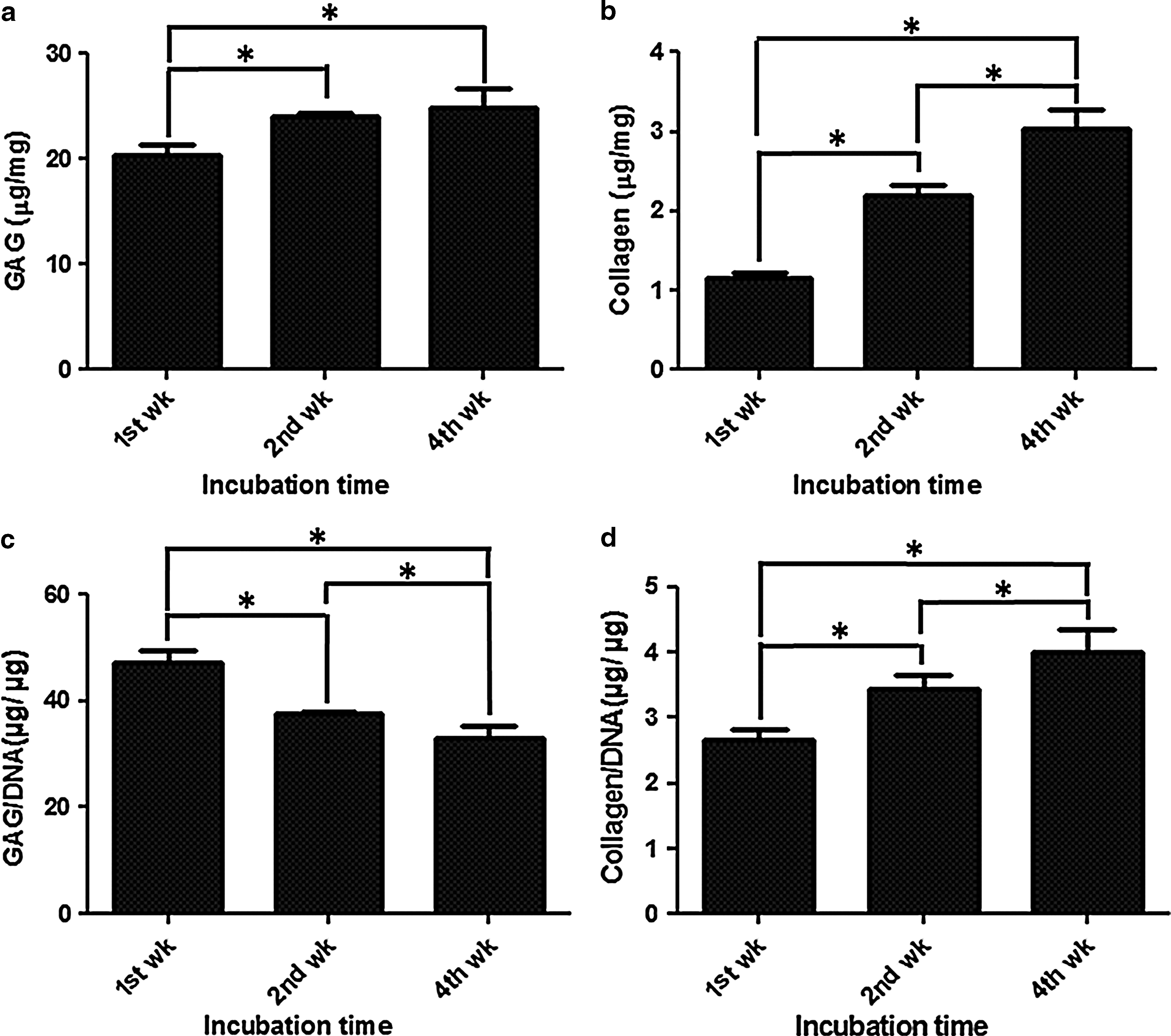
Results of biochemical assays: (
SEM analysis on chondrocyte-laden PEG cryogels
After 1 and 4 weeks of in vitro cultivation, we performed SEM analysis of the chondrocyte-laden PEG cryogels to investigate cell distribution and extracellular matrix (ECM) accumulation. SEM analysis indicated the presence of chondrocyte secreted ECM within the cryogels (Fig. 5). The extent of ECM accumulation appeared to increase from week 1 to 4. A closer observation of the ECM indicates the presence of fibrous material, possibly collagen (Fig. 5b, d) within the cryogel section.
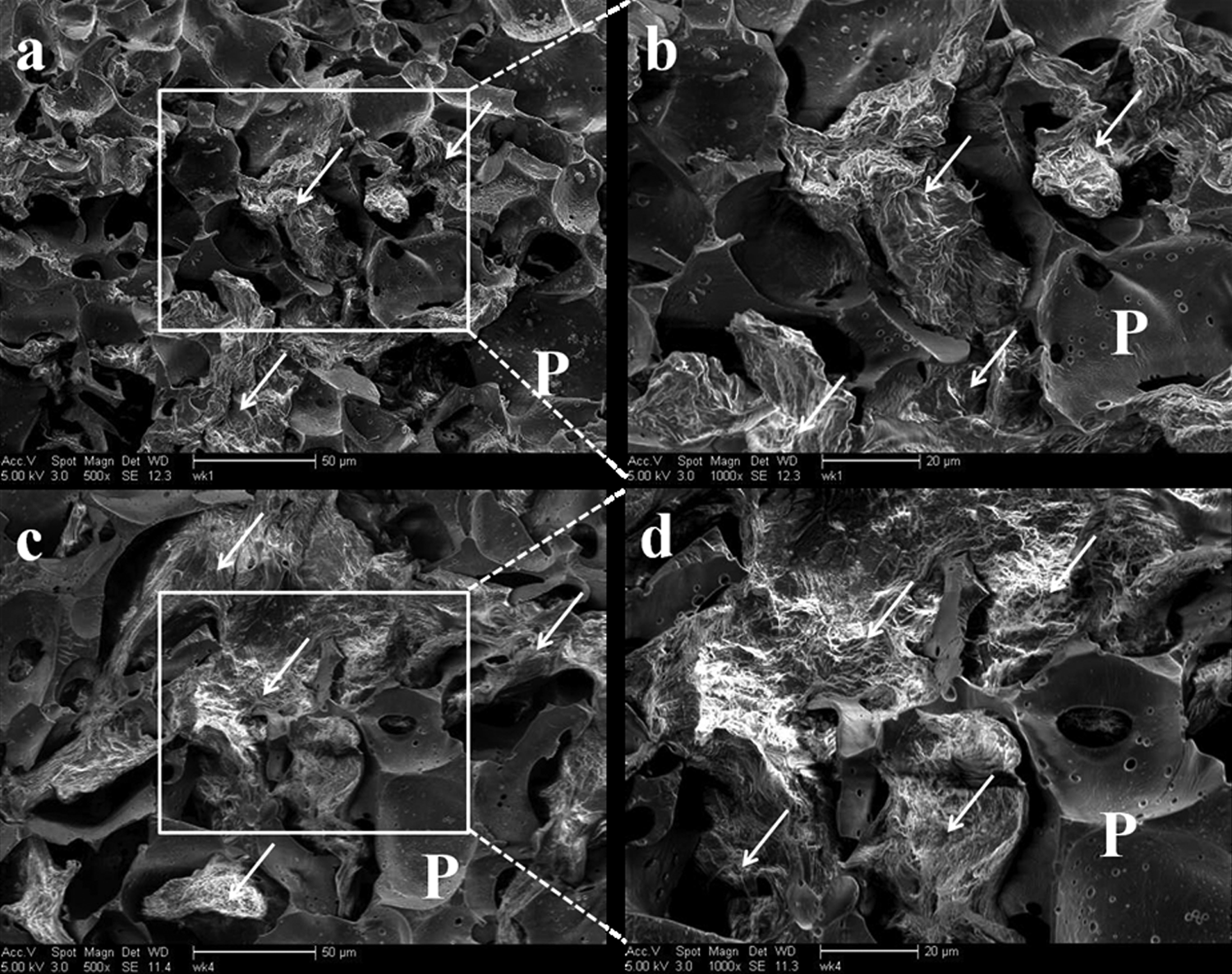
Scanning electron microscopy images of chondrocytes seeded on poly(ethylene glycol) cryogels at different magnifications (scale bar: (
Histological and immunohistochemical analysis
In addition to biochemical analysis, we also examined the cartilage tissue formation by histochemical and immunohistochemical studies to observe newly synthesized proteoglycans and collagen type II after the first and fourth week of cultivation. Hematoxylin and eosin staining of the cell-laden cryogels after week 1 and 4 clearly indicated cell proliferation, confirming the data obtained through DNA quantification (Fig. 6). Basophilic ECM surrounding the chondrocytes was notably increased at week 4 of culture. Moreover, it was observed that chondrocytes within the interconnected macroporous networks had rounded or somewhat elongated morphology (Fig. 6a, b). Safranin-O staining for negatively charged proteoglycans showed matrix production by the chondrocytes within the cryogels. Areas of larger and more intense Safranin-O staining were observed after 4 weeks compared to 1 week of culture (Fig. 6c, d). A similar trend was observed for collagen type II production for the chondrocyte-laden cryogels with culture time (Fig. 6e, f). The cell-secreted ECM was found to be distributed throughout the cryogel. Immunostaining for collagen type I showed absence of collagen type I proteins after week 1 and 4 of culture, indicating that the chondrocytes cultured within the cryogels maintained their phenotype (Fig. 6g, h).

Results of hematoxylin and eosin staining (
Discussion
Interconnected macroporous network structures have been known to support cell growth and matrix production.10–13 Previously, we have reported the development and characterization of interconnected macroporous PEG cryogels using cryogelation techniques. Such cryogels can withstand larger deformation, exhibit higher fracture stresses, and undergo rapid re-swelling upon immersion in aqueous solution. 27 Similar to hydrogels, cryogels also have hydrophilic networks; however, they have interconnected porous network structures unlike conventional hydrogels that exhibit closed pores. Although the studies we reported previously showed the ability of PEG cryogels to support cell culture, 27 their ability to function as tissue engineering scaffolds remains to be explored. In the present study, we have investigated the potential of PEG cryogel scaffolds for cartilage tissue engineering using primary bovine chondrocytes.
The dropwise addition of cell suspension along with the interconnected macroporous network structures and the ability of cryogels to rapidly imbibe aqueous solutions (Figs. 1 and 2) promoted the homogeneous distribution of cells within the cryogel upon seeding with minimal detrimental effects to cells. The high cell survivability observed is in agreement with previously reported studies involving dropwise seeding of cells.
37
The histological analysis further supported the infiltration of cells and their homogenous distribution within the cryogels (Fig. 6). In addition to promoting the adhesion of chondrocytes, cryogels supported their proliferation. Interestingly, the chondrocytes exhibited different growth rates: an initial rapid growth for 1 week, followed by a period of slower growth between 1 and 2 weeks, which then almost plateaued. This trend in proliferation could be attributed to the large pore size available at initial culture times, which decreases with culture time because of ECM matrix accumulation. The decrease in proliferation could also be due to the increased biosynthetic activity of chondrocytes. The proliferation is similar to a previously reported trend describing the three-stage proliferation of chondrocytes on porous poly(
The pore size and interconnectivity of macroporous three-dimensional scaffolds have been shown to play important roles in cell adhesion, migration, viability, metabolism, and growth of cells. 39 For instance, Griffon et al. have demonstrated a pore-size-dependent distribution, attachment, and proliferation of the chondrocytes within the three-dimensional hydrogels. 40 In another study, Oh et al. have demonstrated that the chondrocytes, osteoblasts, and fibroblasts cultured in polycaprolactone scaffolds with varying pore sizes exhibited enhanced cell growth in scaffolds having larger pore sizes. 41 The increased matrix accumulation could adversely affect the effective mass transport associated with the large pore size as with increasing matrix accumulation the pore size is decreasing. Hence, incorporating degradable moieties along with macroporous network would be highly advantageous for engineering large tissues, where the space created by scaffold erosion compensates the decrease in scaffold pore size due to matrix accumulation.40,42 However, there are few reports showing long-term cell survival in interconnected macroporous nondegradable scaffolds as well. 37
In addition to proliferating, chondrocytes secreted cartilage-specific ECM components. Interconnected macroporous networks in the cryogels enabled uniform distribution of cell-secreted GAG molecules throughout the entire scaffold as observed in histological and immunuhistological evaluation (Fig. 6c, d). This is in agreement with previous findings that showed the effect of crosslink density of hydrogels on diffusion and the distribution of GAG molecules. 43 Due to their high solubility in water, GAG can easily diffuse out of the gel before their polymerization into larger molecules.44,45 This, along with the large pore size of croygels, could also explain the observed reduction in GAG content (normalized to the corresponding DNA values) from week 1 to 2. However, with increasing culture time the diffusion of GAG molecules from the cryogels would decrease as the seeded chondrocytes deposit more matrices. Previously reported studies have also shown that cell-secreted GAG diffuses out of loosely crosslinked hydrogels. 46 The ability of GAG molecules to diffuse within the cryogel has been also seen from Safranin-O staining at week 1 to 4; the larger Safranin-O-stained regions were observed in week 4 as compared with week 1.
In contrast to GAG, collagen content within the cryogel was found to increase with culture time. This may be due to the fact that the high molecular weight of collagen molecules along with their intermolecular forces significantly increases their confinement within the cryogel network.47,48 This confinement could also be attributed to the collagen type I coating of cryogels, even though collagen content of the acellular cryogels was found to be below the detectable range by hydroxyproline assay. Although the hydroxypoline assay does not distinguish specific collagen type, findings from the immunohistological staining (for collagen type I and II) indicate that the majority of the cartilage matrix was collagen type II specific, further supporting the maintenance of chondrocyte phenotype on the matrix.
The accumulation of ECM components within the cryogels was also confirmed by SEM analysis, which showed enhanced matrix accumulation at week 4 as compared with week 1. At 4 weeks of culture, accumulation of ECM synthesized by the cells appeared to fill the cryogel macropores. In sum, the macroporous cryogels with significant dimensions and interconnectivity were found to support chondrocyte culture and matrix accumulation in vitro.
Conclusion
This study demonstrates the potential of macroporous PEG cryogels as a cell scaffold for cartilage tissue engineering using primary bovine chondrocytes. We evaluated the highly interconnected and macroporous internal morphology of cryogels by various microscopic analyses. The seeded chondrocytes were found to be successfully distributed within the macroporous network. The seeded chondrocytes proliferated, maintained chondrocyte phenotype, and secreted cartilage-specific matrix within the cryogels. Our findings described in this article demonstrate that the PEG cryogels can successfully support cell proliferation and cartilage-specific ECM production, showing their promising application as scaffolds for cartilage tissue engineering.
Footnotes
Acknowledgments
The authors acknowledge Ryan Anderson for his assistance with the use of the SEM facility in Calit2 at University of California, San Diego. We also thank Ameya Phadke for his critical evaluation.
Disclosure Statement
No competing financial interests exist.