
Abstract
Most tissue engineering studies use human bone marrow mesenchymal stem cells for differentiation into desirable lineages. We derived a novel stem cell from the human umbilical cord Wharton's jelly (hWJSC) that has numerous advantages over other stem cell types in that they can be harvested in abundance very efficiently and painlessly with no risk of patient morbidity, have prolonged stemness properties in vitro, are hypoimmunogenic, and can be differentiated into many tissue types in two-dimensional culture. We compared four different three-dimensional nanofibrous scaffolds (polycaprolactone [PCL], PCL/collagen [PCL/Coll], PCL/hydroxyapatite [PCL/HA], and PCL/Coll/HA) for the attachment, proliferation, differentiation, and mineralization of hWJSCs into an osteogenic lineage. The collagen-based scaffolds (PCL/Coll and PCL/Coll/HA) showed better cell attachment and proliferation than PCL and PCL/HA, with increases of 41.80% and 38.52%, respectively. hWJSCs cultured on PCL/Coll/HA in the osteogenic medium up to 21 days demonstrated increased alkaline phosphatase activity and greater expression of osteocalcin, mineralization, and osteogenic-related genes compared to controls. Given the advantages of hWJSCs over other stem cell types, we propose that hWJSCs may be efficiently differentiated into an osteogenic lineage on a three-dimensional PCL/Coll/HA nanofibrous scaffold for the treatment of bone defects.
Introduction
Human embryonic stem cells (hESCs) and human bone marrow mesenchymal stem cells (hBMSCs) have been differentiated into osteogenic lineages and the derived tissues used for transplantation.1–5 However, the use of hESC-derived tissues is fraught with the problems of immunorejection and teratoma formation, and hBMSCs with the limitations of painful invasive harvest, donor-site morbidity, and infection in the patient, declining cell numbers with age, and short duration of stemness properties in vitro.
We isolated a novel stem cell population from the Wharton's jelly of the human umbilical cord (hWJSCs) with 100% derivation efficiency, short population doubling time, and prolonged duration of stemness properties in vitro. These hWJSCs are noncontroversial, can be harvested painlessly in abundance from discarded cords, have their unique signature of MSC markers, and can be differentiated into many tissues such as neurons, 6 pancreatic islet-like cells, 7 chondrocytes, 8 adipocytes, 9 and endothelial-like cells. 10 These hWJSCs, in addition to their multilineage differentiation potential, are also hypoimmunogenic and do not produce teratomas,6,11 making them an attractive candidate in tissue engineering and regenerative medicine. Recently, Hou et al. 12 successfully differentiated hWJSCs into osteogenic lineages in two-dimensional (2D) cultures in plastic dishes using bone morphogenetic protein 2. They reported that the similar characteristics of osteogenic differentiation they observed in their comparative studies between hWJSCs and hBMSCs support the substitution of hBMSCs with hWJSCs in bone tissue engineering.
However, engineering hWJSCs into bone using three-dimensional (3D) scaffolds to mimic the structural and biochemical cues of the tissue's own in vivo extracellular matrix (ECM) will allow better engraftment and healing compared to differentiation in 2D planes, but not all 3D support systems are made of natural or synthetic polymers, ceramics, or composites that would efficiently support cell attachment and proliferation. Recently, hWJSCs were differentiated on collagen scaffolds (collagen gels) into bone, 13 and as such we thought that it would be interesting to examine the interaction of hWJSCs with nanofibrous substrates (nanoscaffolds) containing collagen, for osteogenic differentiation. Nanoscaffolds have the advantages of good porosity that allow sufficient exchange of nutrients and growth factors and allow the fabrication of nanoscale patterns that mimic the in vivo stem cell niche. Polycaprolactone (PCL), which is a nontoxic synthetic aliphatic polyester, was developed as a bone graft substitute in bone tissue engineering because it undergoes slow hydrolytic degradation into natural metabolites after being fabricated into nanofibers. 14
Since collagen plays a positive role in osteogenic differentiation 13 and hWJSCs secrete large amounts of collagen, we comparatively evaluated the efficiency of four different unpatterned nanofibrous scaffolds (PCL, PCL-collagen [PCL/Coll], PCL-hydroxyapatite [PCL/HA], and PCL/Coll/HA) with or without osteogenic supplements as support systems for hWJSC attachment, proliferation, differentiation, and mineralization into bone. The novelty of our studies was the evaluation of a combination of a novel stem cell with nanoscaffolds and osteogenic supplements for bone differentiation in vitro.
Materials and Methods
Derivation and propagation of hWJSCs
hWJSCs were derived from umbilical cords donated with informed patient consent and ethics approval from the institutional Domain Specific Review Board, Singapore. Derivation of hWJSCs was according to a method developed in our laboratory. 6 The hWJSCs were seeded into sterile plastic Petri dishes (100 mm; Becton Dickinson [BD]) in the hWJSC culture medium. The hWJSC culture medium comprised 80% DMEM supplemented with 20% knockout serum replacement (Invitrogen Life Technologies), 16 ng/mL basic fibroblast growth factor (Millipore Bioscience Research Agents), 1% nonessential amino acids, 2 mM L-glutamine, 0.1 mM β-mercaptoethanol, 1% insulin-transferrin-selenium, and antibiotic mixture of penicillin (50 IU) and streptomycin (50 μg/mL) (Invitrogen Life Technologies). Pure populations of uncontaminated hWJSCs were obtained. The stemness properties of the hWJSCs were confirmed using a battery of MSC marker tests and all studies were carried out on early passages (<7P).
CD marker and cytogenetic analysis of hWJSCs
Briefly, cultured hWJSCs were exposed to trypsin (TrypLETM Express; Invitrogen) for 2 min and then washed in phosphate buffered saline without calcium and magnesium (PBS−) to obtain single cells. The single hWJSCs were then blocked with 10% normal goat serum (NGS) to prevent nonspecific binding. The cells were then incubated with primary antibodies (1:20) for a battery of CD markers viz., CD10, CD13, CD14, CD29, CD34, CD44, CD105, CD117, CD146 (Biolegend), and CD90 (Millipore Bioscience Research Agents), for 30 min, followed by incubation with Alexa Fluor-488 (1:750) secondary antibody for 30 min (Invitrogen Life Technologies). The cells were finally washed in (PBS−), re-suspended in 10% NGS and filtered using a 70 μm nylon strainer to remove any cell clumps, and analyzed using a CyAn™ ADP Analyzer (Beckman Coulter). The chromosome makeup of the hWJSCs was analyzed from karyotypes prepared by conventional cytogenetic analysis.
Fabrication of PCL, PCL/Coll, PCL/HA, and PCL/Coll/HA nanofibrous substrates
PCL (molecular weight, 80,000), gelatin, 1,1,1,3,3,3-hexafluoropropanol (HFP), and 2,2,2-trifluroethanol were purchased from Sigma-Aldrich and bovine collagen type I was purchased from Koken Company. Crystalline HA was a gift from the Department of Metallurgical and Materials Engineering, Indian Institute of Technology, Chennai, India. PCL (10% w/w) was dissolved in methanol and chloroform (1:3) and collagen type I (Coll, 80 mg/mL) was dissolved in HFP by stirring overnight. The molecular chains and conformations of collagen I were not changed by the HFP as confirmed by Fourier Transform InfraRed spectroscopy. Three different solutions were prepared for fabricating the three different nanofibers for bone tissue engineering. These were PCL/Coll (1:1), PCL/HA (1:1), and PCL/Coll/HA (10:60:30 mg/mL dissolved in HFP). We tried different ratios of PCL, collagen, and HA for preparing scaffolds for bone tissue engineering and finally selected the ratio of PCL/Coll/HA (10:60:30) as a substrate for the culture of hWJSCs because our preliminary results showed better cell proliferation and mineralization on these ratios in PCL/Coll/HA than all other scaffolds we fabricated. These ratios of PCL/Coll/HA produced highly porous nanofibrous structures with high surface areas, flexibility for surface modification, and sufficient mechanical strength for handling. Such a fibrous architecture mimicked the natural ECM and assisted in maintaining the normal phenotype of the cells.
Nanofibers were fabricated by separately electrospinning the solutions at 1 mL/h at a voltage of 13 kV using a high-voltage power supply (Gamma High Voltage Research). A ground collection plate (aluminum foil) containing glass cover slips (diameter 150 mm) on which the nanofibers were electrospun was located around 13 cm from the electrospinning needle tip. A positively charged jet was produced from the Taylor cone of the electrospinning apparatus and the nanofibers were sprayed onto the glass cover slips resting on the aluminum foil target. The electrosprayed nanofibers were then dried in a vacuum at room temperature and sputter coated with gold (JEOL JFC-1600 Auto Fine Coater). They were then observed and characterized by field emission scanning electron microscopy (FESEM) (FEI–QUANTA 200F) at an accelerating voltage of 10 kV. The diameters of the nanofibers and HA crystal sizes were measured with scanning electron microscope images using Image J Software (National Institutes of Health).
Osteogenic differentiation of hWJSCs
Four different unpatterned nanofibrous scaffolds, (a) PCL, (b) PCL/Coll, (c) PCL/HA, and (d) PCL/Coll/HA, on separate glass cover slips were housed in the wells of 24-well tissue culture dishes (BD). Sterilized steel rings were placed on top of the glass cover slips containing the nanofibers as a mechanical weight to prevent the nanofibers from lifting off the cover slip. hWJSCs were seeded at 2 × 10⁴ cells/well in a 24-well plate and initially cultured on the four different nanoscaffolds in the presence of the hWJSC medium (without osteogenic supplements) at 37°C in a 5% CO2 in air atmosphere for 24 h. This seeding efficiency was adequate to allow priming and attachment of the cells to the scaffolds. After this, to facilitate differentiation of the hWJSCs toward an osteogenic lineage, the hWJSC medium was supplemented with 100 nM dexamethasone, 50 μM ascorbic acid 2-phosphate, and 10 mM β-glycerophosphate (Sigma) (osteogenic medium) and the cells cultured for 21 days with fresh changes of the medium every 48 h (treatment group). Negative control groups were dishes with hWJSCs from the same passage grown on the same nanoscaffolds but in the presence of the hWJSC medium only without osteogenic supplements. Positive controls were ongoing osteoblast cultures (CRL-11372, American Type Culture Collection, ATCC) on the same nanoscaffolds. Treatment and control groups were compared for cell morphology, cell proliferation, FESEM, alkaline phosphatase (ALP), mineralization, and quantitative real-time polymerase chain reaction (qRT-PCR). The efficiency of osteogenic differentiation was also compared with ongoing 2D native bone and hWJSC cultures grown on plastic in our laboratories.
Cell morphology: Phase contrast microscopy
To evaluate the attachment and morphological behavior of the hWJSCs on the nanofibers, the hWJSCs (2 × 10⁴ cells/well) were cultured on the four different nanoscaffolds in 24-well tissue culture plates (BD) and the morphological changes during culture were monitored daily and photographed using inverted phase-contrast optics (Nikon Instruments) on days 7, 14, and 21.
Cell proliferation assay
The cell proliferation rates of the hWJSCs on the four different nanofibrous scaffolds were evaluated by culture of the hWJSCs (2 × 10⁴ cells/well) both in the hWJSC medium (nonsupplemented) and in the osteogenic medium for 7, 14, and 21 days. The cell proliferation assay was performed using an MTT reagent kit (3-(4,5-dimethylthiazolyl-2)-2,5-diphenyltetrazolium bromide; Sigma) according to the manufacturer's instructions. Briefly, 10 μL MTT reagent (final concentration of 0.5 mg/mL) was added to the medium in the culture dishes and the dishes were incubated for 4 h until a purple precipitate was visible. The medium was then removed and 100 μL of the detergent reagent was added into the dishes and incubation carried out in the dark for 2 h. Absorbance at 570 nm was spectrophotometrically measured using a microplate ELISA reader (Quant; BioTek) with a reference wavelength of 650 nm. After deduction of the blank cell absorbance, the treated cell absorbance over the control cell absorbance was expressed as a percentage of cell survival or proliferation.
Field emission SEM
The hWJSCs were cultured on the four different nanofibrous scaffolds in the osteogenic medium for 7, 14, and 21 days. After these respective culture periods, the nanoscaffolds containing the attached hWJSCs were gently washed with PBS to remove any dead cells and debris. The cells on the nanoscaffolds were then fixed in 3% glutaraldehyde for 3 h at room temperature, dehydrated through a series of graded alcohol solutions, and finally dried in hexamethyldisilazane overnight. The dried cellular constructs were sputter coated with gold and observed under FESEM at an accelerating voltage of 10 kV.
Alkaline phosphatase
The hWJSCs were cultured on the four different nanofibrous scaffolds in the osteogenic medium for 14 days and their ALP activity was assayed by measuring the release of p-nitrophenylphophate (Sigma) according to the method of Fang et al. 15 The ALP levels were normalized to the total protein content determined from an aliquot of the same sample using the Bradford assay (Bio-Rad) as suggested in Ref. 15 .
Mineralization of hWJSCs
The hWJSCs were cultured on the four different nanofibrous scaffolds in the osteogenic medium for 21 days and the production of cell mineralization was evaluated using Alizarin red-S (ARS) staining. Briefly, after the culture period, the hWJSCs on the different nanoscaffolds were washed thrice in PBS, fixed in ice cold ethanol for 1 h, and then washed thrice in distilled water before staining with ARS (40 mM) for 15 min at room temperature. The stained nanoscaffolds were then observed under an optical microscope and after further washes with distilled water they were desorbed in 10% cetylpyridinium chloride (Sigma) for 1 h and absorbance read at 570 nm in a spectrophotometer (Thermo Spectronic).
Von Kossa staining
The hWJSCs were cultured on PCL/Coll/HA nanoscaffolds in the osteogenic medium for 21 days and the degree of mineralization was also evaluated by Von Kossa staining. Briefly, the cells on the scaffolds were rinsed with PBS and fixed in 3.7% formaldehyde solution for 10 min at room temperature. They were then washed with distilled water and stained in 1% silver nitrate solution under ultraviolet light for 60 min. The cells were then washed again with distilled water, treated with 3% sodium thiosulfate for 5 min, and counterstained with 1% nuclear fast red for 5 min. The stained cells were washed with distilled water and photographed under inverted phase-contrast optics (Nikon Instruments).
Immunohistochemistry
The hWJSCs cultured on PCL/Coll/HA nanoscaffolds in the osteogenic medium for 21 days were analyzed for osteocalcin (OCN) by immunohistochemistry. Briefly, the cells were fixed with 4% buffered paraformaldehyde solution, washed with PBS, and treated with 10% NGS. The cells were then incubated with mouse monoclonal anti-OCN primary antibody (1:100) (Invitrogen) for 1 h and goat anti-mouse secondary antibody (Alexa Fluor 488; 2 μg/mL) for 30 min. After PBS washes, the cells were incubated with 4'-6-Diamidino-2-phenylindole (DAPI; 0.5 μg/mL; Molecular Probes, Invitrogen Life Technologies) for 5 min at room temperature, washed with PBS, and analyzed using fluorescence microscopy.
Quantitative real-time polymerase chain reaction
The hWJSCs were cultured on the four different nanofibrous scaffolds in both the osteogenic medium and the hWJSC medium (nonsupplemented) for 21 days. Total RNA was extracted using TRIzolTM reagent (Invitrogen). RNA quality and quantity were measured using a NanodropTM spectrophotometer (Nanodrop technologies), and all samples were treated with DNase-I before first-strand cDNA synthesis with random hexamers using the SuperScriptTM first strand synthesis system (Invitrogen). Primer sequences were taken from earlier published studies and are summarized in Table 1. qRT-PCR analysis was performed with the ABI PRISM 7500 Fast Real-Time PCR System (Applied Biosystems) using SYBR green as previously described 16 and relative quantification was performed using the comparative CT (2-ΔΔCT) method.
F, forward primer; R, reverse primer; ALP, alkaline phosphatase; BSP, bone sialoprotein; CBFA1, core binding factor alpha 1; OCN, osteocalcin; OPN, osteopontin; RUNX2, Runt-related transcription factor; GAPDH, glyceraldehyde 3-phosphate dehydrogenase.
Statistical analysis
The differences observed in hWJSC behavior between treatment and control groups and between nanoscaffolds within the treatment group were compared and analyzed using either one-way analysis of variance with Bonferroni's multiple comparisons post hoc analysis or Students t-test using the statistical package for Social Sciences (SPSS 13). The results were expressed as mean ± standard error of the mean from three different replicates for individual assays and a value of p < 0.05 was considered to be statistically significant.
Results
FESEM of the nanoscaffolds
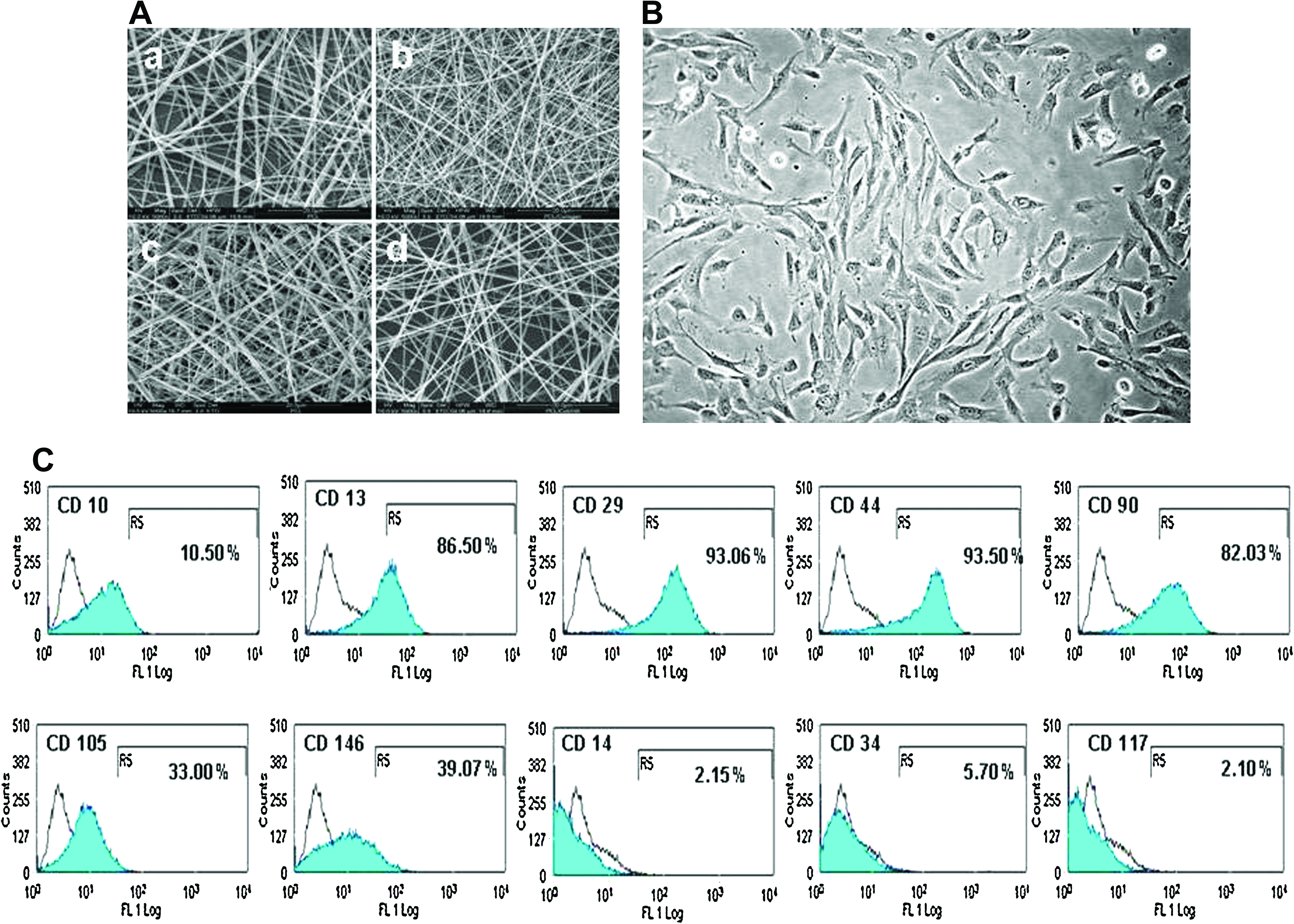
Under FESEM, the nanofibers on all four nanoscaffolds were bead-free, without breaks, and intertwined in a 3D unpatterned topography, producing pores to allow growth and migration of the hWJSCs (Fig. 1A). The diameters of the PCL, PCL/Coll, PCL/HA, and PCL/Coll/HA nanofibers were 510 ± 0.24 nm, 310 ± 0.09 nm, 650 ± 0.16 nm, and 450 ± 0.14 nm, respectively (Fig. 1A). The pore size among the different scaffolds ranged between 5 and 20 μm with a porosity of 80% to 90%.
Morphology, growth, and characterization of hWJSCs
hWJSCs grown on plastic (2D) in the hWJSC medium alone produced epitheliod islands in primary culture, which in subsequent passages transformed to short fibroblasts (Fig. 1B). The hWJSCs had proliferative growth on the plastic surfaces with population doubling times of 24 h.
Flow cytometric analysis of these hWJSCs showed that they were positive for several MSC markers, namely, CD10, CD13, CD29, CD44, CD90, CD105, and CD146 (Fig. 1C). Of these markers, CD13, CD29, CD44, and CD90 were highly expressed (86.50%, 93.06%, 93.50%, and 82.03%, respectively) and CD105, CD146, and CD10 (33.0%, 39.07%, and 10.50%, respectively) moderately expressed. The hWJSCs were negative for CD14, CD34, and CD117 (Fig. 1C).
The same hWJSCs cultured in the osteogenic medium attached and proliferated on the four different nanofibrous scaffolds (PCL, PCL/Coll, PCL/HA, and PCL/Coll/HA) without changing their morphological characteristics. The cells maintained their short fibroblastic morphology, grew into the porous niches of the nanoscaffold, became confluent, and formed a sheet of cells covering the entire scaffold (Fig. 2). The hWJSCs remained firmly attached to the nanoscaffolds even after confluence, in contrast to the conventional 2D plastic dishes where the cell sheets detached after becoming confluent. Although we were evaluating surface differentiation, after removal of the nanoscaffolds from the cover slips the cells were seen attached to the cover slips, indicating that the nanoscaffold pore size was large enough to allow cellular ingrowth. When the hWJSCs on the nanoscaffolds were tested for stemness, they continued to be positive for their respective CD markers.
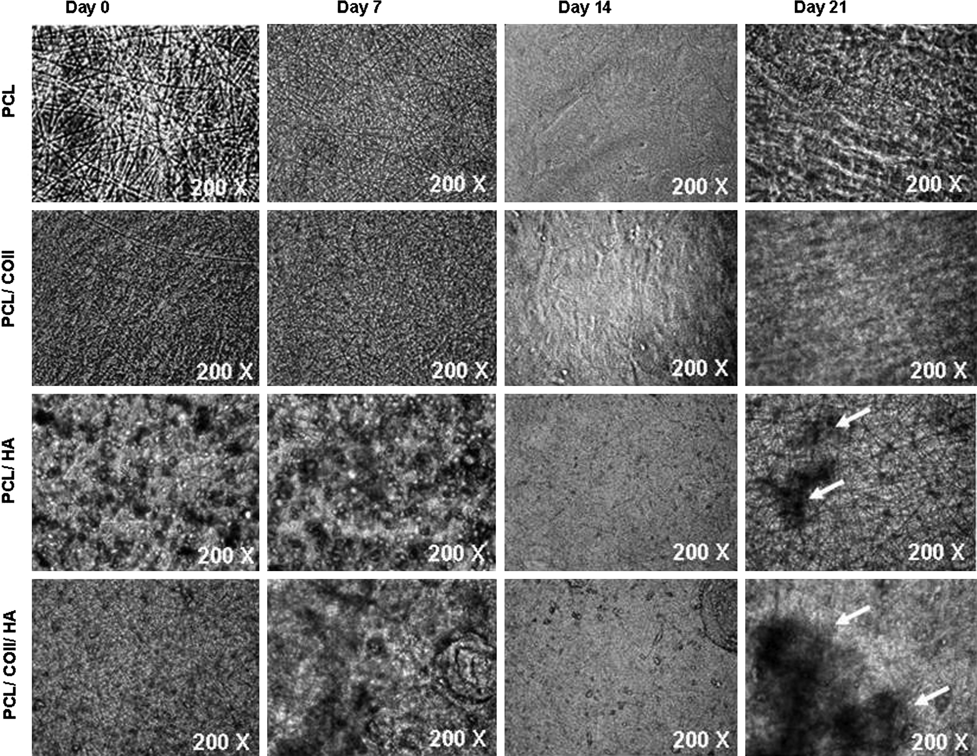
Phase-contrast image of hWJSCs cultured in the hWJSC medium on different nanofibrous scaffolds, namely, PCL, PCL/Coll, PCL/HA, and PCL/Coll/HA until 21 days (Magnification 200×). On day 0, the random mesh topography of the different scaffolds is clearly seen with the integrated HA particles in the PCL/HA scaffold. Proliferation of hWJSCs eventually produced a sheet of cells on the surface by day 14 in all four scaffolds. By day 21, cell piling (arrows) that underwent mineralization was seen, which was more prominent on PCL/HA and PCL/Coll/HA.
Cell proliferation: MTT assay
The cell proliferation rates of the hWJSCs on all four different nanofibrous scaffolds (PCL, PCL/Coll, PCL/HA, and PCL/Coll/HA) in the hWJSC medium increased with time in culture (7, 14, and 21 days). At 7 days the increases in cell proliferation over PCL were 25.71%, 17.14%, and 31.43% for PCL/Coll, PCL/HA, and PCL/Coll/HA nanoscaffolds, respectively. At 14 days the increases in cell proliferation over PCL were 31.71%, 41.46%, and 58.54% for PCL/Coll, PCL/HA, and PCL/Coll/HA nanoscaffolds, respectively. At 21 days the increases in cell proliferation over PCL were 7.69%, 13.85%, and 41.54% for PCL/Coll, PCL/HA, and PCL/Coll/HA nanoscaffolds, respectively. However, only the increase observed for PCL/Coll/HA nanoscaffold over PCL at day 21 was statistically significant (Fig. 3A).
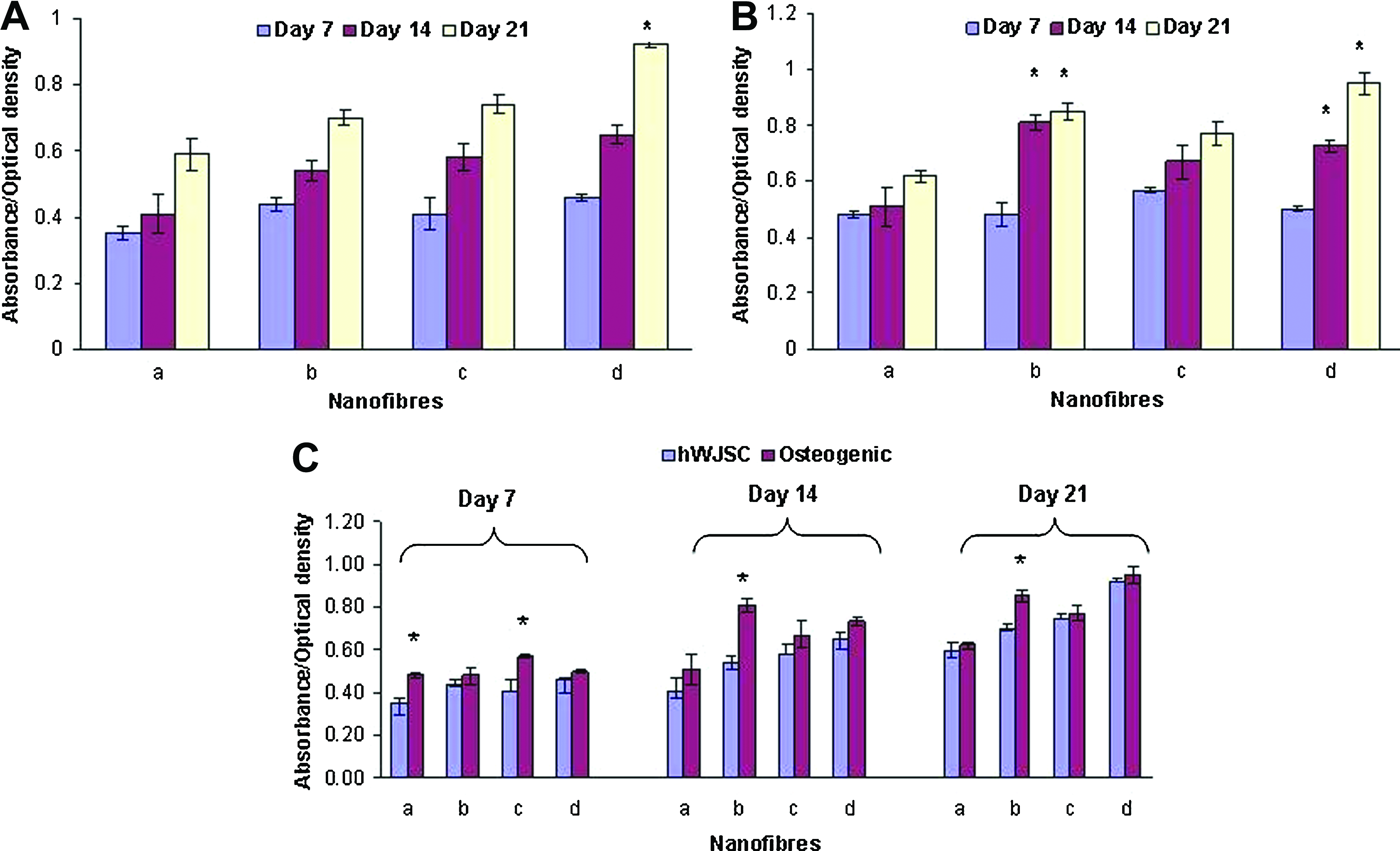
hWJSC proliferation after culture on different nanofibrous scaffolds (a, PCL; b, PCL/Coll; c, PCL/HA; d, PCL/Coll/HA).
When the hWJSCs were exposed to the osteogenic medium for 7, 14, and 21 days, similar increases in cell proliferation were observed with time. At 7 days the increases in cell proliferation over PCL were 18.75% and 4.17% for PCL/HA and PCL/Coll/HA nanoscaffolds, respectively. At 14 days the increases in cell proliferation over PCL were 58.82%, 31.37%, and 43.14% for PCL/Coll, PCL/HA, and PCL/Coll/HA nanoscaffolds, respectively. At 21 days the increases in cell proliferation over PCL were 37.10%, 24.19%, and 53.23% for PCL/Coll, PCL/HA, and PCL/Coll/HA nanoscaffolds, respectively. However, only the increases observed for PCL/Coll and PCL/Coll/HA nanoscaffolds at days 14 and 21 were statistically significant (Fig. 3B).
The hWJSC proliferation rates on all four nanoscaffolds were comparatively better in the osteogenic medium (treatment) than those in the hWJSC medium (controls), and some of these increases were statistically significant (Fig. 3C).
Mineralization of hWJSCs: Alizarin red, Von Kossa, and immunohistochemistry
ARS staining of the hWJSCs cultured on the four different nanofibrous scaffolds in the osteogenic medium for 21 days showed nodules on the scaffold surfaces as early as day 14 that was suggestive of mineralization. The mineral nodules produced from hWJSCs on the PCL/Coll/HA nanoscaffold in the osteogenic medium were the largest in size, greatest in number, and had the most intense ARS staining compared to PCL, PCL/Coll, and PCL/HA (Fig. 4A). Nodule formation was also evident in the FESEM images of the cells cultured on all four nanoscaffolds at days 7, 14, and 21 (Fig. 4B). Calcium mineralization was increased in all four nanoscaffolds in the treatment group compared to controls after elution of the ARS stain and spectroscopic analysis. The increases were 15.79%, 22.54%, and 31.37% for PCL; 36.73%, 44.23%, and 49.51% for PCL/Coll; 34.88%, 28.30%, and 46.03% for PCL/HA; and 33.93%, 38.50%, and 51.96% for PCL/Coll/HA. Only the increases observed for PCL/Coll at 14 and 21 days, PCL/HA at 21 days, and PCL/Coll/HA at 14 and 21 days were statistically significant in the treatment group compared to controls (Fig. 4C).
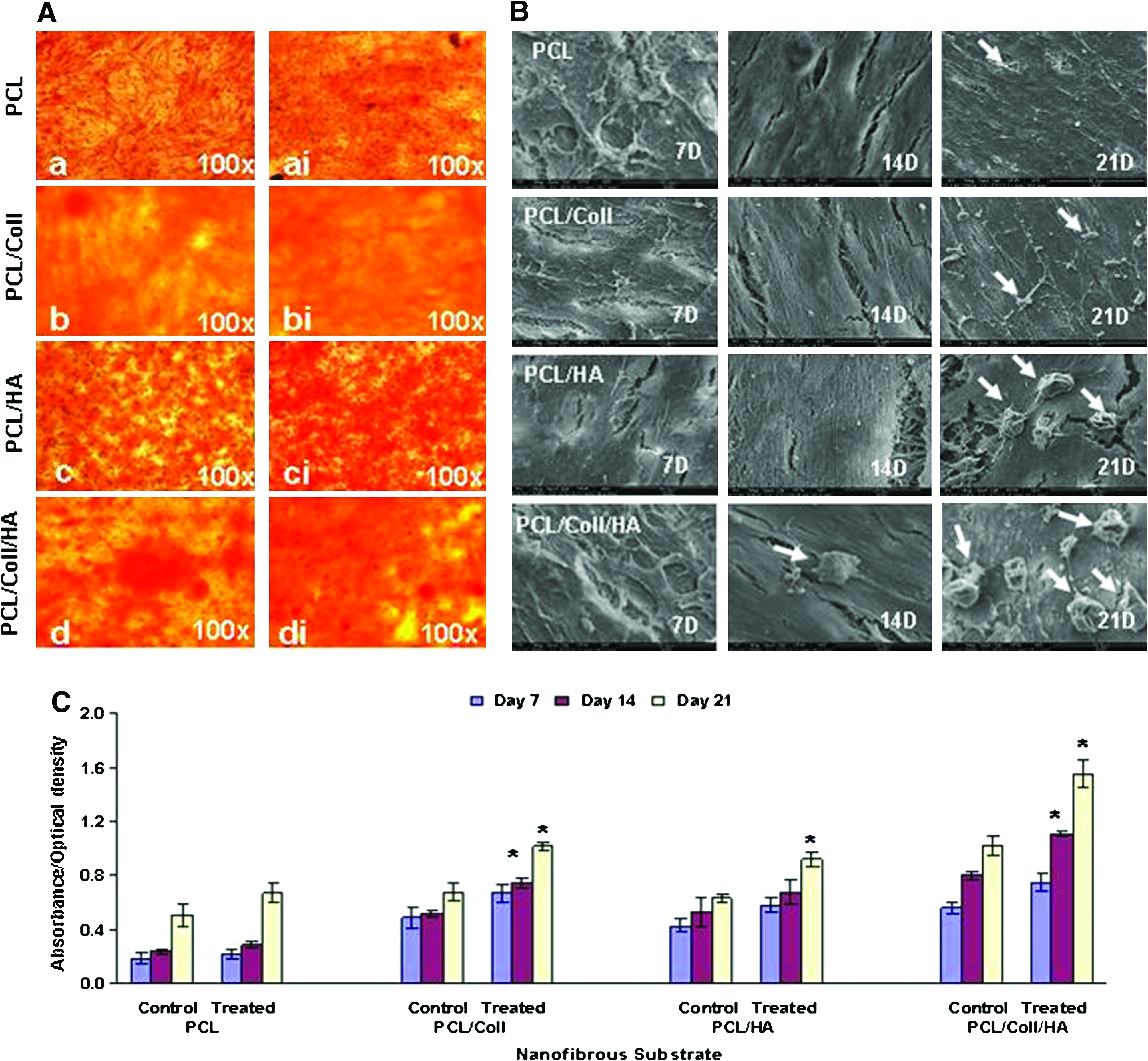
hWJSCs undergoing mineralization after culture on PCL, PCL/Coll, PCL/HA, and PCL/Coll/HA nanofibrous scaffolds in the osteogenic medium for 21 days.
The hWJSCs cultured on PCL/Coll/HA nanoscaffolds in the osteogenic medium for 21 days demonstrated the most intense Von Kossa staining of the nodules compared to other scaffolds in the treatment and control groups (Fig. 5a, ai).

hWJSCs cultured on PCL/Coll/HA nanofibrous scaffolds in the osteogenic medium for 21 days.
The hWJSCs cultured on PCL/Coll/HA nanoscaffolds in the osteogenic medium for 21 days demonstrated the most intense positive staining for OCN using immunohistochemistry with anti-OCN antibodies (Fig. 5b, bi).
Alkaline phosphatase
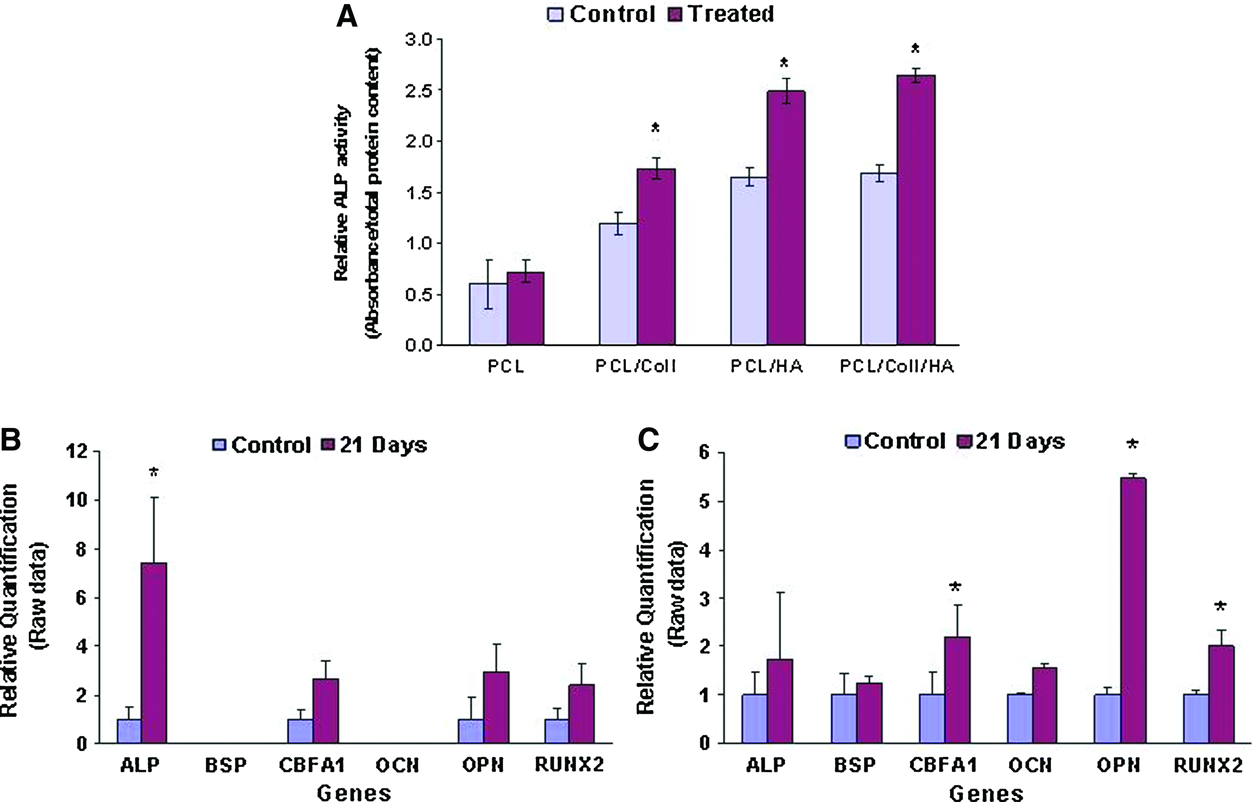
The ALP activity of hWJSCs on the four different nanofibrous scaffolds (PCL, PCL/Coll, PCL/HA, and PCL/Coll/HA) in the osteogenic medium for 14 days was greater by 20.45%, 44.78%, 51.20%, and 59.70%, respectively, compared to controls. The increases observed for PCL/Coll, PCL/HA, and PCL/Coll/HA over controls were statistically significant (Fig. 6A).
Quantitative real-time polymerase chain reaction
qRT-PCR analysis showed expression of osteogenic related genes in cells grown only on the collagen-based scaffolds (PCL/Coll and PCL/Coll/HA). Such gene expression was not significant for the noncollagen-based nanoscaffolds (PCL and PCL/HA). Although most of the osteogenic-related genes studied were expressed in cells grown on the PCL/collagen nanoscaffolds cultured in the osteogenic medium for 21 days, only ALP was significantly increased compared to controls (Fig. 6B). The qRT-PCR analysis of hWJSCs cultured on PCL/Coll/HA nanoscaffolds in the osteogenic medium for 21 days showed increased expression of the osteogenic-related genes compared to controls (ALP by 1.7-fold, bone sialoprotein [BSP] by 1.2-fold, core binding factor alpha 1 [CBFA1] by 2.2-fold, OCN by 1.6-fold, osteopontin [OPN] by 5.5-fold, and Runt-related transcription factor 2 [RUNX2] by 2.0-fold) and the expression levels of CBFA1, OPN, and RUNX2 were significantly different from controls (Fig. 6C).
Discussion
Recently, Hou et al. 12 demonstrated that hWJSCs were a promising alternative stem cell source to bone marrow-derived MSCs (hBMSCs) for bone tissue engineering in 2D planes due to their ready availability and potential to proliferate and differentiate. They reported that after 14 days of bone morphogenetic protein-2 treatment the overall expression level of several osteogenic-specific phenotypes such as collagen type I, OPN, and OCN was similar for hWJSCs and hBMSCs. The signaling pathways for both cell types involved membrane receptor-initiated signals, including smooth muscle actin drosophila (SMADs), P38, and extracellular regulated kinase.
However, to translate such 2D findings to clinical situations, the process of cell culture and expansion on plastic surfaces in vitro followed by the transplantation of single osteocytes in vivo may not provide full restoration of anatomical and physiological balances when it comes to sustained engraftment and functional outcome. The tissue that is to be transplanted needs to have mechanical support that could hold the cultured cells in position for sufficient periods of time to integrate, adapt, and recover, as well as stimulate the natural in vivo tissue repair process via stem cell niches. This can best be achieved through tissue engineering by developing a 3D construct where a combination of a scaffold and the stem cell-derived osteocytes are involved in the repair, restoration, and regeneration of tissue damaged by the injury.
The results of the present study clearly demonstrated that hWJSCs cultured on the different nanoscaffolds readily attached to their surfaces, proliferated, migrated into the depths of the pores created by the unpatterned topography of the nanofibers, and differentiated readily into bone. The osteodifferentiation of the hWJSCs on the nanoscaffolds was as efficient as native bone cultures in our laboratory. The cell numbers did not increase significantly with time (14–21 days) on most of the nanoscaffolds probably because the cells were entering the differentiation phase and attempting to slow down their growth. The PCL/Coll/HA nanoscaffold appears to have the properties of a suitable nanoscaffold because PCL is a nontoxic, biodegradable, cost-effective substance that has been used as a bone substitute, 17 the mineral composition of HA closely resembles natural bone and has osteoconductive properties, 14 and collagen I is the main organic component of the bone ECM that aids in mineralization. 18
PCL is nonhydrophilic while collagen is hydrophilic. Although our earlier air-plasma treatment of the PCL and PCL/HA scaffolds rendered them more hydrophilic and facilitated cell attachment, those nanoscaffolds that contained collagen demonstrated significantly higher cell proliferation, irrespective of whether the hWJSCs were cultured in the hWJSC or osteogenic medium. Collagen, being a natural ECM component, probably provided more focal adhesions and also imparted the necessary biomimetic cues for these cells to attach and proliferate.
The hWJSCs cultured in the osteogenic medium for 21 days on all four nanoscaffolds underwent mineralization and the mineral nodules were more pronounced in the PCL/HA and PCL/Coll/HA nanoscaffolds, thus indicating the role of HA in the mineralization process. The HA content in biocomposites have been shown to enhance the bioactivity of the material. 19 In earlier studies using BMSCs it has been shown that incorporation of HA into nanoscaffolds increased mineralization of cells cultured in the presence of osteogenic growth factors.20,21 The bone cortex comprises approximately of 70% HA and 30% collagen and it is known that inorganic crystals precipitate onto organic matrix surfaces in the biomineralization process. However, the interaction of various proteins such as BSP, OCN, and OPN with HA in its nucleating activities dictate the final biological formation of crystals. 22
ALP is an early marker of osteogenic differentiation and mineralization.23–26 The hWJSCs cultured in the osteogenic medium on the four nanofibrous scaffolds for 14 days in the present study showed increased ALP activity. This ALP activity was comparatively higher in nanoscaffolds containing HA, probably because of the nucleating properties of HA and the ensuing mineralization contributing to the increase in ALP activity.
The increase in ALP gene expression, which is an early marker of osteogenic differentiation, 23 together with expression of RUNX2 and CBFA1, clearly indicates that the hWJSCs were being directed along bone lineage. CBFA1 belongs to the runt-domain gene family 27 and is restricted to osteoblasts. 28 It is also known to induce osteoblast differentiation of nonosteoblastic cells although it mainly regulates expression of other osteoblast-related genes such as OCN, OPN, and BSP during embryonic development.29–30 Activated RUNX2 induces downstream osteoblast-specific markers OCN and OPN. 31 In the present study, the hWJSCs cultured on nanoscaffolds in the osteogenic differentiation medium demonstrated increased expression of OCN and OPN. Detection and association of OPN in the calcification prosthetic heart valves 32 clearly indicate the role of OPN in mineralization and the expression of highly phophorylated OPN is thought to regulate the size and shape of the mineral crystals. 33 The bone gla protein or OCN is exclusively found in bone tissue forming 10%–20% of the noncollagenous protein in bone and is thought to play a role in ossification and mineralization. 34 It is a late marker of osteogenesis and is also involved in bone resorption after bone mineralization. 35 The OCN expression observed after immunohistochemistry of hWJSCs cultured on the PCL/Coll/HA nanoscaffold for 21 days in the osteogenic medium in the present study thus clearly confirms the production of osteogenesis.
The bone matrix is composed mainly of type-I collagen, HA, calcium, and phosphorous in a crystalline form 36 together with other components such as glycosaminoglycans, OCN, and osteonectin. 37 Human Wharton's jelly is a rich source of proteoglycans and hyaluronic acid (which constitutes about 70% of glycosaminoglycan content) immobilized in an insoluble collagen fibril network 38 and are secreted by the hWJSCs residing in the jelly helping the umbilical cord to be well hydrated and together with the connective tissue matrix protects the umbilical blood vessels within the umbilical cord. Thus, these secretory properties of hWJSCs as well as the recent evidence that clearly highlights other advantages of hWJSCs over other MSC types and hESCs6,39 make them the more beneficial and ideal stem cell for differentiation of bone and future clinical applications compared to other stem cell types.
Considering the several advantages hWJSCs possess over other stem cell types such as their noncontroversial nature, ease of derivation, abundant supply, painless harvest, proliferative growth in vitro, wide multipotency, long-lasting stemness characteristics in vitro, hypoimmunogenecity, and inability to produce teratomas in vivo, they may be a useful stem cell for tissue-engineered constructs. Nanoscaffolds may be an optimal 3D platform for hWJSCs to attach, proliferate, and differentiate in vivo and have the advantage of being biodegradable in a specific time once the hWJSC-derived bone cells have integrated into the bone defect, thus bringing about repair.
Footnotes
Acknowledgments
The authors acknowledge the grant support provided by the National University of Singapore (R-174-000-089-133) and National Medical Research Council, Singapore (R-174-000-103-213) and the technical assistance provided by Mr. Arjunan Subramanian.
Disclosure Statement
No competing financial interests exist.