
Abstract
Application of nanofibers for the purpose of tissue mimicking and regeneration has become widespread in the field of biomedicine. In this study, polyethersulfone (PES) electrospun nanofibrous membranes were fabricated, modified, and loaded with unrestricted somatic stem cells (USSC) to mimic the natural structure of bone. Untreated PES, plasma-treated PES, and collagen-grafted PES (COL-PES) nanofibers were characterized via Brunauer-Emmett-Teller method, attenuated total reflection Fourier transform infrared, contact angle measurements, and scanning electron microscopy. Their capacity to support proliferation, infiltration, and osteogenic differentiation of USSC was investigated using MTT assay, real-time reverse transcriptase–polymerase chain reaction, histologic staining, alkaline phosphatase activity, and calcium content assay. All the scaffolds had nanofibrous and highly porous structure with large surface area. After surface treatments, hydrophilicity of scaffolds increased intensively and their biocompatibility improved. During osteogenic differentiation of stem cells, alkaline phosphatase activity and calcium content exhibited the highest level in cells on COL-PES. Real-time reverse transcriptase–polymerase chain reaction showed significant difference between the expression levels of osteoblast-related genes on COL-PES compared to other scaffolds. Excellent infiltration of USSC was observed in nanofibrous membranes especially COL-PES. It can be concluded that COL-PES nanofibrous scaffold has potential for bone grafting because of its three-dimensional structure and bioactivity which enhance proliferation, differentiation, and infiltration of USSC.
Introduction
Synthetic or natural polymers are considered as raw materials of nanofibers. Biocompatibility, biodegradability, and mechanical properties of polymers are the criteria upon which they are selected and processed in electrospinning.4,15 Fabricated nanofibrous mats may not have the proper characteristics desired for cell–scaffold interactions. Surface modification of nanofibers is performed to improve biocompatibility and functionality of scaffolds, which makes them more suitable as a tissue-mimicking environment. Surface plasma treatment and protein immobilization on biomaterials are two appropriate techniques that were previously shown to improve cell behavior on engineered scaffolds.16–19
Polyethersulfone (PES) is a biocompatible and nonbiodegradable polymer that is widely used in hemodialysis and different types of membranes.20–22 PES nanofibrous membrane was fabricated by gas/jet electrospinning, 23 and its potential application in medical devices and tissue reconstruction has been investigated in a few recent studies. Chuaa et al. 24 studied the expansion and adhesion of hematopoietic stem cells on PES nanofibers modified with different surface chemical groups. In another study, the effect of PES nanofiber diameter on its biocompatibility and differentiation of neural stem cells was also evaluated. 25
One of the major challenges in the application of nanofibers in tissue engineering is their low potential to attract cells for infiltration and migration into the nanofibrous structure. 26 Many efforts have been performed to improve the characteristics of electrospun nanofibers like porosity and pore size to create three-dimensional (3D) scaffolds, but little success achieved.27–29 Also in our previous study, infiltration of stem cells was enhanced into surface-modified PES. 30
So far, many attempts have been made to design and develop appropriate nano-structured biomaterials to restore the structure and function of damaged hard tissues like bone.31–38 Bone has a rigid and complex structure that is composed of cells and ECM. Most of the ECM components are collagen nano-sized fibrils that have nonwoven organization in which mineral salts have been incorporated. Bone-related cells located inside ECM are responsible for production of ECM components, mineralization, and intercellular communication. These specialized cells are thought to be derived from bone marrow mesenchymal stem cells under microenvironment clues and differentiation signals. 39
Lately, a new type of multipotent stem cells, unrestricted somatic stem cells (USSC), were isolated from umbilical cord blood. They have the ability to differentiate into mesenchymal and nonmesenchymal cells. Comparing the source, isolation of USSC is easier and less invasive than mesenchymal stem cells. 40 USSC were also shown to have a strong capacity for differentiation into osteolineage accompanied by induction of angiogenesis and vasculogenesis.41,42 These characteristics make USSC an ideal candidate to be used in bone regeneration.
An ideal bone graft substitute (BGS) should have the following properties: (1) osteoconductivity, (2) osteoinductivity, (3) containing osteogenic cells, and (4) structural integrity. Osteoconductivity is provided by the nonviable matrix such as ceramics in the graft substitute. Growth factors such as bone morphogenetic protein and transforming growth factor serve as osteoinductive agents that give rise to differentiation of osteogenic cells into mature bone cells.43–45 Following this pattern, we studied the potential of PES nanofibrous membrane as a new BGS. Collagen I was grafted onto the surface of nanofibers to promote the osteoconductivity of PES scaffold. USSC were used as osteogenic cells that were differentiated into osteolineage under the induction medium. This complex ideally provided a structure to improve healing at bone defect sites in further in vivo studies.
Materials and Methods
Isolation, culture, and differentiation of unrestricted somatic stem cells
Collection, isolation, and expansion of human USSC were performed as described previously. 40 Briefly, cord blood was collected from the umbilical cord vein with informed consent of the mother, and the mononuclear cell fraction was separated by density centrifugation over a Ficoll-Hypaque gradient (Pharmacia-Amersham, d = 1.077 g/mL). Growth and expansion of the cells were performed in low-glucose Dulbecco's modified Eagle medium (DMEM; GIBCO-BRL) supplemented with 30% fetal bovine serum (FBS; Gibco), dexamethasone (100 nM, Sigma-Aldrich), penicillin (100 U/mL, Gibco), streptomycin (0.1 mg/mL, Gibco), and L-glutamine (2 mM, Gibco). USSC were cultured in a humidified atmosphere of 95% air with 5% CO2 at 37°C and extensively propagated. After almost 2 weeks, USSC colonies were appeared, and then the cells were detached with 0.25% Trypsin-EDTA (Gibco) and replated, and 10% FBS was used for further culture. Passage 2 (P2) cells were used for this study. In addition, MG-63 cells (human osteosarcoma cell line) were obtained from Pasteur institute of Iran and were used as control. For osteogenic differentiation, the cells were cultured in DMEM supplemented with 10% FBS, 10−8 M dexamethasone, 0.2 mM ascorbic acid 2-phosphate (Sigma), and 10 mM β-glycerophosphate (Sigma).
Scaffold preparation
Nanofibrous PES scaffolds were prepared via electrospinning. Briefly, a 25 wt% solution of PES (BASF) in dimethylformamide was placed in a 10 mL syringe. An extension tube connected the syringe to a 21-gauge needle that was placed in a 15 cm distance from a steel grounded collector. The solution fed through the tube into the needle by a syringe pump. Application of a 20 kV voltage between the needle and collector forced the solution droplet to leave the needle and deposit on the collector as fibers with nano-sized diameters.
Surface treatment
Treatment of nanofiber surface was performed in two stages: (a) plasma treatment and (b) collagen grafting. Surface plasma treatment was performed by low frequency plasma generator of 40 kHz frequency with a cylindrical quartz reactor (Diener Electronics). Pure oxygen was introduced into the reaction chamber at 0.4 mbar and then the glow discharge was ignited for 5 min. For collagen grafting, plasma-treated sheets were cut into 1.5-cm-diameter punches and immersed in 1-ethyl-3-(3-dimethylaminopropyl)carbodiimide/N-hydroxysuccinimide (Merck) solution (5 mg/mL) for 12 h. After rinsing with distilled water, the scaffolds were immersed in 1 mg/mL collagen I solution (Nutacon BV) overnight. The scaffolds were then rinsed with distilled water and divided into three groups: PES as untreated nanofibers, PL-PES as plasma-treated PES nanofibers, and COL-PES as collagen-grafted PES nanofibers. In addition, tissue culture polystyrene (TCPS) and collagen-coated TCPS (COL-TCPS) were used as control. Further studies were performed on these five surfaces seeded with USSC and MG-63.
Cell seeding
Before cell seeding, circular scaffolds were immersed overnight in the following solutions: (1) 70% ethanol for sterilization, (2) penicillin, streptomycin, and amphotericin B to prevent from yeast growth, and (3) culture medium to ensure sterilization and enhance cell attachment after seeding. An initial cell density of 5 × 10⁴ per cm2 of scaffolds was suspended in 75 μL medium and seeded onto the top of the circular scaffolds, TCPS and COL-TCPS and incubated for 2 h. To immerse the scaffold completely, 500 μL medium was used and replaced every 2 days and the cell culture was performed in 24-well plates. The cells on these scaffolds were incubated under basal and osteogenic medium (OM) for 2 weeks.
Scanning electron microscopy
The surface morphology of scaffolds was characterized using a scanning electron microscope (SEM; Philips XL30) after specimens were coated with gold using a sputter coater. The fiber diameter was determined from SEM images using image analysis software. Morphology of USSC on the scaffolds during osteogenic differentiation was also investigated by SEM. The cell-loaded scaffolds were rinsed with phosphate buffered saline after 7 and 14 days of osteogenic differentiation and fixed in gelutaraldehyde 2.5% for 1 h. For dehydrating, the scaffolds were placed in a series of gradient of alcohol concentration and then dried.
Contact angle measurement
To study the wettability of the nanofiber surface after surface treatment, water contact angle was measured by the sessile drop method with a G10 Kruss contact angle goniometer at room temperature. A water droplet is placed on the scaffold surface and contact angle was measured after 10 s.
Attenuated total reflection Fourier transform infrared spectroscopy
Surface chemical modifications after plasma treatment and collagen grafting were investigated by attenuated total reflection Fourier transform infrared (ATR-FTIR). The spectra were recorded using an Equinox 55 spectrometer (Bruker Optics) equipped with a deuterated triglycine sulfate (DTGS) detector and a diamond ATR crystal.
Porosity and surface area measurements
For porosity determination, four randomized circular samples with the diameter of 20 mm were used and the estimated porosity of each sample was calculated by the following equation: Porosity = 1 − (calculated membrane density/known material density) × 100.
Surface area of nanofibers in scaffolds was measured through Brunauer-Emmett-Teller (BET) method by BELSORP-mini apparatus (BEL Japan, Inc.). Nitrogen adsorption-desorption isotherms of nanofibers were obtained and surface area was calculated from BET plot of isotherms by BELSORP-mini software.
MTT assay
Proliferation of USSC on different scaffolds was evaluated via 3-(4,5-dimethylthiazol-2-yl)-2,5-diphenyltetrazolium bromide (MTT) assay. Sterilized nanofibrous membranes were placed in a 24-well culture plate, seeded with a cell density of 5 × 10³ cells per cm2, and incubated at 37°C, 5% CO2. After 1, 2, 3, 4, and 5 days of cell seeding, 50 μL of MTT solution (5 mg/mL in DMEM) was added to each well (n = 4). For conversion of MTT to formazan crystals by mitochondrial dehydrogenases of living cells, the plate was incubated at 37°C for 3 h. For dissolution of the dark-blue intracellular formazan, the supernatant was removed and constant amount of an appropriate solvent was added. The optical density (OD) was read spectrophotometrically at a wavelength of 570 nm. The same procedure was performed for cultured USSC on TCPS as control.
Real-time reverse transcriptase–polymerase chain reaction
To quantify the difference between the mRNA levels of osteogenic markers, gene expression in USSC on scaffolds was analyzed using real-time reverse transcriptase–polymerase chain reaction (PCR). Total RNA was extracted and random hexamer primed cDNA synthesis was carried out using Revert Aid first strand cDNA synthesis kit (Fermentas). cDNA was used for 40 cycle PCR in Rotor-gene Q real-time analyzer (Corbett). Real-time PCR was performed using Maxima™ SYBR Green/ROX qPCR Master Mix (Fermentas) followed by melting curve analysis to confirm PCR specificity. Each reaction was repeated three times and threshold cycle average was used for data analysis by Rotor-gene Q software (Corbett). Genes and related specific primers are illustrated in Table 1. Relative expression was quantified using ΔΔCt method. Target genes were normalized against glyceraldehyde 3-phosphate dehydrogenase and calibrated to USSC P2.
GAPDH, glyceraldehyde 3-phosphate dehydrogenase; ALP, alkaline phosphatase.
Alkaline phosphatase activity
For alkaline phosphatase (ALP) activity assay, total protein of cells on TCPS and scaffolds was extracted using 200 μL radioimmunoprecipitation assay (RIPA) lysis buffer. The lysate was then centrifuged at 14,000 g at 4°C for 15 min to sediment cell debris. Supernatant was collected and ALP activity was measured using p-nitrophenyl phosphate as substrate and a standard curve for calcium concentration in soloution. The activity of enzyme (IU) was normalized against total protein.
Calcium content assay
The amount of calcium deposited on TCPS and different scaffolds by USSC during osteogenic differentiation, was measured using cresolphthalein complexone method. Calcium extraction was performed by homogenization of the scaffolds in 0.6 N Hydrochloric acid (Merck) followed by shaking for 4 h at 4°C. OD was measured at 570 nm after the addition of the reagent to calcium solutions. Calcium content was obtained from the standard curve of OD versus a serial dilution of calcium concentrations.
Histologic staining
Infiltration of USSC into scaffolds was investigated during osteogenic differentiation via histologic staining. Cell-loaded scaffolds were fixed in formalin (10% buffered, Merck), embedded in paraffin, crossly sectioned, and stained with hematoxylin and eosin (H&E). The sections were then analyzed under light microscope (Labomed).
Statistical analysis
All experiments were conducted at least three times. Data are expressed as mean ± standard deviation. One-way analysis of variance was used to compare results. A p-value of <0.05 was considered statistically significant.
Results
Scaffold characterization
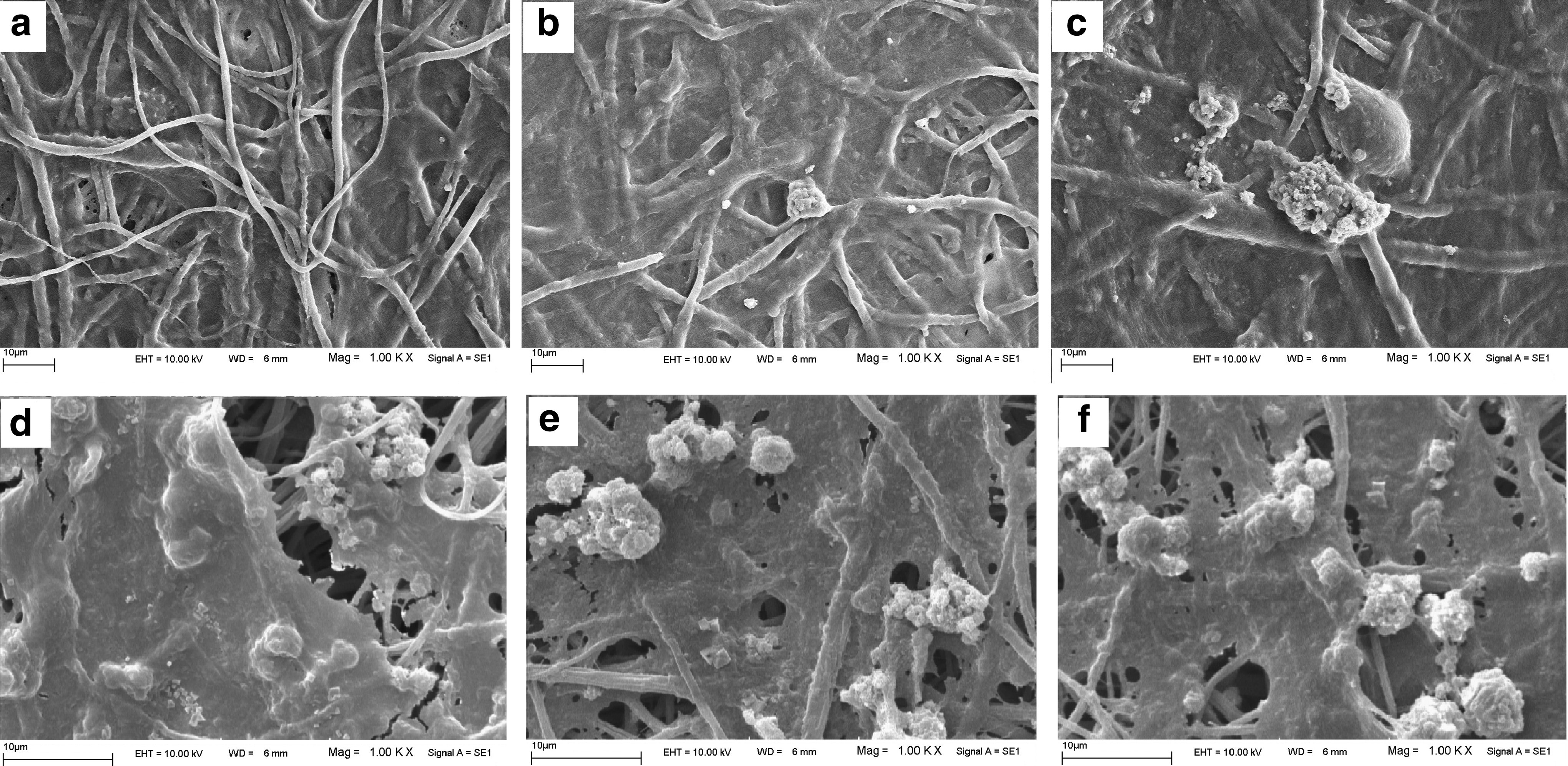
Fabricated nanofibers were characterized morphologically by SEM images (Fig. 1). PES electrospun membrane had nanofibrous and porous structure (Fig. 1a) which did not affected significantly after plasma treatment (Fig. 1b) and subsequent collagen coating (Fig. 1c).

Morphology of fabricated nanofibers: PES
Treated and untreated scaffolds were shown to have ultrathin fibers and high porosity (Table 2). BET analysis also revealed that electrospun membranes had a high surface-to-volume ratio which remained constant after treatments. Surface hydrophilicity of nanofibers was strongly increased after plasma treatment and collagen grafting.
PES, polyethersulfone; PL-PES, plasma-treated PES; COL-PES, collagen-grafted PES.
Grafting of collagen onto the surface of PES nanofibers was shown by ATR-FTIR spectroscopy (Fig. 2). Difference between PES and COL-PES spectra was observed in two peaks at 1649 and 3325 cm−1. These peaks were related to amide A (1649 cm−1) and amid II bands (3325 cm−1) which exist in collagen backbone. Another predicted peak at 1565 cm−1 was overlapped by PES peaks in that region. This peak is also common in proteins and arises from molecular vibrations of amide I bands.
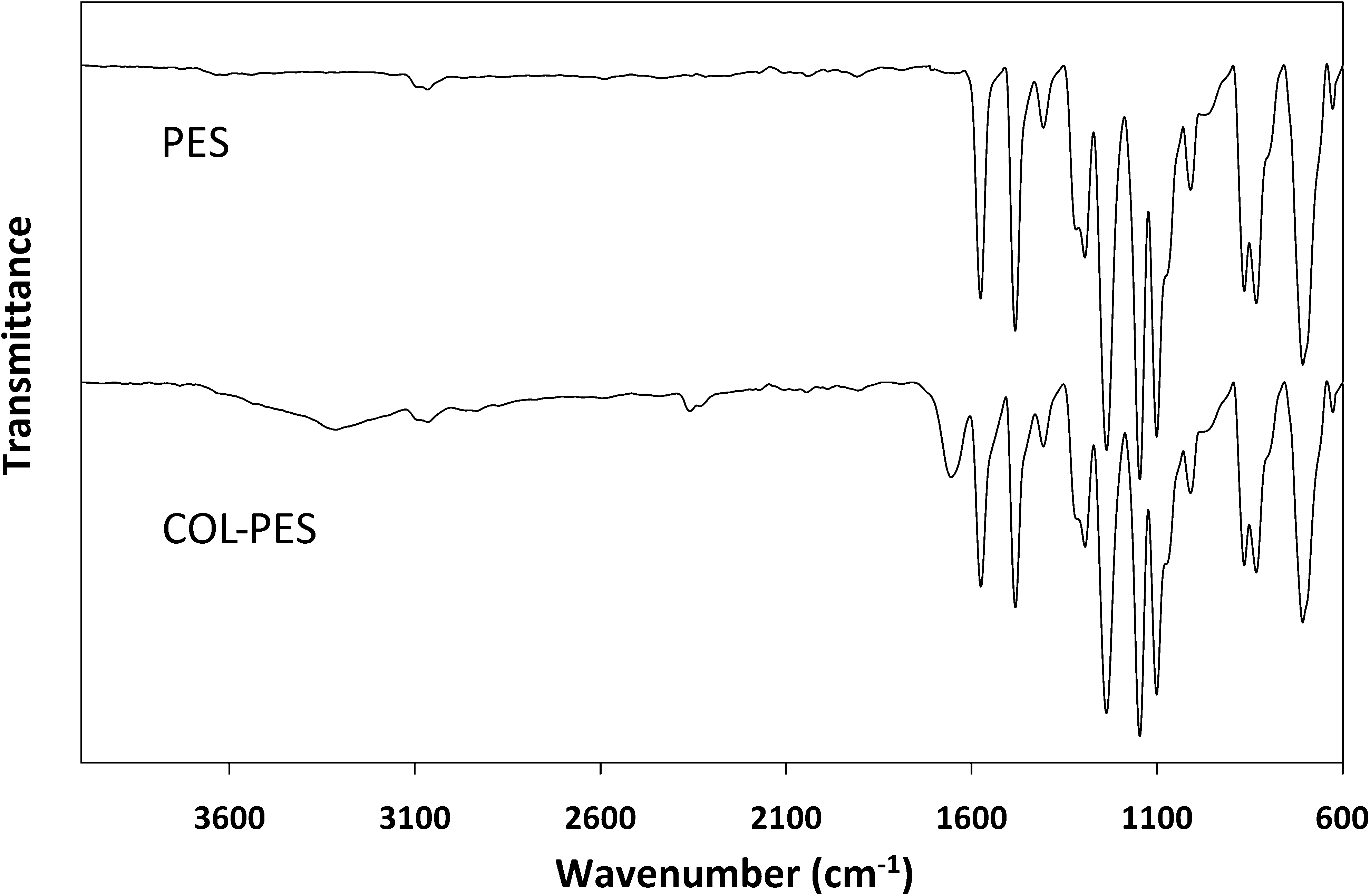
Attenuated total reflection Fourier transform infrared spectra of PES and COL-PES.
Cell proliferation and morphology on scaffolds
Viability and the rate of USSC proliferation on different scaffolds were compared during period of culture (Fig. 3). With an equal initial cell density on day 1, a significant difference between cell viability on COL-PES and other scaffolds was observed on day 3. It was also shown that COL-PES supported cell proliferation as much as TCPS except day 5, on which the highest cell viability was observed on COL-PES. However, there was no significant difference between cell viability on PES, PL-PES, and TCPS on this day. According to the assessment of USSC proliferation during time, it was revealed that, contrary to nanofibrous membranes, the rate of proliferation decreased on TCPS after day 3.
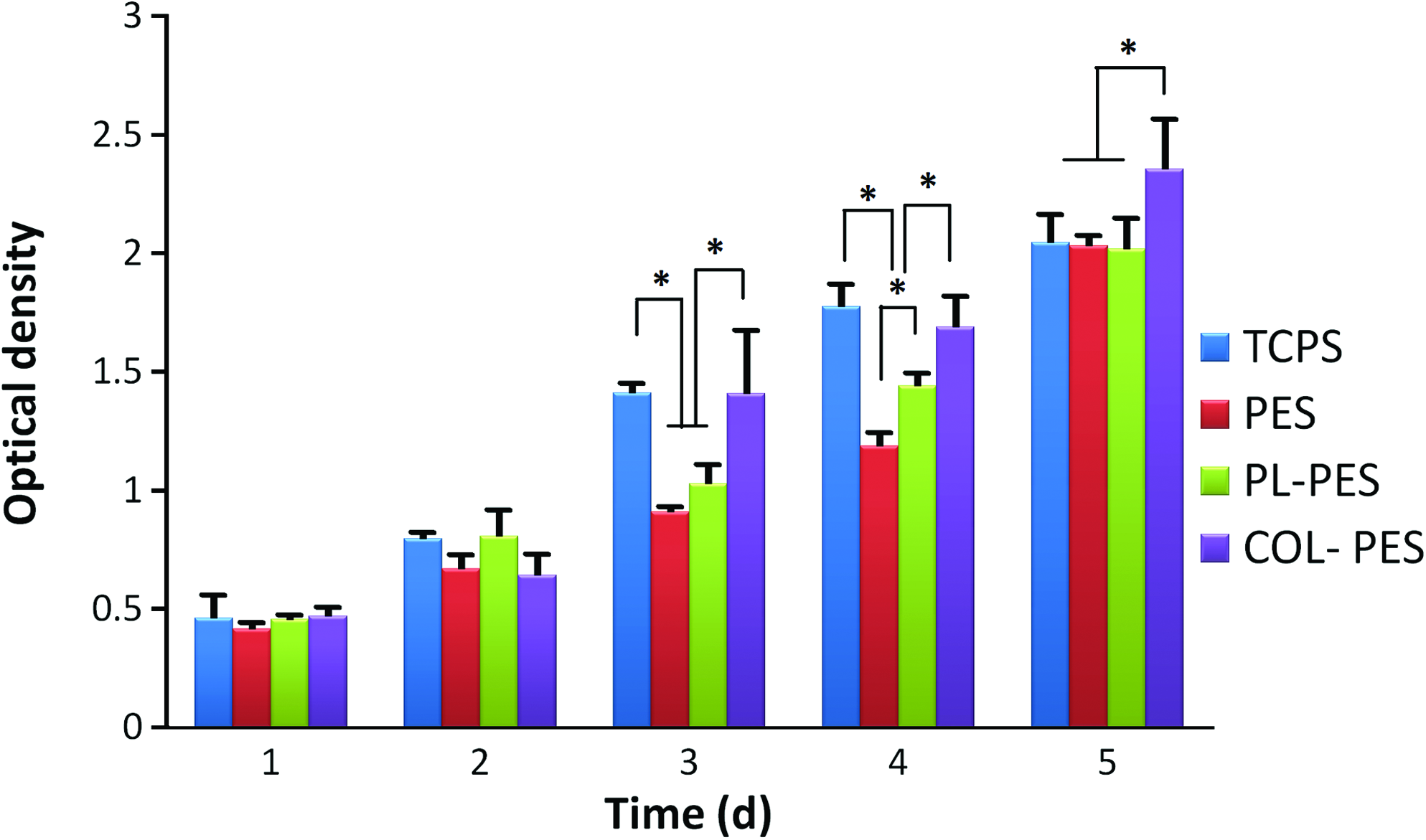
The proliferation of USSC on scaffolds and TCPS during a 5-day culture period; asterisk shows significant difference with p < 0.05. USSC, unrestricted somatic stem cells; TCPS, tissue culture polystyrene. Color images available online at www.liebertonline.com/tea
USSC exhibited flattened normal and typical morphology during osteogenic differentiation (Fig. 4). Biomineralization of USSC was also obvious on day 14 under osteogenic culture.
Morphology of USSC during osteogenic differentiation on nanofibrous PES
Gene expression analysis
Expression of three bone-related genes was analyzed in USSC and osteoblasts on different scaffolds, TCPS and COL-TCPS on days 7 and 14 of osteogenic differentiation (Fig. 5). In USSC, runx2 was expressed continuously on scaffolds and TCPS during time but upregulated in the second week and exhibited higher values on COL-TCPS compared to others. There was also significant difference between ALP transcripts on COL-PES compared to other surfaces on day 14. Higher values of ALP transcripts were also observed on COL-PES compared to other groups on day 7. Osteonectin was expressed in a higher level on COL-PES compared to other scaffolds on day 14. However, this gene showed a higher expression on TCPS and COL-TCPS compared to nanofibers on this day. In osteoblasts, significant higher values of runx2 transcripts were observed on COL-PES compared to other groups on day 7. On this day, ALP was expressed much higher on COL-PES and PL-PES compared to others. On day 14, higher expression of osteonectin was observed in osteoblasts on TCPS and COL-TCPS compared to nanofibers. However, this gene showed a higher amount of expression on PL-PES and COL-PES compared to others on day 7.

Relative expression of ALP
ALP activity and mineralization of stem cells
Activity of ALP was measured as a marker of osteogenic differentiation (Fig. 6). A similar pattern of an increased ALP activity of stem cells in the first week followed by a decrease on day 14 was observed in all groups under OM. However, the increase of ALP activity in the cells cultured under basal medium was observed during the time. In OM, USSC on COL-TCPS and COL-PES showed significant higher values on days 7 and 14 while these cells did not have significant different levels of ALP activity on PES and PL-PES compared to TCPS. The ability of these surfaces to support the osteogenic differentiation of MG-63 osteoblasts was also shown as control. The capacity of cells to deposit calcium minerals was measured during induction (Fig. 7). In a global aspect, both USSC and MG-63 cells showed much higher mineralization in all groups under OM compared to basal medium. Interestingly, mineralization of stem cells on COL-PES in the second week was significantly higher than other groups under OM. An increase in calcium content during osteogenic differentiation of the cells was observed in all groups. On day 7, there was no significant difference between mineralization of USSC on COL-TCPS and COL-PES that had the higher values in comparison to other groups. Higher calcium deposition of stem cells was also observed on nanofibrous scaffolds compared to TCPS on days 7 and 14. In basal medium, a significant higher mineralization of USSC and MG-63 cells was observed on COL-PES compared to other surfaces on day 14. Mineralization of osteoblasts was higher than that of stem cells in basal medium on all surfaces. In OM, the amount of calcium deposition of osteoblasts was significantly higher on COL-PES compared to other groups on day 14. An accurate look to the mineral deposits revealed their granular shape with porous structure formed from aggregated smaller globular deposits that were secreted into cell surface during osteogenic differentiation (Fig. 8).

ALP activity of USSC

Biomineralization of USSC

Calcium deposition on COL-PES scaffold on day 14 of USSC osteo-lineage differentiation at 5000×
Infiltration of USSC into nanofibers
Ingrowth of stem cells was studied via H&E-stained cross sections of nanofibrous scaffolds on day 14 (Fig. 9). At the end of the differentiation period, USSC were shown to reach the bottom of the scaffold and a uniform distribution of USSC was observed through all thickness of COL-PES with an excellent view of 3D cell–nanofiber structure. On the contrary, USSC on PES and PL-PES infiltrated only through the upper layers of nanofibers and could not passed the distance as much as the cells on COL-PES.

Hematoxylin and eosin staining of cross sections of cell-seeded PES
Discussion
Every year, millions of implants and protheses are used to cure bone defects and fractures caused by trauma or diseases. These BGS must have common characteristics such as biocompatibility and structural integrity. Recently, ideal BGS have been introduced with properties like osteoconductivity and osteoinductivity. Beside this, incorporation of appropriate cells allows bone healing and osteointegration in an accelerated manner.43–45 In the strategy for bone reconstruction using engineered scaffolds, bone structure should be mimicked to achieve the highest efficiency. Similar to all other tissues, bone is composed of cells and ECM in a complex, rigid, and highly porous structure. Bone ECM contains organic and mineral phases that are mostly in a nanoscale structure. Nanofibrils of collagen I are the most abundant component in organic phase and nano-crystalline hydroxyapatite form inorganic part of ECM in bone. Osteoblasts are the major cells in bone which are responsible to secretion of ECM proteins and further mineralization. These cells are active during bone generation and healing. 39
In the present study, a nanofiber-based approach was selected to mimic bone structure. Collagen I synthetic nanofibers have not shown desired mechanical properties 46 so PES was electrospun and grafted with collagen I to fulfill the mechanical requirements and bioactivity simultaneously. We postulated that incorporation of osetoblast-like cells into COL-PES nanofibers would assimilate it more to bone tissue. In natural bone, osteoblasts are derived from bone marrow mesenchymal stem cells. 47 Here, cord blood-derived stem cells were incorporated in nanofibers, and under OM, they gave rise to osteoblast-like cells in 14 days. Cord blood-derived USSC are a newly isolated stem cells that have been shown to have proper characteristics such as low immunogenicity, easy accessibility, and rapid proliferative capacity. 40 They also have strong potential for differentiation toward osteoblasts and intrinsically have the potential for contribution to vasculogenesis.41,42
High porosity and surface area are among the requirements that should be fulfilled in tissue engineering scaffolds. 48 Here, fabricated PES nanofibers before and after modification showed porosity >70%, which is considered as a proper amount of porosity for nanofibrous scaffolds. Intrinsically, because of nano-scale nature of electrospun fibers, they had a large surface-to-volume ratio, which was showed via BET analysis. It was also observed that surface modification did not have a significant effect on average fiber diameter of PES. Biocompatibility of PL-PES and COL-PES was predicted to be more than PES because of their high surface hydrophilicity. Existence of collagen was determined via ATR-FTIR through its specific IR peaks and also observed in junctions of PES nanofibers after grafting (Fig. 1c). Biocompatibility of nanofibers was studied and compared to TCPS. All PES, PL-PES, and COL-PES showed proper biocompatibility, and COL-PES had the highest ability to support cell proliferation. According to the MTT results, the rate of cell proliferation decreased after day 3 on TCPS contrary to nanofibers. This observation confirmed our previous report on improved infiltration of stem cell through PES nanofibers. 30 During cell proliferation, the more they cover a surface, the higher their proliferation rate decreases because of their contact inhibition. After they become confluent and form a monolayer on a two-dimensional matrix like TCPS, they stop proliferating. 49 Here, this was observed after day 3 of USSC culture on TCPS contrary to nanofibers on which the rate of USSC proliferation did not decrease. Since the surface of nanofibrous membrane was gradually covered with cells during culture, USSC in surface monolayer started to grow through the pores of scaffold, attached to underneath nanofibers, proliferated, and formed a 3D organization of cells and nanofibers. This hypothesis was confirmed with H&E staining of the cross sections of COL-PES during osteogenic culture, which cell infiltration was observed. A 3D form of cell–nanofiber combination was formed on day 14. On this day, high cell viability revealed that infiltrated cells inside the scaffold were metabolically active and was introduced to sufficient amount of nutrients transported into inner spaces. This is a result of high porosity of fabricated PES nanofibrous membranes. Although the largest pores in the membrane were about 2 μm (Fig. 1), but contrary to other nanofibrous membranes, PES electrospun nanofibers are not densely packed. The intrinsic characteristic of PES electrospun membranes can be the major reason for high cell infiltration. This structure is essential in efficient bone generation in vivo. Collagen was the possible reason for enhanced infiltration of stem cells in COL-PES scaffold because of the highest viability on day 5, on which cell density on PES, PL-PES, and TCPS became equal. Effect of collagen to improve cell infiltration within poly(L-lactide) (PLLA) nanofibrous scaffold was previously reported. 50
There are reports on the potential of electrospun nanofibers applied as bone grafts in vitro34,51 and recently in vivo.52,53 These studies have proposed electrospun matrices as proper candidates to be used in bone tissue engineering. In most of these researches, there was an approach to improve the characteristics of electrospun scaffolds via utilization of composite, hybridized, or surface-functionalized nanofibers. It was shown that inclusion of some bone natural components such as calcium phosphate minerals or collagen I enhanced bone generation in vitro and in vivo. Collagen I is known as 95% of protein content in bone and act as an osteoconductive macromolecule in bone. 54 In the present study, COL-PES was predicted to serve as a proper scaffold for bone regeneration. It was incorporated with cord blood USSC and was compared with PES and PL-PES for in vitro bone formation assays. Under osteogenic stimulation, the potential of stem cells on nanofibers was compared with TCPS. In bone tissue engineering, it was shown that application of stem or progenitor cells incorporated in proper scaffolds improved healing of bone defects more efficiently than the engraftment of scaffolds alone. 55
Osteogenic markers during differentiation of USSC on nanofibers, TCPS, and COL-TCPS were analyzed to evaluate the potential of PES, PL-PES, and COL-PES to be used for bone graft. ALP is generally considered as an osteoblast-related phenotype that is likely involved in mineralization process in osteoblasts. Its activity shows the amount of stem cell differentiation into osteo-lineage. 56 The pattern of ALP activity in USSC during osteogenic induction is consistent with a previous report on the biology and characteristics of USSC. 40 ALP activity in stem cells on COL-PES is significantly higher than other scaffolds on days 7 and 14 of differentiation. Higher activity of ALP was consistent with its higher expression on COL-PES at mRNA level. This shows that collagen in COL-PES has enhanced differentiation of USSC toward osteoblasts. This was confirmed with the results of gene expression on COL-TCPS. This enhancement is also observed in calcium deposition of USSC on COL-PES compared to PES, PL-PES, TCPS, and COL-TCPS. Since calcium minerals form a major content of bone, the ability of an engineered bone tissue to efficiently deposit calcium is believed an important and essential requirement. This task is performed by bone cells after migration into bone graft or by preincorporated cells after engraftment into bone defect. 39 Here, higher calcium deposition on COL-PES scaffold showed the effect of collagen to signal USSC to enhance mineralization. Promotion of osteogenic differentiation of USSC on COL-PES compared to other scaffolds was also confirmed on the gene expression levels. ALP was expressed in a higher amount on COL-PES on day 14. Osteonectin is also involved in the initial crystal growth and mineralization. Compared to PES and PL-PES, USSC on COL-PES showed higher transcripts of osteonectin on day 14. The expression of runx2 and osteonectin was higher on COL-TCPS compared to other groups. This can show the enhancement effect of collagen I on the expression of these genes. However, the main osteogenic markers such as ALP activity and mineralization confirmed a synergistic effect of collagen I and nanofibers on the osteogenic differentiation of stem cells on COL-PES scaffold.
To evaluate the osteoinduction of COL-PES nanofibers, the fabricated scaffolds were implanted subcutaneously into Balb/c mice. After 8 weeks, the scaffolds were retrieved, sectioned, and stained with H&E and Von Kossa (data not shown). However, no significant signs of ectopic bone formation were observed in implants in such a time period. There are some publications that have shown the osteoinduction of scaffolds implanted in nonosseous tissues.57–60 Of interesting in those studies, the most of scaffolds were composed of calcium phosphates or were loaded with osteoinductive growth factors like bone morphogenetic protein-2 in contrast to our scaffolds. In a recent publication, we also showed that PLLA nanofibers coated with nano-hydroxyapatite could induce ectopic bone formation after 10 weeks of implantation in mice. 12 Although COL-PES scaffolds showed no significant capacity for osteoinduction in an 8-week time period in vivo, this novel material with interesting in vitro ability holds promising potential in this field for further applications in preclinical studies to treat critical-sized bone defects.
Taking together, bone tissue engineering-related behavior of USSC was improved on COL-PES nanofibers. This behavior is defined as arranging in a 3D combination with scaffold accompanied with a high capacity for differentiation toward osteo-lineage. Plasma treatment of PES did not affect the ability of USSC for differentiation into osteoblasts and only contribute to the rate of proliferation on nanofiber surface and improved its biocompatibility. In this study, COL-PES scaffold was introduced as new potential BGS that in in vitro interaction with USSC confirmed its high capacity for application in bone tissue engineering.
Conclusion
Collagen I and nanofibrous structure showed an efficient contribution to a new potential BGS. COL-PES had the highest capacity to support osteogenic differentiation and infiltration of stem cells, which was confirmed via assessment of osteogenic markers and histologic examination. USSC in COL-PES formed a 3D distribution through nanofibers. It can be concluded that COL-PES is a suitable potential 3D bone graft with high capacity for bone healing and regeneration in vivo.
Footnotes
Acknowledgment
This work was financially supported by Stem Cell Technology Research Center (Tehran, Iran).
Disclosure Statement
No competing financial interests exist.