
Abstract
Growth factor (basic fibroblast growth factor or vascular endothelial growth factor)-immobilized polycaprolactone (PCL)/Pluronic F127 porous beads were prepared as an injectable bulking agent for effective treatment of urinary incontinence. The growth factor-immobilized porous beads may stimulate smooth muscle cell (SMC) differentiation of muscle-derived stem cells or defect tissues around urethra to improve the sphincter function (bioactive therapy) as well as to provide a bulking effect (passive therapy). The porous PCL/F127 beads were fabricated by an isolated particle-melting/melt-molding particulate-leaching method. The growth factors were easily immobilized onto the surfaces of the PCL/F127 porous beads via heparin binding and were continuously released for up to 28 days. Both growth factor-immobilized porous beads had a positive effect for the SMC differentiation of muscle-derived stem cells, as were demonstrated by the analyses of quantitative polymerase chain reactions, Western blot using SMC-specific markers, and immunohistochemical staining. In particular, the basic fibroblast growth factor-immobilized porous beads showed desirable SMC differentiation behavior that can be applied as an injectable bulking agent for the treatment of urinary incontinence.
Introduction
Thus, the main aim of this study was the development of a bioactive bulking agent that can induce MDSC differentiation. We prepared growth factor (basic fibroblast growth factor [bFGF] or vascular endothelial growth factor [VEGF])-immobilized polycaprolactone (PCL)/Pluronic F127 porous beads as an injectable bulking agent. This may stimulate the smooth muscle cell (SMC) differentiation of MDSCs or defect tissues around urethra to improve the sphincter function (bioactive treatment by growth factors) as well as to provide a bulking effect (passive treatment by porous beads) for effective treatment of urinary incontinence. MDSCs are found in the urethral sphincter and are believed to be involved in sphincter regeneration. 18 Both bFGF (18 kDa) and VEGF (46 kDa), which are the heparin-binding growth factor family, are well known as those that induce smooth muscle differentiation19,20 as well as angiogenesis. During the angiogenesis, the growth factors stimulate the capillary plexus formation/maturation, endothelial cell assemblage, and SMC recruitment. 21 The growth factors were immobilized onto the surfaces of the PCL/F127 porous beads by specific interactions between Pluronic F127 and heparin (hydrogen bonding) and the following heparin and growth factors (ionic interaction), which can preserve the biological function of growth factors without denaturation. Figure 1 demonstrates schematic diagrams for the successive binding of the heparin and the growth factor onto the pore surface of the Pluronic F127-entrapped PCL (PCL/F127) porous bead. The behavior of growth factor release from the growth factor (bFGF or VEGF)-immobilized porous beads and their SMC differentiation of the MDSCs were investigated.
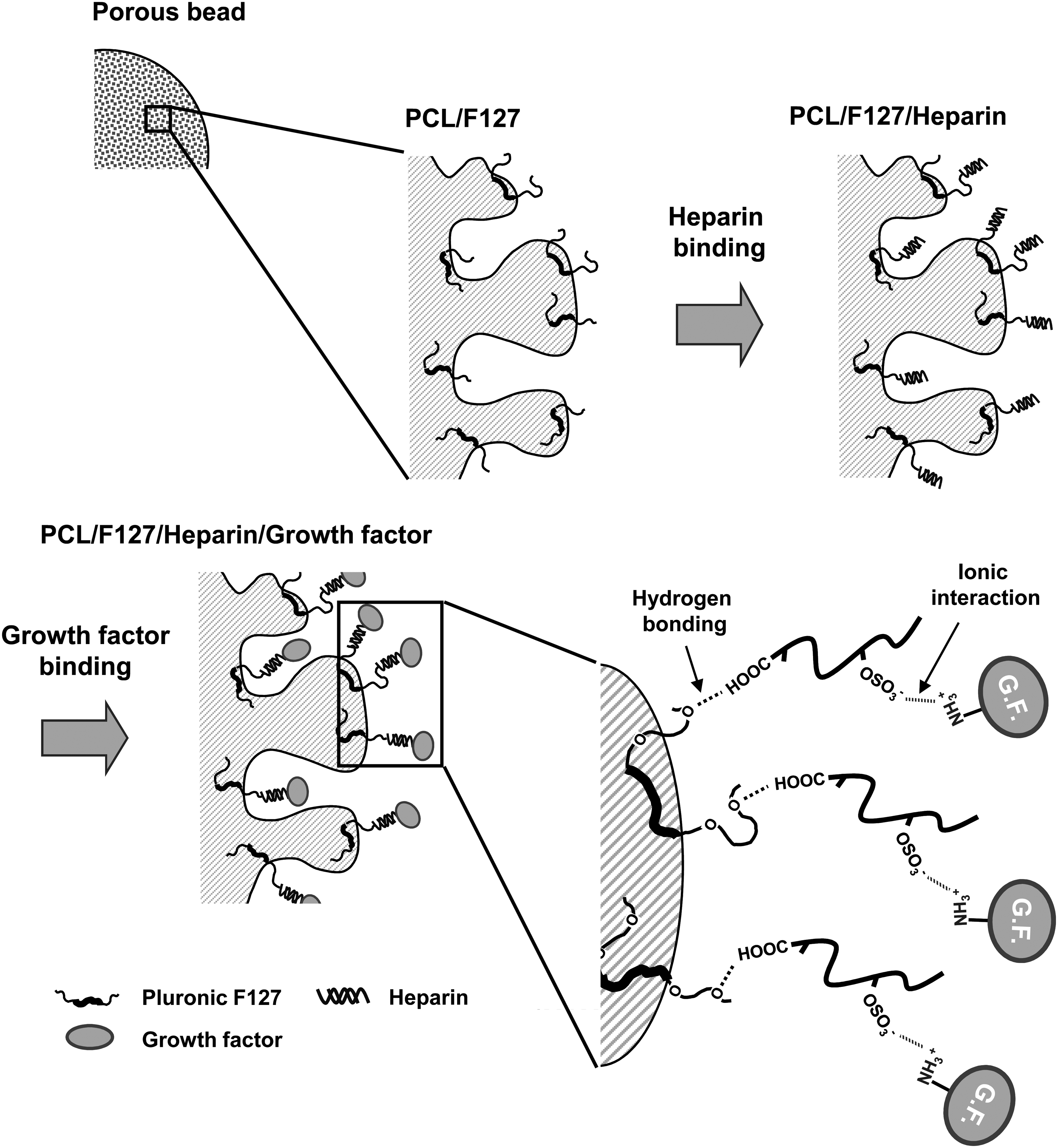
Schematic diagrams of the successive binding of heparin and growth factor onto the pore surface of the PCL/F127 porous bead. PCL, polycaprolactone.
Materials and Methods
Materials
PCL (molecular weight 43,000–50,000; Polyscience) and Pluronic F127 (EG99PG65EG99, molecular weight 12,500; Sigma) were used to fabricate porous beads. Heparin and growth factors (bFGF and VEGF) were purchased from Celsus Laboratories and R&D Systems, respectively. All other chemicals were analytically graded and were used as received. Water was purified (<18 mΩ) using a Milli-Q purification system (Millipore Co.).
Fabrication of PCL/Pluronic F127 porous beads
PCL/Pluronic F127 porous beads were fabricated by the modification of an isolated particle-melting method (for nonporous beads) and the following melt-molding particulate-leaching method (for porous beads) described elsewhere. 22 To fabricate the nonporous beads, PCL pellets were frozen and crushed into microsized particles (random-shape) using a freezer mill (SPEX 6750; Metuchen). The crushed particles were separated by microsieving using standard testing sieves (Chunggye Industrial Co.), and then the particles (a size range of 200–300 μm) were evenly dispersed in a cold Pluronic F127 aqueous solution and induced the gelation of Pluronic F127 at ∼25°C for 1 h (20 wt%; sol-gel transition temperature, ∼20°C; PCL particles/Pluronic F127 solution, 1/50 [w/v]). The PCL particles dispersed in the gel matrix were then stored in a prewarmed water bath at 65°C for 30 min. At this step, the random-shape PCL particles were melted (melting point of PCL, ∼60°C) and transformed to spherical shapes individually in the gel matrix without the aggregation of particles. After this treatment, the PCL bead (spherical, nonporous)/Pluronic F127 gel mixture was cooled down to ∼4°C and then centrifuged. After removing the supernatant, the PCL beads were then freeze-dried to obtain beads coated with Pluronic F127.
The PCL/Pluronic F127 porous beads were fabricated using the Pluronic F127–coated PCL nonporous beads and sodium chloride particles (NaCl; size range of 50–100 μm). The Pluronic F127–coated PCL nonporous bead and salt particle mixtures (1/40 w/w ratio; 2.5 g) were placed in a brass mold (diameter, 18 mm; thickness, 2.5 mm), and the mold was thermally compressed at 80°C under 10 MPa for 3 min followed by another compression under 15 MPa for 1.5 min using a compression molding press. After removing them from the mold, the beads were then immersed in excess water for 1 day with mild shaking (the water was changed every 2–4 h) to leach out salts and free Pluronic F127 molecules from the beads. Eventually, PCL/Pluronic F127 porous beads (size range of 400–550 μm) were obtained after vacuum-drying overnight. The PCL porous beads (without Pluronic F127) were also fabricated using the same crushed PCL particles and salt particles for the purpose of comparison.
The morphologies of the PCL-crushed, nonporous, and porous beads were observed by a scanning electron microscope (Model S-3000N; Hitachi). The porosity of the porous beads was estimated by the volume change of the beads before (nonporous beads) and after (porous beads) the melt-molding particulate-leaching process. 22
Growth factor immobilization and release test
Two different growth factors, bFGF and VEGF, were incorporated onto the surfaces of the PCL/F127 porous beads via heparin immobilization. To immobilize heparin, the porous beads were soaked in the heparin solution (1 mg/mL [in 2 wt% NaCl solution]) at 4°C for 3 h. Then, the heparin-immobilized beads were thoroughly rinsed with 2 wt% NaCl solution and water successively, and then freeze-dried. The amount of immobilized heparin was determined using toluidine blue assay. 23 To observe the heparin distribution in the PCL/F127 porous beads, the fluorescein isothiocyanate–heparin (Polyscience) was also coupled onto the beads using the same procedure above. Then, the crossly sliced porous bead sections were observed using a fluorescent microscope (Model BX50F4; Olympus). To estimate the effect of Pluronic F127 on heparin binding onto the surfaces of the PCL/F127 porous beads, the heparin was also immobilized onto the PCL porous beads without Pluronic F127, and their heparin immobilization behavior was compared with that of the PCL/F127 porous beads.
To incorporate the growth factors onto the heparin-immobilized PCL/F127 porous beads, the beads were soaked into two different growth factor solutions (bFGF and VEGF, each 200 ng/mL), respectively, at room temperature for 3 h. The growth factor-immobilized beads were washed with phosphate-buffered saline (PBS; pH ∼7.4) three times and the amount of immobilized growth factors was quantified by a direct ELISA technique, 24 in which the standard ELISA procedure in the kit (Duoset®; R&D Systems) was followed using the porous beads as the primary substance. To investigate the role of heparin immobilized onto the surfaces of the PCL/F127 porous beads for the growth factor binding and release behavior, the growth factor-adsorbed PCL/F127 porous beads without the heparin immobilization were also prepared using the same procedure. Growth factor (bFGF or VEGF)-immobilized porous beads (10 mg) were incubated in 1 mL PBS supplemented with 1% bovine serum albumin at 37°C for a period of up to 28 days under mild shaking (∼50 rpm) to perform the release study. At preset time intervals, the incubation solutions were collected and replaced with fresh PBS. The amount of released growth factor in the collected medium was determined by the ELISA kit. The PCL and PCL/F127 porous beads did not show any degradation or swelling symptom during the growth factor release test periods.
In vitro cell culture in growth factor-immobilized porous beads
To investigate the potential of SMC differentiation by the growth factor-immobilized porous beads, MDSC was used as a model cell. The MDSCs were harvested from the hindlimb muscles of a rat and purified using the preplate technique. 25 In brief, the isolated muscle was minced into a coarse slurry using smaller needles. The muscle was enzymatically dissociated by the addition of collagenase type XI (0.2%; Sigma) for 1 h at 37°C, dispase (240 units; Gibco) for 45 min, and trypsin (0.1%; Gibco) for 30 min. After enzymatic dissociation, the cells were centrifuged, resuspended in a culture medium (Dulbecco's modified Eagle's medium [Gibco] containing 10% fetal bovine serum [Gibco], 10% horse serum [Gibco], and 1% penicillin/streptomycin [Gibco]), and plated on collagen and poly-D-lysine-coated flasks (PP 1). After 2 h, nonadherent cells in PP 1 were transferred into other flask (PP 2). The following day, the nonadherent cells were transferred to another flask and the same procedure was repeated up to PP 5 (no adherent cells in the flasks) and the adherent cells in the last flask (PP 6) were trypsinized and collected to use in this study as MDSCs. The proliferation and SMC differentiation behaviors of MDSCs seeded in the bFGF or VEGF-immobilized PCL/F127 porous beads were compared with those in the same beads without the growth factor immobilization. For the seeding of the MDSCs (passage no. 14; the phenotype of MDSC can be maintained for up to 30 passages 26 ) into the porous beads (with and without growth factor immobilization; 10 mg), the beads were immersed in a needle tip-stopped syringe filled with 1 mL of the cell suspension (cell density, 1.0 × 10⁶ cells/mL). The cell suspension was infiltrated into the porous beads under negative pressure when the syringe piston was pulled out. The beads were maintained for 2 h at 37°C for cell adhesion to the beads. Then, the beads were carefully transferred to 24-well polystyrene dishes (Corning), and the culture medium was added to the dishes (1 mL/well). Then, the cells in each bead group were cultured in an incubator in a humidified atmosphere of 5% CO2 for up to 28 days with mild shaking (∼50 rpm). The culture medium was exchanged with a fresh medium every 3 days during the cell culture periods. The cell proliferation behavior in the beads during the cell culture periods was estimated by the measurement of DNA content. For this, the cells from each bead group at predetermined time intervals (0, 7, 14, and 28 days) were digested overnight in a Papain buffer at 60°C. The DNA content was determined using a DNA-binding fluorochrome, Hoechst 33258 (Sigma-Aldrich), and purified calf thymus DNA as the standard.
Quantitative polymerase chain reaction analysis
After the cell culture for 14 and 28 days in each bead group, RNA was extracted using a monophasic solution of phenol and guanidine isothiocyanate (Trizol, Invitrogen). The extracted RNA samples were converted to complementary DNA using reverse-transcriptase (SuperScript III®; Invitrogen), according to the manufacturer's instructions. All of the polymerase chain reactions (PCRs) were performed for the detection of SMC-specific transcripts using ABI StepOne plus (Applied Biosystems) with SYBR Green® gene expression assay (Invitrogen). Expression of the following genes were examined: α-smooth muscle actin (α-SMA), calponin, caldesmon, and myosin heavy chain, which are commonly used as molecular markers to assess for SMCs. 27 Glyceraldehyde-3-phosphate dehydrogenase was used as a housekeeping gene. To guarantee the reliability of the results, all of the samples were processed in triplicate. The data were analyzed using a Sequence Detection Software. The comparative CT method (User bulletin #2; Applied Biosystems) was used to standardize the data for all of the samples of the housekeeping glyceraldehyde-3-phosphate dehydrogenase values.
Western blot
After the cell culture, the total protein was extracted from each bead group, using the cell lysis buffer (Cell signaling). Lystates were resolved by 12% sodium dodecyl sulfate–polyacrylamide gel electrophoresis and transferred onto nitrocellulose membranes (Amersham). The membranes were blocked with 5% skim milk (in 20 mM Tris-HCl, 150 mM NaCl, and 0.1% Tween 20). The membranes were then probed with the antibodies to α-SMA (Abcam), reacted with the bound antibody, and observed using the horseradish peroxidase-conjugated secondary antibody (GenDEPOT). Immunoreactivities were detected using an enhanced chemiluminescence. Images were obtained using an X-ray developer (Daesung).
Immunohistochemistry
After the cell culture for 0, 7, 14, and 28 days in each bead group, immunohistochemical staining was also conducted to observe α-SMA, which is one of the representative smooth muscle-specific proteins. For immunohistochemical staining, the cells adhered on the porous beads were fixed in 4% paraformaldehyde in PBS for 10 min and permeabilized with PBS containing 0.1% Triton X-100 (0.1% PBST). They were then incubated with α-SMA monoclonal antibody (diluted to 1:100; Sigma) for 12 h (4°C) and Alex Fluor 568–conjugated secondary antibody (goat anti-rabbit IgG; Invitrogen) for 1 h (room temperature). The nuclei were stained with 4′6-diamidino-2-phenylindole (Vector Laboratories). Images were observed with confocal microscopy (LSM510 META; Carl Zeiss).
Statistical analysis
The data obtained from each bead group were averaged and expressed as mean ± standard deviation. The Student's t-test was used to determine the significance of the differences between the bead groups. The differences were considered statistically significant at p < 0.05.
Results
Characterization of PCL/Pluronic F127 porous beads
The PCL/Pluronic F127 porous beads were fabricated by the modified isolated particle-melting and subsequent melt-molding particulate-leaching methods. 22 The random-shaped PCL-crushed particles were transformed into spherical nonporous beads through the isolated particle-melting method (Fig. 2). The PCL/F127 porous beads exhibited a highly porous surface and interior pore structures (Fig. 2), which can provide a large surface area for growth factor immobilization. The pore sizes in the beads were almost the same as the salt particle sizes used (salt particle size range of 50–100 μm). The porosities of the beads determined by the total volume change between nonporous and porous beads were ∼90%.

Scanning electron microscope photographs of crushed PCL particle, F127-coated nonporous PCL bead, and porous PCL/F127 bead. Upper right image indicates cross-sectional view of the porous PCL/F127 bead. The porous beads were fabricated by sequencing processes from the crushed particles (isolated particle-melting method) and then the nonporous beads (melt-molding particulate-leaching method).
Growth factor immobilization and release behavior
Figure 3 shows the amount of heparin immobilized on the surfaces of the PCL and PCL/F127 porous beads, as determined by the toluidine blue assay. It was observed that the PCL/F127 porous beads allow for much larger heparin immobilization on the surfaces of the PCL/F127 porous beads (0.47 ± 0.04 μg/mg) compared with the PCL ones (0.08 ±0.02 μg/mg). From the fluorescence microscopy images (Fig. 3), it was also observed that the fluorescein isothiocyanate–heparin (expressed by the color green) was more brightly and evenly expressed in the PCL/F127 porous beads than the PCL ones, indicating greater and even heparin immobilization on the PCL/F127 porous beads.
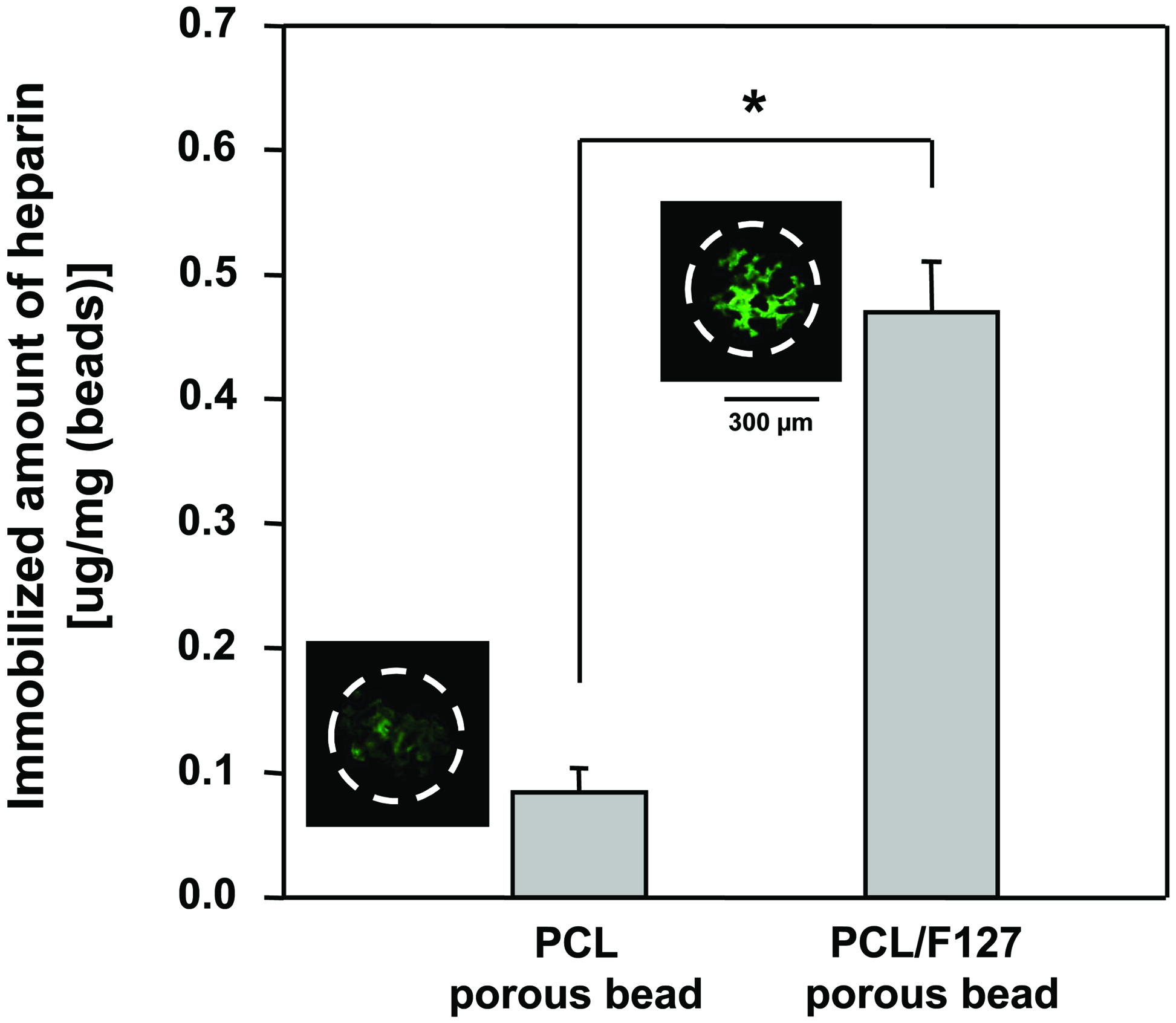
Amount of heparin immobilized onto the PCL and PCL/F127 porous beads (toluidine blue assay; n = 3, *p < 0.05). Fluorescence microscope images show the heparin distribution onto the porous bead. The fluorescein isothiocyanate–heparin was expressed as green color. Color images available online at www.liebertonline.com/ten.
The quantitative amount of growth factors, bFGF and VEGF, immobilized on the porous beads was determined by the direct ELISA technique. 24 The loading amounts of the growth factors on the PCL/F127 porous beads with (+Hep) and without (−Hep) heparin immobilization were determined as follows: VEGF (−Hep), 17.4 ± 2.1 ng; VEGF (+Hep), 36.4 ± 4.0 ng; bFGF (−Hep), 13.2 ± 3.4 ng; bFGF (+Hep), 22.2 ± 2.8 ng (Fig. 4A). The heparin-immobilized porous beads had significantly higher loading amount of both growth factors than the porous beads without the heparin. The release profiles of the growth factors from the porous beads were shown in Figure 4B. The heparin-immobilized porous beads showed a moderate initial burst release of growth factors, and then the growth factors were continuously released up to 35.1 ± 0.8 ng (VEGF) and 21.8 ± 1.8 ng (bFGF) over 28 days. On the contrary, the porous beads without heparin immobilization showed a much higher initial burst for the initial 7 days (time for 50% release of total growth factors released for 28 days; VEGF, ∼1 day [−Hep] vs. ∼3 days [+Hep]; bFGF, ∼1 day [−Hep] vs. ∼5 days [+Hep]). The most growth factors loaded on the porous beads without heparin were released within 7 days.

In vitro SMC differentiation of MDSCs in growth factor-immobilized porous beads
To investigate the in vitro proliferation and SMC differentiation potential of MDSCs by continually releasing growth factors from the porous beads, the MDSCs were seeded into the PCL/F127 porous beads with and without the growth factor immobilization. The cell proliferation in the porous beads was assessed through the measurement of DNA contents at the preset periods (0, 7, 14, and 28 days) after seeding. Figure 5 shows that the cells grew fast within the initial 7 days, and then the cell numbers for all groups were not significantly different in culture. The cells grew to a higher density in the presence of growth factors because of the stimulation of cell migration/survival/growth and the protection of cell apoptosis by the growth factors. 28 The difference of cell growth between bFGF and VEGF-immobilized porous beads was not significant, even though the bFGF-immobilized beads showed a little better cell growth.
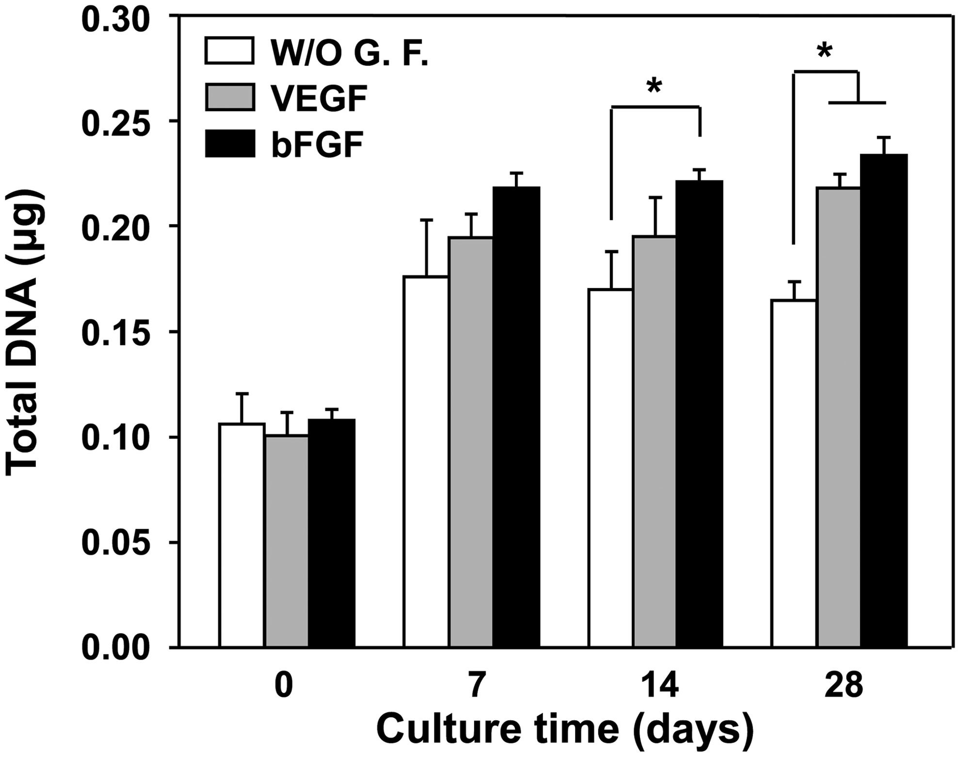
Total DNA content with time after in vitro MDSC culture in the PCL/F127 porous beads with and without growth factor immobilization (n = 3; *p < 0.05). MDSC, muscle-derived stem cell.
To estimate the effect of bFGF and VEGF released from the porous beads on SMC differentiation of the MDSCs, a quantitative PCR analysis for some genes was conducted. Four molecular markers for SMC differentiation, α-SMA, calponin, caldesmon, and myosin heavy chain, which are commonly used as markers to assess for phenotype of SMCs, were analyzed. The results showed that both growth factor-immobilized porous beads produced higher gene expression than the beads without the growth factors in all markers (Fig. 6), suggesting that the continually released bFGF and VEGF can be a good environment for SMC differentiation of MDSCs. We also recognized that the bFGF induced better SMC differentiation than the VEGF in our system, even though there was a slower release of bFGF than VEGF (see the Fig. 4B).
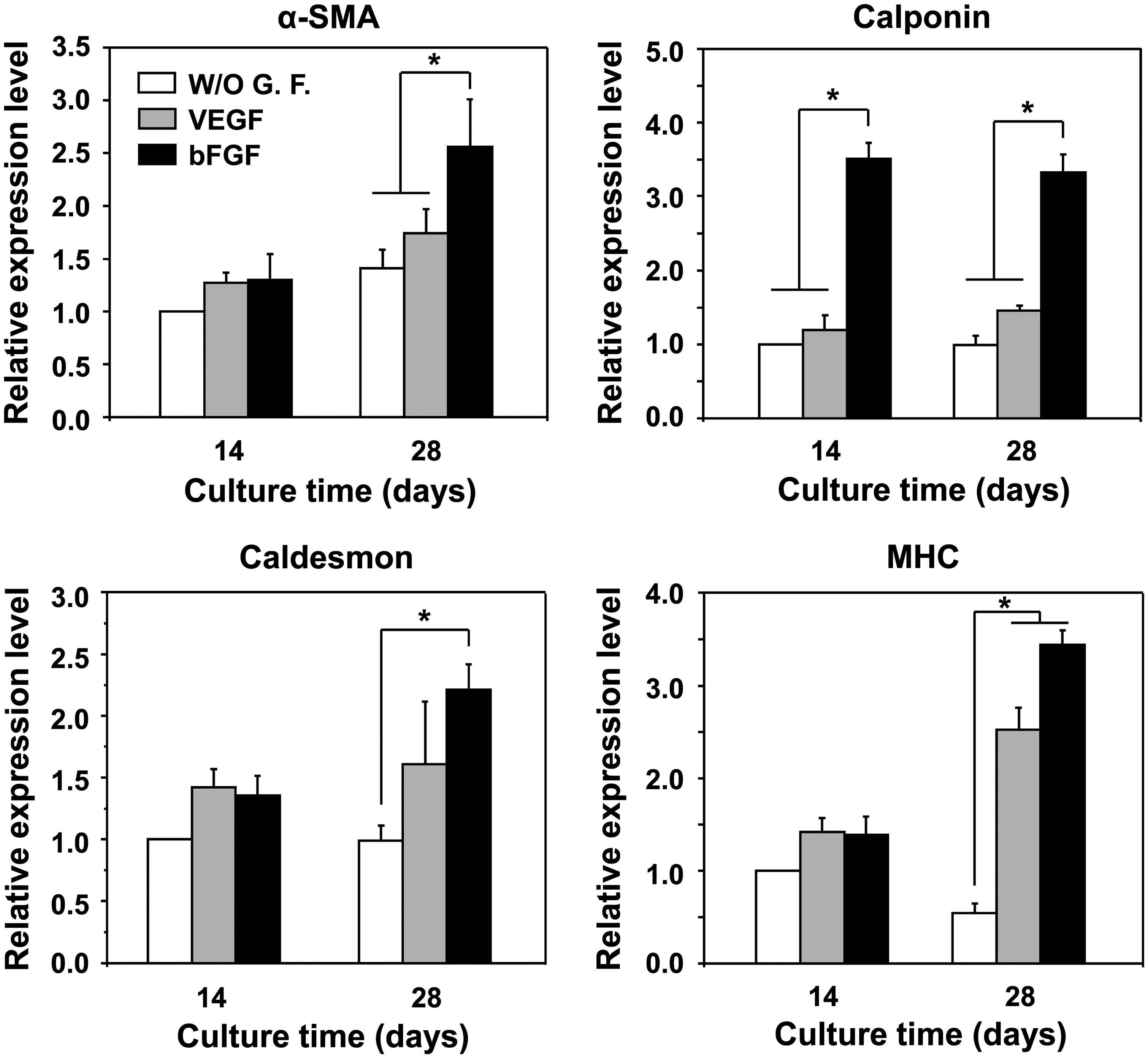
Quantitative polymerase chain reaction after in vitro MDSC culture in the PCL/F127 porous beads with and without growth factor immobilization for 14 and 28 days (n = 3; *p < 0.05). mRNA expression was measured and normalized against glyceraldehyde-3-phosphate dehydrogenase.
To investigate whether the differentiated MDSCs contain smooth muscle-specific proteins, we probed the cellular contents with the α-SMA monoclonal antibody. α-SMA is considered the most intensely expressed marker for SMCs. 29 Figure 7 shows immunohistochemical observations of MDSCs cultured in the PCL/F127 porous beads with and without growth factor immobilization. The cells were uniformly distributed in all porous beads, indicating that the porous beads fabricated by the isolated particle-melting and the following melt-molding particulate-leaching methods satisfied the requirements as cell carriers, that is, highly porous and interconnected pore structures. Weak α-SMA expression (red color) was appeared at 7 days in all bead groups, and thereafter expression gradually increased during the 28-day time period. In the Western blot analysis, it was also observed that α-SMA expression gradually increased over time (Fig. 8). In particular, the bFGF-immobilized porous beads showed greater α-SMA expression than the VEGF-immobilized one as well as the beads without growth factors, indicating that the continually released bFGF provides efficient environment for MDSC differentiation into SMCs, as also discussed earlier.

Confocal microscope images showing the smooth muscle cell differentiation of the MDSCs by the growth factor (bFGF or VEGF) continuously released from the PCL/F127 porous beads (blue, cell nucleus [4′6-diamidino-2-phenylindole]; red, α-SMA; dotted circle, porous bead). α-SMA, α-smooth muscle actin. Color images available online at www.liebertonline.com/ten.

Western blot analysis on α-SMA protein production of the MDSCs cultured in the PCL/F127 porous beads with and without growth factor immobilization for 14 and 28 days. Color images available online at www.liebertonline.com/ten.
Discussion
It is well understood that the controlled storage and timely disposal of urine could be provided by the appropriate function of bladder/sphincter, pelvic organ support, and neuronal command. 2 From this point of view, the functional recovery of these damaged tissues may be a fundamental therapeutic technique for urinary incontinence. In this study, two different growth factor (bFGF or VEGF)-immobilized PCL porous beads were prepared to estimate their potential as a bioactive bulking agent that can stimulate the defect tissues (particularly smooth muscles) around the urethra by the sustained release of growth factors for the differentiation of MDSCs and thus improve the functional recovery of the tissues for the effective treatment of urinary incontinence. It is well known that the MDSC has a multidifferentiational potential 30 and a variety of growth factors could direct its differentiation to specific cell types, such as bone cells (by bone morphogenetic protein), 31 fibroblasts (by transforming growth factor-β), 32 Schwann cells (by nerve growth factor), 26 endothelial cells (by VEGF), 26 and SMCs (by VEGF/coculture with SMC). 20 Also, the plethora of soluble factors found at various surgical sites in vivo results in the contribution of MDSCs in the regeneration of numerous tissues.33,34 Many researchers also reported that the MDSCs could stimulate the restoration of various tissues.15,16,35,36
Spherical nonporous PCL beads were fabricated by the modification of an isolated particle-melting method. 22 In this method, each crushed particle isolated by Pluronic F127 gel matrix is molten above the melting temperature of PCL, and then the molten PCL particles were constrained into spherical shapes to minimize the surface area (surface/volume ratio) by interfacial forces in the gel matrix 37 without the aggregation of the particles. The predeterminate-sized spherical nonporous beads were obtained by crushed bead size without the loss of the polymers. During the porous bead fabrication step using the melt-molding particulate-leaching method, the salt particles and Pluronic F127 were uniformly infiltrated into the molten PCL beads by thermal compression, and therefore the volume of the PCL beads increased (later, the increased volume becomes pore volume in the porous bead after salt leaching) and the Pluronic F127 molecules are physically entrapped within the molten pore surface region of the PCL beads. Therefore, the PCL beads can provide the hydrophilicity by surface exposure of the hydrophilic polyethylene glycol chains of Pluronic F127. 38 The Pluronic F127 molecules exposed on the pore surface of the bead can also act as an intermediary between the PCL pore surface and heparin that can interact with the growth factors (see Fig. 1).39,40
To incorporate growth factors, the PCL/Pluronic F127 porous beads were first immobilized with heparin via intermolecular hydrogen bonding between the ether oxygen of Pluronic F127 exposed on the pore surfaces of the bead and carboxylic acid group of heparin. 39 On the basis of the results of Figure 3, we can speculate that the Pluronic F127 chains are sufficiently exposed onto the surfaces of the porous beads to effectively interact with the heparin. The heparin immobilization on the PCL porous beads without Pluronic F127, even if the amounts were small, may be explained by the physical adsorption (nonspecific binding) of the heparin on the pore surfaces. The large amount of heparin immobilized on the surfaces of the PCL/F127 porous beads can allow for the effective interaction with growth factors.
Two different heparin-binding growth factors, bFGF and VEGF, as potential bioactive molecules were separately incorporated onto the heparin-immobilized PCL porous beads to stimulate the SMC differentiation of MDSCs (in vitro) or defect tissues around the urethra to improve sphincter function (in vivo). The heparin-immobilized porous beads had a significantly greater loading amount of both growth factors than the porous beads without the heparin immobilization, caused by the ionic interaction between both O-sulfate and N-sulfate groups of heparin molecules and certain lysine and arginine residues in the growth factors. 40 The growth factors detected on the porous beads without heparin immobilization can also be explained by the physical adsorption of growth factors on the pore surfaces of the bead, which may lead to the initial burst release of them in the medium. From the Figure 4A, it was observed that the bFGF has a weaker binding efficiency with heparin immobilized onto the porous bead than the VEGF. This may be attributed to the differences in the structure of bFGF and VEGF heparin-binding sites as well as their respective molecular weights (bFGF, 18 kDa; VEGF, 46 kDa). 41 The heparin-immobilized porous beads showed a moderate initial burst release of growth factors, and then the growth factors were released continuously for up to 28 days. These initial bursts may be explained by the fast desorption of nonspecifically bound growth factors onto the porous beads. 42 The growth factors specifically bound (via ionic interactions) on the porous beads may be slowly released by the gradual dissociation of ionic bonds in PBS solution. However, the porous beads without heparin had a much higher initial burst and the growth factors were rapidly released within 7 days, owing to the desorption of growth factors adsorbed onto the porous beads without any specific interactions. Even though the timely release of growth factors and their effects to control MDSC differentiation toward SMCs are not exactly known and the in vitro and in vivo results may not be the same, the sustained release of growth factors from the heparin-immobilized PCL porous beads, which can prolong their biological effects, 43 may be very helpful to recover the sphincter muscle function caused by the induction of the SMCs.
To estimate the effect of growth factors released from the porous beads on SMC differentiation of the MDSCs, quantitative PCR and Western blot analyses were conducted. We recognized that the bFGF and VEGF being continuously released from the beads could provide a good environment for SMC differentiation of MDSCs. In particular, the bFGF induced a better SMC differentiation than the VEGF, even though there was a slower release of bFGF than VEGF (see the Fig. 4B). From these results, the bFGF-immobilized PCL/F127 porous beads seem to provide an optimum environment for SMC differentiation of MDSCs in our system. The exact mechanism why the bFGF released from the porous beads leads to the better SMC differentiation efficacy than the VEGF is not yet clear. However, we can speculate that the bFGF-immobilized PCL/F127 porous beads may be a promising bioactive bulking agent system that can improve the sphincter muscle function around the urethra (by the induction of SMCs) and thus effectively treat urinary incontinence. Takahashi et al. 19 also reported that the sustained release bFGF (incorporated gelatin hydrogel) could increase the urethral smooth muscle layer and thus improve the contractility of the sphincter. On the basis of our findings and literature, we recognize that the hydrophilized PCL/F127 porous beads could be also a good candidate as a bioactive carrier for the entrapment of drugs and cells as well as growth factors for their target delivery.
Conclusion
We prepared a growth factor (bFGF or VEGF)-immobilized PCL/Pluronic F127 porous beads as an injectable bioactive bulking agent that can stimulate the defect tissues around the urethra (by the sustained release of growth factors), as well as provide a bulking effect (by porous beads) for the effective treatment of urinary incontinence. Both growth factors were easily immobilized onto the surfaces of the PCL/F127 porous beads via heparin binding without using any toxic chemicals. The growth factors immobilized on the heparin-bound porous beads were released with a sustained manner for up to 28 days. From the study of in vitro SMC differentiation of MDSC as a model cell, both growth factor-immobilized porous beads showed a positive effect for SMC differentiation. This may promote the regeneration of damaged smooth muscle around the urethra, and thus can effectively treat urinary incontinence. In particular, the bFGF-immobilized porous beads showed better SMC differentiation behavior than the VEGF-immobilized one. An in vivo study using an urinary incontinent animal model to evaluate the bFGF and VEGF-immobilized PCL/F127 porous beads for their effects on SMC induction, as well as sphincter muscle function recovery around the urethra to treat urinary incontinence is now in progress.
Footnotes
Acknowledgment
This work was supported by the Pioneer Research Center Program through the National Research Foundation of Korea funded by the Ministry of Education, Science and Technology (2010-0002176).
Disclosure Statement
No competing financial interests exist.