
Abstract
Stem cell therapy has been used to repair ischemic tissues in the limbs, in myocardial infarctions, and in the brain. To understand the mechanisms of healing, a contrast agent capable of inducing sufficient magnetic resonance (MR) contrast would be useful in providing fundamental information about the cell migration and incorporation into the ischemic tissue. A magnetic resonance imaging contrast agent composed of dextran and gadolinium chelate was synthesized. Hydroxyl groups of dextran were activated with 1,1′-carbonylbis-1H-imidazole and reacted with propanediamine to obtain aminated dextran. This modified polymer was then reacted with mono-N-succinimidyl 1,4,7,10-tetraazacyclododecane-1,4,7,10-tetraacetate, then with fluorescein isothiocyanate, and finally reacted with gadolinium chloride solution (Dex-DOTA-Gd3+). Endothelial progenitor cells (EPCs) were selected as a stem cell model for magnetic resonance imaging tracking. Cells were isolated from the bone marrow harvested from the femurs and tibias of rats. Dex-DOTA-Gd3+ was then introduced into the EPCs by electroporation. The intracellular stability and cytotoxicity of Dex-DOTA-Gd3+ were evaluated in vitro. Dex-DOTA-Gd3+-labeled EPCs were transplanted into a rat model of ischemic limb, and MR images were acquired. Dex-DOTA-Gd3+ was found to efficiently label EPCs over a long duration without significant cytotoxicity. This provides an MR signal sufficient for tracking the EPCs intramuscularly injected into the limb.
Introduction
MRI tracking of cells labeled with iron oxide-based magnetic contrast agents has been used for visualization of many aspects of angiogenesis in different cell types such as the hematopoietic and neural progenitor cells.10–12 One of the advantages of labeling cells with superparamagnetic iron oxide particles (SPIO) is the high sensitivity obtained compared with that obtained with other contrast agents. 13 Wilhelm et al. reported that SPIO-labeled cells can be detected in small quantities (30–60 cells in agarose gel) by high-resolution MRI. 14 Other studies reported stem cell homing to the organs and the BM, 15 differentiation and migration of oligodendrocyte progenitors into brain parenchyma, migration and incorporation of labeled cells to the sites of tumor angiogenesis, 16 and magnetic guiding of the endothelial progenitor cells (EPCs) to a target site. 14 However, despite the great advantages provided by SPIO labeling, some studies have reported certain disadvantages. Magnetic resonance (MR) signals in iron oxide-labeled cells transplanted into a mouse hind limb did not show significant differences from the day 1 to 28 post-transplantation; however, there is evidence for a progressive decrease in the cell number when monitored using genetically introduced bioluminescent signals.
Histological analysis confirmed that macrophages loaded with iron oxide particles were located between muscle bundles but no transplanted cells were detected near the macrophages. 15 Therefore, one of the problems when labeling with iron oxide is the instability to remain in the cells in vivo, which can probably be termed as leakage. It is possible that the free SPIO are subjected to endocytosis by macrophages. A similar situation was described in iron oxide-labeled mesenchymal stem cells that were transplanted into a rat model of infarcted myocardium. 16 In addition, iron oxide induces negative contrast in MRI, which, in some cases, can be difficult to attribute to the signal loss of the labeled cells, because of many other sources of negative contrast of MRI in the body.
Our group recently reported the development of a polymeric MRI contrast agent based on a gadolinium-chelated poly(vinyl alcohol). 17 In this study, NIH-3T3 cells were labeled with the contrast agent via electroporation. Cell viability and proliferation were not affected by polymer labeling, and MR measurements showed that labeled cells could be clearly tracked in vivo. In this study, we selected EPCs as a stem cell tracking model. The EPCs isolated from the peripheral blood and BM have been used in the therapeutic angiogenesis of ischemic limbs, in stroke, and in myocardial infarction.18–25
Advances in the techniques for visualizing EPCs, which are activated by cytokines at the site of injury, are imperative for understanding the mechanisms of proliferation, recruitment, mobilization, and incorporation of EPCs into the foci of vasculogenesis. Although the detection sensitivity of gadolinium chelates is generally lower than that of iron oxide, their positive contrast is easy to detect in a determinant tissue.26,27 Therefore, we synthesized a water-soluble gadolinium chelate as an alternative MRI contrast agent. This agent, called Dex-DOTA-Gd3+, was designed for labeling EPCs. Dex-DOTA-Gd3+ was delivered into EPCs by electroporation to investigate its feasibility for cellular imaging and its capability for tracking the fate of the cells in vivo over long periods. In addition, we defined a procedure for properly visualizing labeled EPCs transplanted into a rat model of ischemic limbs.
Materials and Methods
H-NMR measurements
Proton nuclear magnetic resonance (H-NMR) spectra were recorded using a 300-MHz 7.1-T NMR spectrometer (Gemini 2000/300; Varian Inc.). The concentration of paramagnetic species Gd(III) was measured by inductively coupled plasma atomic emission spectroscopy (Model 7510; Shimadzu Co., Kyoto, Japan).
Synthesis of Dex-DOTA-Gd3+
Amination of dextran (MW, 40 kD) was conducted as follows: dextran (10 mmol sugar unit) was dissolved in 60 mL of anhydrous dimethylsulfoxide followed by addition of 1,1′-carbonylbis-1H-imidazole (7.5 mmol). The reaction was allowed to proceed under a nitrogen atmosphere at room temperature for 4 h. 1,3- propanediamine (75 mmol) was then added to the resulting reaction mixture, which was then stirred overnight at room temperature. The reaction product was subsequently purified by dialysis (Spectra/Pore membrane; MW cut-off=10 kDa) in distilled water. The remaining solution was lyophilized, and dextran-diamine was obtained. About 1% of the total amino groups were reacted with fluorescein-5-EX, succinimidyl ester (FITC; Invitrogen, Molecular Probes®, Eugene).
1 H-NMR (D2O): δ 4.99 (br,CHO2), 3.605 (br,CHOH), 3.769 (br,CHOH), 5.2 (br,CHO), 3.253 (br,C(=O)NHCH2), 2.88 (br,CH2NH2), 1.792 (br,CH2CH2CH2), 3.53 (br,CHOH), 3.922–3.983 (br,CHCH2).
Dextran-diamine was diluted in 60 mL of anhydrous dimethylsulfoxide and reacted with mono-N-succinimidyl 1,4,7,10-tetraazacyclododecane-1,4,7,10-tetraacetate (DOTA, 1 mmol of NH2 in dextran-diamine: 1.2 mmol of DOTA) under a nitrogen atmosphere at room temperature for 1 day. The reaction mixture was purified in distilled water by using a dialysis membrane (Spectra/Pore membrane; MW cut-off=10 kDa). The final solution was lyophilized, and dextran-diamine-DOTA was obtained.
1 H-NMR (D2O): δ 5.02 (br,CHO2), 3.63 (br,CHOH), 3.77 (br,CHOH), 5.2 (br,CHO), 3.3 (br,C(=O)CH2N), 3.51 (br,NCH2C(=O)OH), 3.326 (br,C(=O)NHCH2),br,CH2CH2N), 1.91 (br,CH2CH2CH2), 3.92 (br,CHOH).
Dextran-diamine-DOTA was diluted in 50 mL of distilled water and treated with 2.2 mole equivalents of gadolinium chloride solution, which was added dropwise with stirring. After the stabilization of the solution with addition of 1 M NaOH to obtain a final pH of 6.6–7.0, the reaction product was stirred for 1 day at room temperature. After this, the reaction mixture was purified in distilled water by using a dialysis membrane (Spectra/Pore membrane; MW cut-off=10 kDa). The final solution was lyophilized, and dextran-DOTA-Gd3+ was obtained (Dex-DOTA-Gd3+).
MRI measurements
T1-weighted images were obtained in a 1.5-T compact MRI system (MRmini; Dainippon Sumitomo Pharma) with a TR of 2000 ms and TE of 9 ms (FOV: 4×8 cm; matrix: 126×256; slice thickness: 1 mm; slice gap: 0 mm; number of slices: 35).
Isolation of EPCs
BM was flushed from the femurs and tibias of F344 rats (4-week-old males) after previous cytokine induced mobilization of BM-derived EPCs by subcutaneous injections of granulocyte colony-stimulating factor (Kirin Pharma) at 200 μg/kg/day over 5 days.28,29 CD34 and FLK-1 positive BM cells were isolated by magnetic beads (Streptavidin Microbeads; Miltenyi Biotec GmbH) coated with the antibodies CD34 and FLK-1 (sc-6251 and sc-7324; Santa Cruz Biotechnology, Inc.) and by using the biotin labeling kit NH2 (Dojindo Molecular Technologies, Inc.). Cells were placed in fibronectin-coated dishes and cultured with an endothelial cell basal medium (EBM-2) supplemented with EGM-2 SingleQuots (Clonetics Lonza). 18
Histological identification of EPCs
One week after isolation, the cells were detached using ReagentPack™ (Clonetics Lonza), and 1×10⁵ cells were placed in fibronectin-coated dishes (3.5-mm glass bottom dish; Matsunami Glass IND., Ltd.). Fluorescent staining of adherent cells was used to confirm the EPC phenotype. EPCs were incubated with 1,1′-dioctadecyl-3,3,3′,3′-tetramethylindo-carbocyanine perchlorate-labeled acetylated LDL (DiI-acLDL, 10 μg/mL; Biomedical Technologies; n=3) at 37°C for 4 h.19,21,24 EPCs were further stained with fluorescein Griffonia simplicifolia lectin I, isolectin B4 (10 μg/mL; Vector Laboratories, Inc.; n=3) for 2 h.30,31 Additional staining with endothelial nitric oxide synthase was performed (eNOS; Santa Cruz Biotechnology, Inc.; n=3). 31 After staining, the samples were viewed with a confocal microscope (Nikon Eclipse TE 2000-E; Nikon Corporation). Cells with positive fluorescence were identified as EPCs.19,21,30,31 Six days after implantation of double-labeled EPCs by using Dex-DOTA-Gd3+ and a Qtracker® cell 655 labeling kit (Molecular Probes; Invitrogen Detection Technologies, Eugene; labeling was performed according to the manufacturer's instructions), a rat was sacrificed to demonstrate that the MR images of the contrast agent Dex-DOTA-Gd3+ when inside the cells actually correspond to the EPCs transplanted into the ischemic limb. Adductor muscle was dissected and subsequently embedded in Tissue-Tek for freezing (Sakura Finetechenical Co. Ltd.). Samples were then observed with a confocal microscope (Nikon Eclipse TE 2000-E; Nikon Corporation) (n=20; thickness, 8 μm). Paraffin sections of the excised tissue were then stained for macrophages as follows: Frozen sections were thawed, and the tissue was fixed in acetone. Sections were then washed in TBS buffer (50 mM Tris-HCl buffer containing 0.01% Tween-20 and 0.15 M NaCl) and treated with 0.6% H2O2 in 80% methanol at room temperature for 20 min. After washing in TBS buffer, the samples were incubated with mouse antirat CD68 (AbD Serotec) as a primary antibody at 4°C overnight. Sections were washed in TBST and stained with Histofine® Simple Stain MAX PO (Nichirei Biosciences, Inc.) and a second antibody at room temperature for 30 min. After washing in TBST, the tissue was incubated in 3,3′-diaminobenzidine tetrahydrochloride solution until a brown reaction product appeared.
Determination of capillary density
To quantify the effect of transplanted-labeled cells on neovascularization, an assessment was performed by measuring the number of capillaries highlighted by alkaline phosphatase (AP) staining within 36 randomly chosen fields under a light microscope (Nikon Coolscope II, Nikon corporation) in sections taken from the ischemic hind limb (12 measurements/rat) at day 35. Tissue specimens were taken from the adductor and semi-membranous muscles. Capillary density was compared with the nonischemic limb. Frozen sections of tissue (8 μm) were stained with AP substrate kit III (Vector laboratories, Inc.) to detect capillary endothelial cells.32,33 Additional sections were stained for von Willebrand factor (polyclonal rabbit antihuman) (Dako LSAB System-HRP for use on rat specimens; DakoCytomation) to further confirm the phenotype of the endothelial cells.
Cell labeling by electroporation
After isolation, the EPCs were cultured for 2 months (cells were used in the fourth passage for all experiments) in fibronectin-coated dishes and cultured with EBM-2 supplemented with EGM-2 SingleQuots (Clonetics Lonza) at 37°C in 5% CO2. Afterward, 5×10⁵ cells were counted, placed in 60-mm dishes, and cultured for 1 day. Cells were then washed with phosphate-buffered saline solution (PBS; Invitrogen) and cultured in 3 mL of EBM (phenol red-free; Clonetics Lonza) for 30 min. Dex-DOTA-Gd3+ was subsequently added to the medium at a concentration of 10 mM, and electrical pulses were applied to the cells by using a CUY-21 electroporator (NEPPA GENE) under the following conditions: field strength: 300 V/cm, number of pulses: 10, and pulse duration: 5 ms. Cells were then cultured for 1 h and washed several times with PBS.
Dex-DOTA-Gd3+-labeled EPCs were divided and placed into fibronectin-coated dishes for further microscopy analysis (35-mm dishes, 27-mm quartz bottom, 1.2×10⁵ per dish; n=30). To verify whether Dex-DOTA-Gd3+ leakage from the cells occurred after electroporation, the cells were washed with PBS and treated with 1 mL of lysis buffer (25 mM Tris; pH 7.8, 2 mM 1,2-diamino-cyclohexane-N,N,N′,N′-tetraacetic acid, 10% glycerol, and 1% Triton X-100). After 1 h of incubation at 37°C, 100 μL of the resulting solution was placed in a 96-well plate, and the fluorescence intensity was measured with a fluorometer (n=10; excitation: 430 nm, emission: 540 nm; Wallac 1420 ARVO SX; Perkin-Elmer Life Sciences). With the known quantity of Gd in the polymer, the Gd inside the cells could be determined by linear regression analysis of the known fluorescence intensity of the cell lysate versus the fluorescence intensity of the Dex-DOTA-Gd3+ at different concentrations.
Cytotoxicity
Cytotoxicity was analyzed by a lactate dehydrogenase cytotoxicity test (Wako Pure Chemical Industries, Ltd.; performed according to the manufacturer's instructions) after coculturing 104 EPCs with different concentrations of Dex-DOTA-Gd3+ for 24 h. Additionally, the tetrazolium salt (WST-1) was used to measured cell proliferation and viability (Premix WST-1 cell proliferation assay system; Takara BIO Inc.). Dex-DOTA-Gd3+-labeled and nonlabeled EPCs were placed in 12-well plate fibronectin-coated dishes (4×10⁴ cells/well). The Premix WST-1 ready-to-use solution was added to the wells (100 μL solution/mL of medium), and the cells were incubated at 37°C under 5% CO2 for 24 h. The absorbance was measured, and the cell quantity was calculated using a linear regression analysis of the fluorescence intensity of nonlabeled EPC in a determined number. Samples were collected thrice per week over a period of 10 days (n=6).
Relaxivity
T1 relaxation was estimated for each gadolinium complex. Longitudinal relaxation times (T1) were measured by a combination of measurements made in a large NMR tube (650 μL of distilled water to dilute the polymer to different concentrations) and in a small tube (containing 50 μL of benzene-D6). An attenuator was used to obtain the signal of water protons. Samples were measured in a 300-MHz (7.1-T) NMR spectrometer (Gemini 2000/300; Varian Inc.) by using an inversion recovery technique with 19 inversion times, ranging from 1 to 5000 ms at room temperature (25°C). A typical 180° pulse was for 19 μs. T1 values were calculated by a least-square fitting analysis of the signal intensities measured at 19 inversion times values in an exponential fashion. The relaxivity of the gadolinium complex was determined by a linear regression of 1/T1 versus the concentration of the gadolinium complex.
Rat ischemic limb model
Male F344 rats (8-week-old) were anesthetized with isoflurane (1.5% in the air). The left femoral artery and vein and their branches were ligated and excised completely through a skin incision. The femoral artery and vein were excised from their proximal origin as a branch of the external iliac artery to the distal point where it bifurcates into the saphenous and popliteal arteries. 34 Rats (n=8) were injected in 3 places with a total of 150 μL of Bolheal® containing Dex-DOTA-Gd+3-labeled EPCs inside the muscle at the inguinal region where the femoral artery and vein were excised. Injections were applied as follows: To allow normal movement of the cell through the muscle, 6.3×10⁶ labeled cells in 50 μL of Bolheal component A (thrombin, 250 units/mL) were intramuscularly injected in three different places in the abductor and quadriceps (total, 1.8–2.0×10⁷ cells), and 50 μL of Bolheal component B (fibrinogen, 80 mg/mL) was then injected at the same sites to temporarily immobilize the cells (gelation occurred in the muscle). Ischemic limb controls (n=8) were injected with 150 μL of Bolheal without cells.
Statistical analysis
All data are expressed as means±SD. Statistical significance was evaluated using an unpaired two-tailed Student's t-test for two variables. Differences were considered significant when p values were less than 0.05.
Results
MRI contrast agent
To develop a suitable MRI contrast agent for EPCs, we selected dextran (MW=40 KDa) because of its biocompatibility, rapid clearance in the body, 35 biodegradability, 36 and approval by the U.S. Food and Drug Administration (Fig. 1). Inductively coupled plasma atomic emission spectroscopy revealed that 19.5% of OH groups reacted with DOTA-Gd in the contrast agent structure, which corresponds to m=48 and n=199.
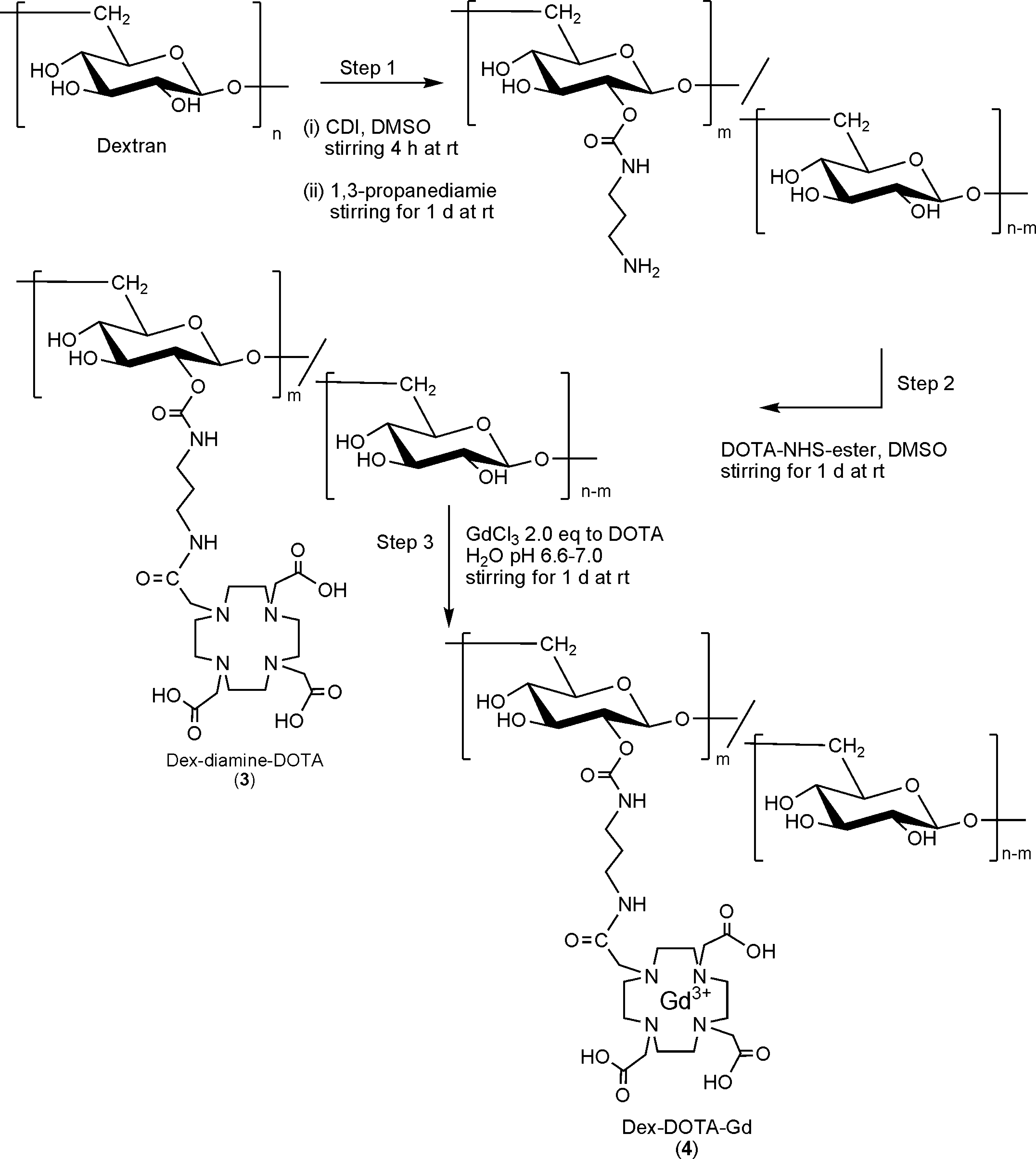
Synthesis of MRI contrast agent. Schematic structure of Dex-DOTA-Gd3+, which consists of a dextran derivative modified with the gadolinium (III) chelate DOTA-Gd3+. MRI, magnetic resonance imaging.
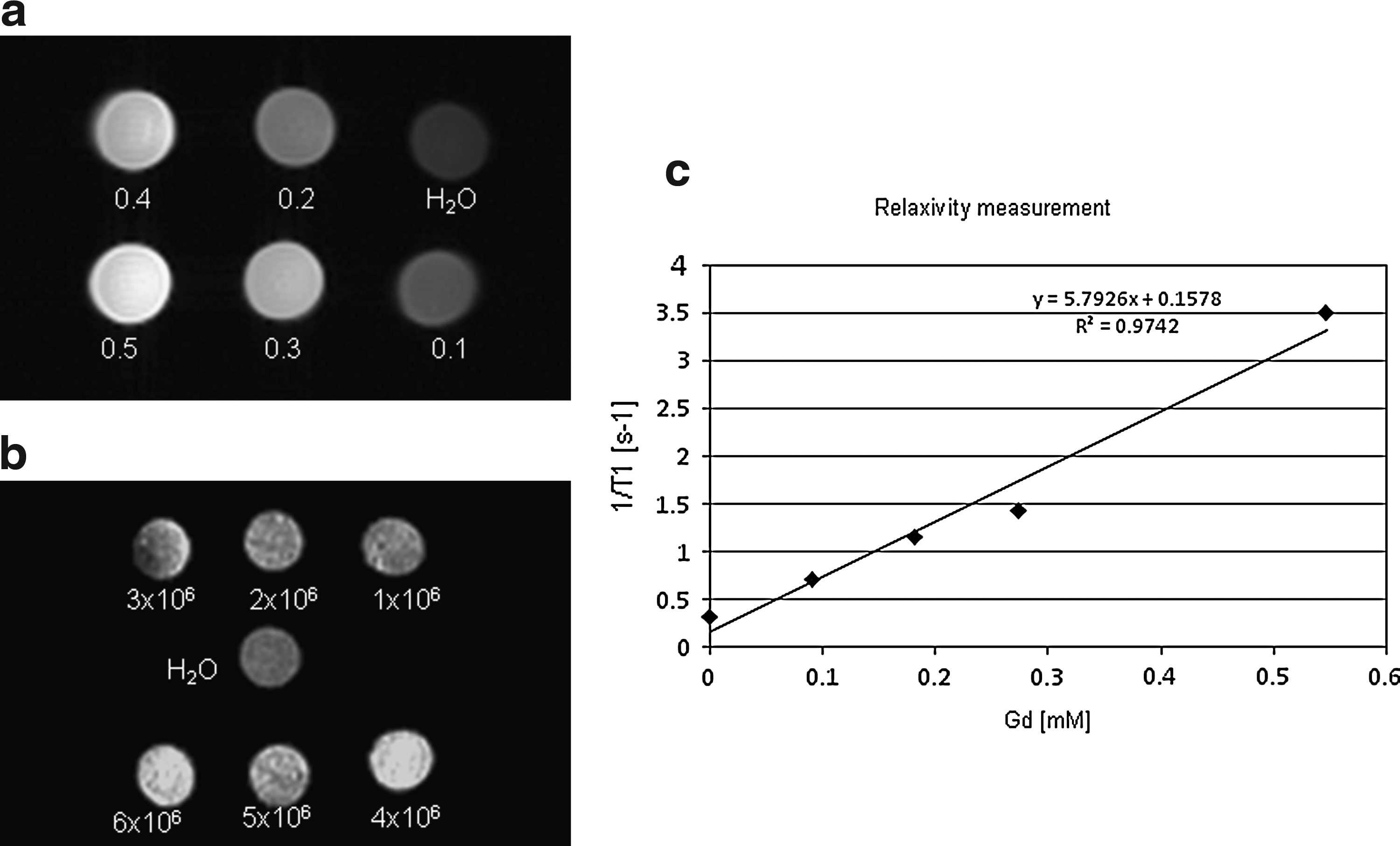
Figure 2a shows the MR images of Dex-DOTA-Gd+3 aqueous solutions at different concentrations. MR contrast agents shorten the longitudinal relaxation time T1 value, which is defined as the time constant of the exponential recovery of proton spins to their equilibrium along an applied field after disturbance. The relaxivity (R1) for Dex-DOTA-Gd3+, which represents the reciprocal of the relaxation time per unit of Gd concentration in s−1/mM, was determined by measuring the longitudinal relaxation time T1 values of several aqueous solutions by NMR and plotting 1/T1 versus the polymer concentration. Dex-DOTA-Gd3+ showed a relaxivity value of 5.8 s−1/mM (Fig. 2c), which, in comparison with approximate values of about 4.2 s−1/mM for DOTA-Gd and 3.8 s−1/mM for gadolinium-diethylenetriaminepentaacetic acid (DTPA-Gd),37,38 represents an increase of 38% and 52.6% over the relaxation value. Therefore, an enhancement of visualization of the resolution in MR images with Dex-DOTA-Gd3+ as a contrast agent is possible in comparison with the standard Gd3+ and Magnevits®. MR images of Dex-DOTA-Gd3+ aqueous solutions show an increase in signal intensity with regard to an increase in the concentration of the polymer (Fig. 2a). This increase is due to a reduction in the longitudinal relaxation time. Moreover, images show that the Gd complex has the capacity to return to its equilibrium state after radio frequency excitation. It can be seen that Dex-DOTA-Gd3+ MR images obtained at a concentration of 0.2 mM are visually different from MR images of distilled water.
Properties of the MRI contrast agent
Labeling of EPCs
The phenotype of the EPCs was then confirmed by immunostaining with DiI-acLDL, and staining with lectin and eNOS (Fig. 3). All cells were confirmed as the EPC phenotype based on their capacity to incorporate acetylated low-density lipoprotein, to bind lectin, and to bind endothelial nitric oxide synthase throughout the process of obtaining images (Fig. 3).30,31 Both FLK-1 and CD34 are expressed by all hematopoietic stem cells and EPCs, but cease to be expressed during hematopoietic differentiation.18,19

Phenotype identification of the isolated EPCs. One week after isolation, fluorescent staining of the adherent cells was used to confirm the EPC phenotype. Cells were incubated with DiI-acLDL, isolectin B4, and eNOS. After staining, the samples were visualized with a confocal microscope. The cells demonstrated positive fluorescence, which indicates the EPC phenotype. Scale bar=50 μm. eNOS, endothelial nitric oxide synthase. Color images available online at www.liebertonline.com/tea
EPC cells were labeled with Dex-DOTA-Gd3+ by means of electroporation and placed into agarose hydrogel. Figure 2b shows MR images of EPCs containing Dex-DOTA-Gd3+ in 100 μL of agarose hydrogel. The amount of labeled cells necessary to obtain differences in contrast with regard to water was found to be 1×10⁶ (1.11×10⁻¹⁰ mmol Gd), and the quantity of gadolinium incorporated into each cell was 0.12 pg of Gd. Cytotoxicity was analyzed by a lactate dehydrogenase cytotoxic test. Percentages of viable cells labeled with Dex-DOTA-Gd3+ at different concentrations are shown in Figure 4a. This clearly shows that for Dex-DOTA-Gd3+ concentrations lower than 15 mM, cell viability is as high as 90%, whereas higher concentrations of Dex-DOTA-Gd3+ were found to be slightly toxic, but with a range between 70% and 80% cell viability.
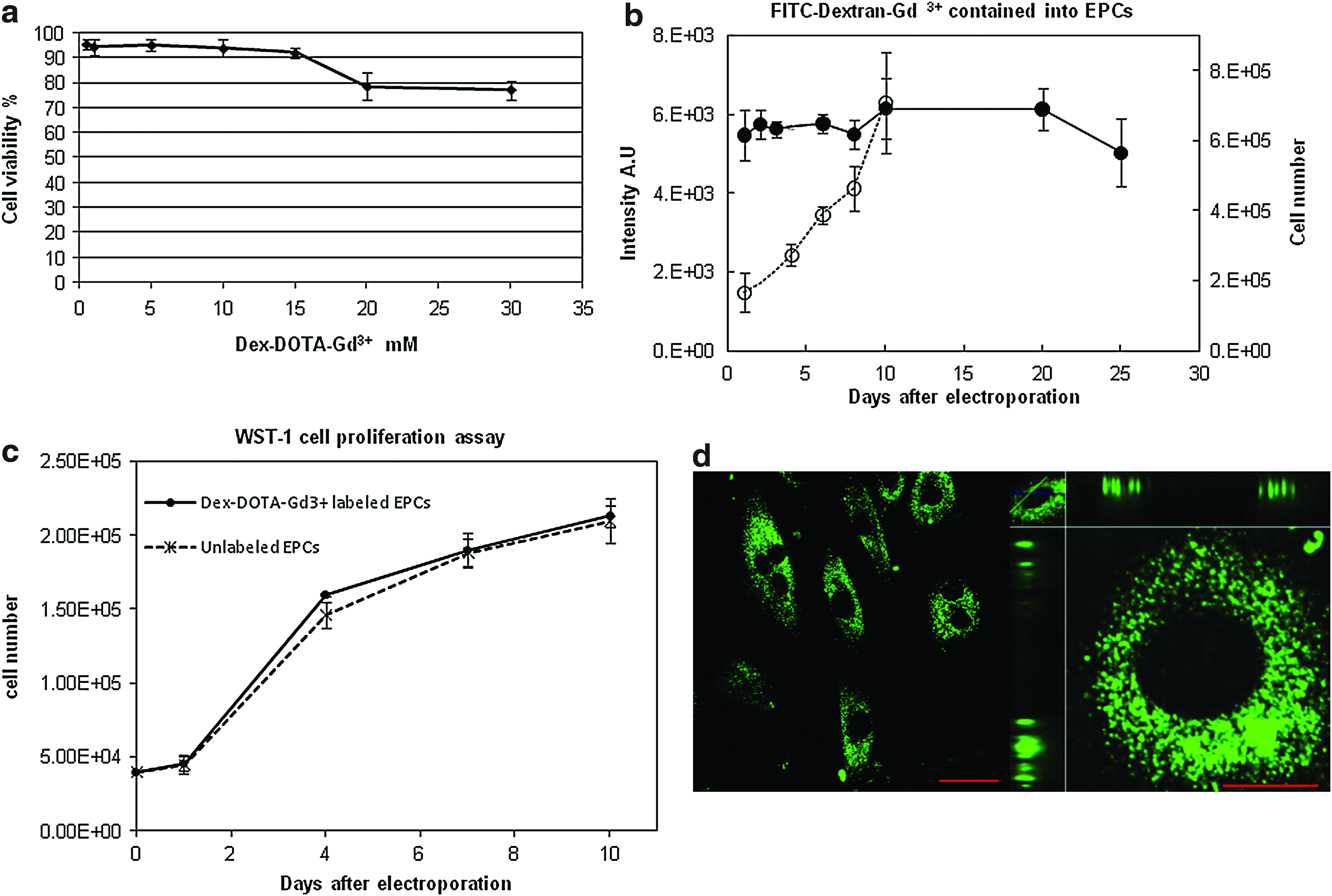
Therefore, we selected a polymer concentration of 10 mM to label the cells, because this concentration has low toxicity while providing an adequate quantity of gadolinium to achieve the required T1 shortening and high resolution for tracking labeled cells. Consistent with the previous results, the WST-1 assay performed with 10-mM polymer concentration revealed that EPCs were not affected by the contrast agent at least within 10 days after electroporation when cells achieved maximum confluence. No statistical differences were identified between the proliferation of nonlabeled EPCs and Dex-DOTA-Gd3+-labeled EPCs.
To analyze the distribution and stability of the contrast agent inside the cells, the Dex-DOTA-Gd3+-labeled cells were cultured for 25 days. As seen in Figure 4b, the EPCs exhibit a remarkably high degree of intracellular labeling with the cytoplasm containing large amounts of contrast agent. Interestingly, no further transport in the nuclei was observed 25 days after labeling. Figure 4c shows the Dex-DOTA-Gd3+ concentration inside cells with regard to the culture period and cellular proliferation rate.
The quantity of polymer remained stable for 25 days after electroporation. This suggests that Dex-DOTA-Gd3+ did not leak out of the cells during the culture period, and that the cells grew well. The differences detected from day 1 to 25 were not statistically significant (p>0.05), demonstrating stability of Dex-DOTA-Gd3+ labeling.
In vivo tracking of EPCs
Having demonstrated successful cellular MR imaging of EPCs by means of Dex-DOTA-Gd3+, we wanted to determine the efficacy of our MRI contrast agent for detecting and tracking EPCs in an in vivo rat model of ischemic hind limb. Immediately after inducing ischemia, we injected rats with Dex-DOTA-Gd3+-labeled EPCs in the adductor and quadriceps muscle in the inguinal region where the femoral artery and vein were excised (Bolheal was used to temporarily immobilize cells, as indicated in the Methods section). Animals were imaged thrice per week to determine the fate of the transplanted cells in the tissue. Figure 5 shows MR images obtained in vivo by using a 1.5-T animal MRI system within 20 days after transplantation. Cells could be clearly detected in the muscles and were observed to migrate for at least 16 days until the cells totally vanished at day 19. A rat was then sacrificed 6 days after transplantation, and the zone that appeared in the MR image was excised. The MR images in Figure 6a show that the cells are located in the adductor muscle. Frozen section slides revealed the presence of Qtracker 655-labeled EPCs in the ischemic area. During the development of the method for tracking EPCs in vivo, we observed that 5–6.3×10⁶ labeled cells diluted in 50 μL of Bolheal were necessary to achieve cell imaging at least 14 days after transplantation.
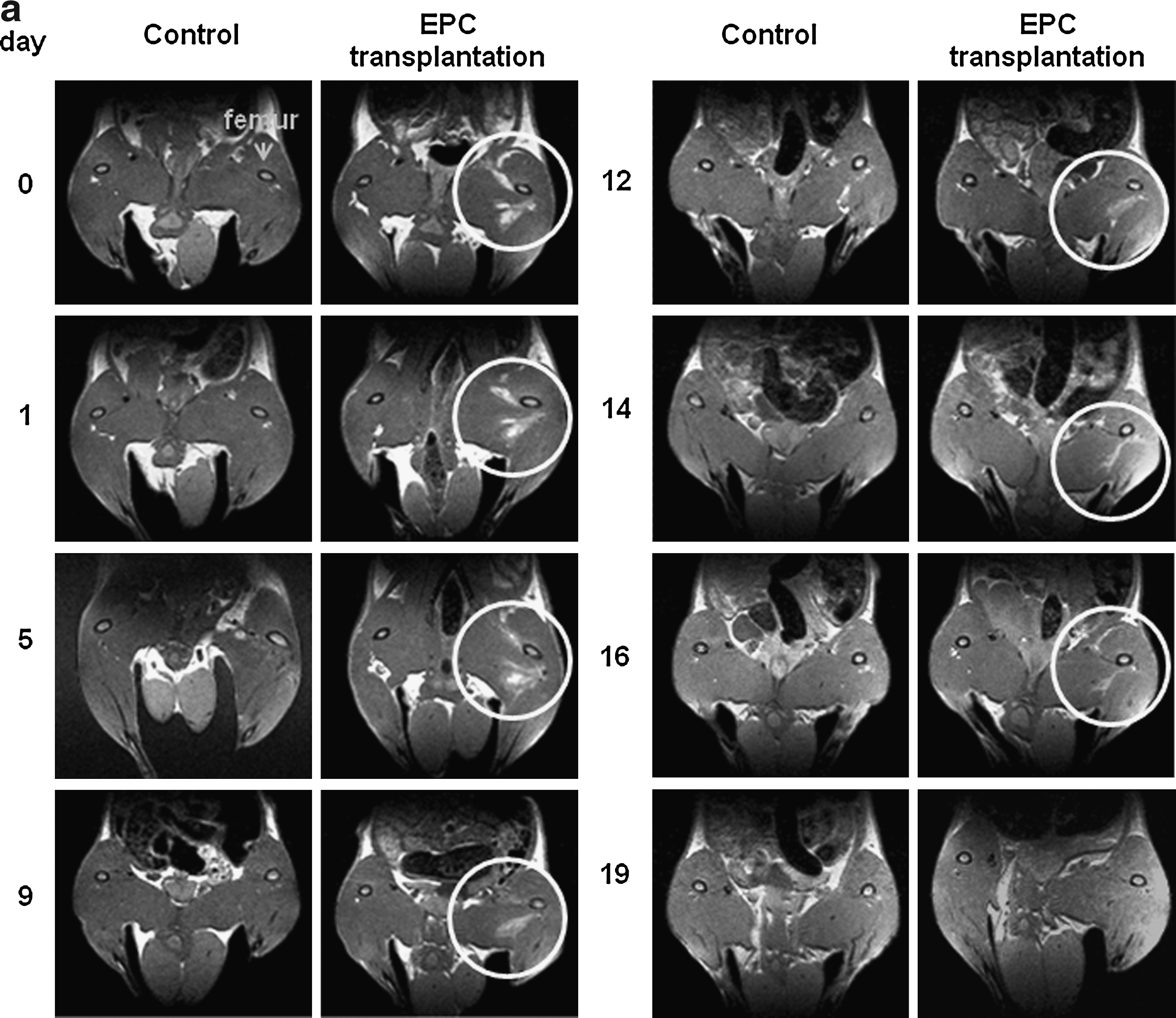
MR images of Dex-DOTA-Gd3+-labeled EPCs (2×10⁷) after intramuscular injection in ischemic limb rat models.

Identification of transplanted EPCs in the ischemic limb.
Immunohistochemical staining for von Willebrand factor and (AP) of the removed muscles at 35 days revealed the presence of numerous capillary endothelial cells in the rats implanted with Dex-DOTA-Gd3+-labeled EPCs (Fig. 7a); capillary/muscle fiber ratios for this rat group markedly increased (two-fold, p<0.001; Fig. 7b) relative to the ratio for the ischemic control group. There were no significant differences in capillary density between the ischemic control and the normal limb (p<0.05; Fig. 7b). This blood flow recovery by the EPC transplantation suggests that our cell labeling system based on Dex-DOTA-Gd3+ electroporation does not affect cell viability, cell growth, or stem cell functions.

Immunohistochemical staining of the nonischemic and ischemic limbs and determination of capillary density Immunohistochemical staining for AP and vWF staining indicates viable endothelial cells. Five fields from two muscle samples from each animal (n=9) were randomly selected.
Discussion
BM-derived EPCs were successfully labeled with Dex-DOTA-Gd3+ by electroporation at 0.12 pg of Gd per cell. This extent of labeling is significantly lower than 12 pg of iron oxide/cell, 16 but is nonetheless sufficient to achieve the signal enhancement required to provide good contrast in the body during MRI measurements. Several investigations on magnetic labeling of cells for MRI tracking in vitro and in vivo have shown that iron oxide nanoparticles are suitable for imaging stem cells.10,12,39 Nevertheless, more recent reports have called into question its use as a contrast agent, because of false-positive MRI signals, which lead to inconsistencies between a persistently positive MRI signal and histologically labeled cells.16,2,40–42
Commonly used Gd complex contrast agents such as DOTA-Gd3+ and Magnevist exhibit relatively slow relaxation times in comparison to Dex-DOTA-Gd3+. This is because these contrast agents are simpler and low-molecular weight structures and need higher doses of contrast agent to accomplish the same resolution. The incorporation of DOTA-Gd3+ into dextran is likely to have caused an increase in the relaxation time due to a steric limitation imposed on the rotational movement of the polymer, which leads to an increase in the rotational correlation coefficient.43,44 In addition, DOTA-Gd conjugated to a biocompatible dextran carrier and the free Dex-DOTA-Gd3+ are expected to be rapidly cleared from the body and to have few interactions with the cell membrane because of their high solubility.
Our results demonstrate that Dex-DOTA-Gd3+ achieves intracellular labeling through electroporation, which is an essential condition for labeling cells. A membrane modified with a polymer will interfere with cell-cell interactions during the recruitment of cells in the mechanism of angiogenesis. A polymer in the cell membrane can be easily detached and may be taken up and transferred to other cells in vivo. The fluorescence intensity of the cell lysis solution suggests that Dex-DOTA-Gd3+ remains stable in the EPCs for at least 25 days (Fig. 4c). A decrease in the fluorescence intensity indicates that the polymer has leaked out of cells during culture or that cells are dying, but no significant change in the fluorescence intensity has been demonstrated in the labeled EPCs. In addition, the cells showed a normal proliferation rate (during 10 days) 45 after Dex-DOTA-Gd3+ labeling by electroporation.
An additional cell proliferation activity and viability analysis was performed by using the WST-1 assay system. The results demonstrated that EPCs were not affected in the first 10 days after labeling by electroporation with Dex-DOTA-Gd3+ as a MRI contrast agent. No statistically significant differences were found between the proliferation of labeled and nonlabeled EPCs. After 10 days, the EPCs stopped proliferating and the quantity of cells remained stable. The viability claims are backed up with evidence for the first 10 days.
In vivo MR images indicate that the Dex-DOTA-Gd3+-labeled EPCs are perfectly detectable and that the fate of the cells can be followed within 16 days after transplantation with a cell density of 5–6×10⁶ cells/0.05 mL (Fig. 5). Labeled cells totally faded after 19 days. This is likely due to cell bio-distribution during cell migration, which is probably caused by cytochemical attraction of EPCs via incorporation into newly formed vessels and may also be influenced by the release of pro-angiogenic factors in a paracrine manner.28,46–48 Moreover, it is likely that cellular proliferation occurs in vivo. This would reduce the concentration of the polymer inside the EPC cytoplasm and cause low contrast in the limb. The proliferation rate of the labeled cells in vitro indicated that after 10 days, the cells increased in number by three-folds, and the fluorescence intensity remained stable. This indicates that the concentration of Dex-DOTA-Gd3+ remained constant but the concentration per cell might be reduced by three-folds. Analogous to the in vitro results shown in Figure 2b, if the image contrast of 6×10⁶ cells is reduced by three-folds, the signal intensity should be similar to the signal provided by 2×10⁶ cells. If this intensity reduction is assumed in vivo, the 6.3×10⁶ labeled cells transplanted into the ischemic limb 10 days after proliferation could then be tracked, because the Dex-DOTA-Gd3+ still has the capacity to produce a signal. At day 14 after implantation of labeled cells, the concentration of Dex-DOTA-Gd3+ underwent a four-fold reduction, which is the limit of the signal intensity provided by this contrast agent.
A rat was sacrificed 6 days after implantation of labeled cells. Muscles were carefully excised with particular attention to the location where MR images showed labeled cells. The presence of Qtracker 655-labeled EPCs localized in the neovascular zones of the ischemic limb ensures that MRI signals correspond to signals generated by the labeled cells (Fig. 6a). In addition, the number of macrophages found in the muscles (Fig. 6b) was insignificant, but was related to the obtained MR images or cells. This ensures that in contrast to cells loaded with iron oxide, the Dex-DOTA-Gd3+-labeled EPCs have not been subjected to endocytosis by the macrophages and are reliable for tracking of labeled cells over a long period.
Preliminary in vivo data suggest that Dex-DOTA-Gd3+-labeled cells were incorporated into sites of neovascularization and arranged into the capillary network. Direct local transplantation of labeled cells into the ischemic limb was found to quantitatively augment the capillary density in the ischemic limb in vivo.
Conclusions
The use of MRI for tracking EPCs by using a novel contrast agent in the therapeutic angiogenesis of ischemic limb models would be extremely useful for the anatomical visualization of localization of the transplanted stem cells over a long period. Dex-DOTA-Gd3+ as an MRI contrast agent for imaging stem cells in vivo has consistently satisfied certain properties such as providing desired MRI contrast properties and ex vivo cell labeling before transplantation, generating high-degree and stable intracellular labeling, ensuring biocompatibility without affecting cell viability backed up with evidence for the first 10 days after electroporation, possessing proliferative and healing capacities, and remaining consistently detectable over long periods of time.
Footnotes
Acknowledgments
This work was supported by grant-in-aid from the Ministry of Health, Labour, and Welfare of Japan (Health and Labour Sciences Research Grants, Research on Nanotechnical Medicine) and a Research Grant for Cardiovascular Diseases (18A-2) from the Japan Association for the Advancement of Medical Equipment. The authors thank Jun-ichiro Enmi, Takayuki Ose, Hajime Fukuda, and Akihide Yamamoto for their cooperation.
Disclosure Statement
The authors declare that there are no competing financial interests.