
Abstract
Cartilage tissue engineering using cells and biocompatible scaffolds has emerged as a promising approach to repair of cartilage damage. To date, however, no engineered cartilage has proven to be equivalent to native cartilage in terms of biochemical and compression properties, as well as histological features. An alternative strategy for cartilage engineering is to focus on the in vivo regeneration potential of immature engineered cartilage. Here, we used a rabbit model to evaluate the extent to which the maturity of engineered cartilage influenced the remodeling and integration of implanted extracellular matrix scaffolds containing allogenous chondrocytes. Full-thickness osteochondral defects were created in the trochlear groove of New Zealand white rabbits. Left knee defects were left untreated as a control (group 1), and right knee defects were implanted with tissue-engineered cartilage cultured in vitro for 2 days (group 2), 2 weeks (group 3), or 4 weeks (group 4). Histological, chemical, and compression assays of engineered cartilage in vitro showed that biochemical composition became more cartilagenous, and biomechanical property for compression gradually increased with culture time. In an in vivo study, gross imaging and histological observation at 1 and 3 months after implanting in vitro-cultured engineered cartilage showed that defects in groups 3 and 4 were repaired with hyaline cartilage-like tissue, whereas defects were only partially filled with fibrocartilage after 1 month in groups 1 and 2. At 3 months, group 4 showed striking features of hyaline cartilage tissue, with a mature matrix and a columnar arrangement of chondrocytes. Zonal distribution of type II collagen was most prominent, and the International Cartilage Repair Society score was also highest at this time. In addition, the subchondral bone was well ossified. In conclusion, in vivo engineered cartilage was remodeled when implanted; however, its extent to maturity varied with cultivation period. Our results showed that the more matured the engineered cartilage was, the better repaired the osteochondral defect was, highlighting the importance of the in vitro cultivation period.
Introduction
In our previous study, we demonstrated the feasibility and superiority of a novel cell-derived extracellular matrix (ECM) scaffold for cartilage tissue engineering over a conventional polyglycolic acid scaffold both in vitro and in vivo in a nude mouse model.13,14 We found that the chemical composition, in terms of glycosaminoglycan (GAG), collagen and DNA content, and compressive strength of the engineered cartilage using the ECM scaffold increased gradually up to certain level with time in culture, but were not comparable to those of the native cartilage. For example, the GAG content and compressive strength reached 66% (in vivo) and 44% (in vitro), respectively, of that of the native cartilage. Therefore, these experiments highlighted certain limitations associated with in vitro manufacture of cartilage.
Recently, Scotti et al. suggested that ideal maturation of the construct before in vivo implantation can affect regeneration of cartilage by optimizing matrix quality and integration. 15 They embedded swine chondrocytes into fibrin glue hydrogel, and found enhancement of chondrocyte synthetic activity, resulting in steady biomechanical properties of the construct. They suggested that an optimal preculture could provide the surgeon with a better engineered cartilage for implantation. However, they did not proceed to evaluate the question of how this more mature tissue could affect regeneration of the articular cartilage especially in vivo model. 15
We hypothesized that the maturity of a cartilage construct cultured in vitro affects its adaptation into the recipient site, impacting biochemical composition, histological remodeling, integration with surrounding cartilage, and ossification of the construct. In this study, tissue-engineered cartilage of varying degrees of maturities were prepared in vitro using ECM scaffolds and allogenous rabbit chondrocytes for 2 days, 2 weeks, and 4 weeks, respectively, and implanted into cartilage defects in the rabbit trochlear groove for evaluation of their remodeling capacity in vivo for 1 and 3 months, respectively.
Materials and Methods
In vitro study
Preparation of the cell-derived ECM scaffold
The cell-derived ECM scaffold was constructed as described in our previous study.13,14 Articular cartilage was harvested from the stifles of 2–3-week-old porcine`s knee joint. Cartilage pieces were carefully dissected from the other tissues and washed with phosphate-buffered saline (PBS), which was followed by treatment with 0.05% (w/v) Pronase (Boehringer, Mannheim, Germany) at 37°C for 1.5 h. They were washed twice with PBS and then subjected to 0.2% (w/v) collagenase (Worthington Biochemical Corp., Lakewood, NJ) treatment for 15 h in Dulbecco's modified Eagle's medium (DMEM; Gibco, Grand Island, NY) supplemented with 5% newborn calf serum (NCS; Hyclone, Logan, UT). Once the porcine cartilage tissues were completely digested, the isolated porcine chondrocytes were centrifuged at 600 g for 10 min. 16 Cell pellets were washed twice and seeded in tissue culture plates (diameter 100 mm) at a density of 1.9×10⁵ cells per plate. Chondrocytes were cultured in monolayers with DMEM supplemented with 10% NCS, 1% antibiotics–antimycotics, and 50 μg/mL L-ascorbic acid. After 3–4 days, the medium was removed and 0.05% trypsin–ethylenediaminetetraacetic acid (EDTA; Gibco) was added briefly to separate the chondrocyte/ECM membrane from the bottom of the culture plate. The membrane was carefully isolated with a wide-bore pipette and transferred individually to a 50 mL conical tube filled with 30 mL DMEM and 5% NCS. The tube was then centrifuged at 600 g for 20 min to consolidate the membrane into a pellet-type construct. It was then incubated overnight at 37°C and transferred to a six-well culture plate for extended culture. The constructs were allowed to grow into the cartilage tissue for 3 weeks, and the culture medium (5 mL) was changed three times a week. 17 After 3 weeks, the newly grown cartilage construct was washed in PBS and then stored at −20°C for 3 days. To fabricate an ECM scaffold, the construct was freeze-dried for 48 h at −56°C under 5 mTorr. The freeze-dry process also removes roughly cellular components. Freeze-dried specimens were split into two parts using a 6-mm punch: a disk-shaped core and a ring-shaped periphery. Due to the dimensional consistency of the core area, the disk-shape was chosen as a preform of the ECM scaffold. The disk-shaped core was further trimmed off the surface layer to be 1 mm in thickness for cell seeding (volume=28.26 μL).
Construction of engineered cartilage using rabbit chondrocytes and the ECM scaffold
Cell-derived ECM scaffolds were soaked in sterile 70% ethanol for l hr, then washed several times in PBS, and immersed overnight in serum-free DMEM before cell seeding. Chondrocytes isolated from articular cartilage of New Zealand white rabbit`s knee joint (2 weeks old) were dynamically seeded at passage 1 in the ECM scaffold for 1.5 h using a nutator at a density of 3×10⁶ cells/mL. 13 The chondrocyte-seeded ECM scaffold was cultivated in 6-well plates for 2 days, 2 weeks, and 4 weeks in vitro before implantation.
Histological and chemical assays
The normal cartilage, ECM scaffold, and tissue-engineered cartilage in vitro were fixed with 4% formalin at least for 24 h, and embedded in paraffin wax. As previously described, the samples were sectioned at 4 μm thickness and stained by Safranin-O. 13
For chemical assay of the implants, the dried samples were digested in papain solution (5 mM L-cysteine, 100 mM Na2HPO4, pH 6.4, 5 mM EDTA, and 125 μg/mL papain type III) at 60°C for 24 h and then centrifuged at 12,000 g for 10 min (n=4). To measure GAG content, the supernatant was subjected to 1,9-dimethylmethylene blue colorimetric assay.13,14 GAG content was determined using chondroitin sulfate from shark cartilage (Sigma, St Louis, MO) as a standard. Collagen content was measured using the hydroxyproline assay. 18 Bovine collagen (0–10 μg/mL of tracheal cartilage; Sigma Chemical Co, St Louis, MO) was used as a standard solution. DNA content was measured using the Quant-iT™ dsDNA BR assay kit (Invitrogen; Eugene, OR). The ECM scaffold alone was used as a control in all assays.
Measurement of mechanical compressive strength
Implants were subjected to a mechanical compressive strength test with a Universal Testing Machine (Model H5K-T; H.T.E., Salfords, England). Specimens (n=5) were cut into a uniform disk shape and then placed transversely on the metal plate, where they were pressed at a crosshead speed of 1 mm/min and 0.001N of preload. Individual compressive strengths were calculated at the break point of 10% strain. The ECM scaffold and native cartilage from 2-week-old rabbits were used as negative and positive controls, respectively.
In vivo study
Experimental design and surgery
The experimental protocol was approved by the Institutional Animal Experiment Committee. Eighteen New Zealand white rabbits with an average weight of 3.0–3.5 kg (about 3 months old) were used in the study. Surgical procedures, including limb preparation and draping, were performed under general anesthesia with katamin and lumpun (ratio 3.5:1.5). Both knee joints were operated on during the same surgery. An arthrotomy was made through a midline longitudinal incision on a medial parapatellar with the patella dislocated laterally to expose the femoral condyles. To create an osteochondral defect of 3 mm in depth and 5 mm in diameter, a 5 mm drill was used at the patella groove. A total of 24 rabbits (three rabbits per group per time point) were assigned to four groups, including the untreated control (group 1) and experimental groups implanted with engineered cartilage cultured in vitro for 2 days, 2 weeks, and 4 weeks (groups 2, 3, and 4, respectively). Implants were inserted into the defects and press-fixed without covers or suture materials.
Macroscopic and histological evaluation
At 1 and 3 months after surgery, the rabbits were euthanized by over-dose injection of Pentobarbital for retrieval of the femoral condyles. A macroscopic image of the condyles was first observed, and the samples were paraffin-embedded, sectioned, and processed for routine Safranin-O staining or immunohistochemical analysis. For immunohistochemical analysis, the sections were incubated with mouse monoclonal anti-rabbit collagen type II antibody (Chemicon, Temecula, CA) at a 1:200 dilution for 1 h at room temperature. The sections were then incubated sequentially with biotinylated secondary antibody at a dilution of 1:200 for 1 h and peroxidase-conjugated streptavidin solution for 30 min at room temperature (DAKO LSAB System, Carpinteria, CA). After counterstaining with Mayer's hematoxylin (Sigma), the sections were mounted with a mount solution before microscopic observation (Nikon E600, Tokyo, Japan).
Histological scoring (International Cartilage Repair Society score)
To evaluate the quality of the repaired articular cartilage in the defects, a modified version of the histological grading scale was used. 19 The scale consists of seven categories and assigns a score ranging from 0 to 18 points (Table 1). Parameters included cell morphology, matrix staining (Safranin-O), structural integrity, surface regularity, thickness of cartilage, regenerated subchondral bone, and integration with adjacent cartilage.
Statistical analysis
Statistical analysis of the experimental data was carried out using one-way analysis of variance for multiple comparison and Tukey`s t-test (two-tail) for pair wise comparison. Statistical significance was assigned as *p<0.05, **p<0.01, and ***p<0.001.
Results
Evaluation of engineered cartilage in vitro
Histological and chemical assay for engineered cartilage
Expression of sulfated proteoglycan was examined by Safranin-O staining of both the ECM scaffold itself and the engineered cartilage cultured in vitro for 2 days, 2 weeks, and 4 weeks (Fig. 1A–D). With time, the porous space within the scaffold gradually became filled with ECM (Fig. 1).

The Safranin-O staining of the ECM alone
Total amounts of DNA, GAGs, and collagen in engineered cartilage were measured at each culture time point by chemical assay (Fig. 2). DNA content of the engineered cartilage significantly increased with time from 2 days, reaching a maximum threefold increase at 4 weeks. They were 0.34±0.02, 2.51±0.86, 5.16±0.75, and 7.36±0.81 μg for the ECM scaffold alone and the cultured samples at 2 days, 2 weeks, and 4 weeks, respectively (Fig. 2A). GAG content increased markedly with time, particularly after 2 weeks (Fig. 2B). In the ECM scaffold alone, the amount of GAG was 276.5±20.6 μg, and increased in the engineered cartilage to 378.5±65.6, 1302.8±65.4, and 1450±30 μg at 2 days, 2 weeks, and 4 weeks, respectively. Collagen content also showed a gradual increase with time, reaching a value at 4 weeks that was more than fourfold higher than that at 2 days (Fig. 2C). The collagen contents were 385.2±7.6 μg in the ECM scaffold alone, and gradually increased in the engineered cartilage to 498.9±22.7, 914.7±59.2, and 1973.1±201 μg at 2 days, 2 weeks, and 4 weeks, respectively. The unit values of DNA, collagen, and GAG contents per mg dry weight were ranging approximately 40%–70% of the normal values of rabbit cartilage and did not change significantly along with time points (data not shown).

The amount of DNAs
Mechanical properties: compressive strength of the implants
The ECM scaffold alone and the engineered cartilage were tested for compressive strength at the break point of 10% strain. The value for ECM scaffold alone was 12.3±0 kPa, whereas the values for engineered cartilage at 2 days, 2 weeks, and 4 weeks were 7±0.08, 16.4±2.2, and 21.5±2.2 kPa (n=5/group), respectively (Fig. 3). Compressive strength of the engineered cartilage at 4 weeks reached a value threefold higher than that at 2 days. Compressive strength of the engineered cartilage at 4 weeks (21.5±2.2 kPa) was about 44% of that of the native cartilage of rabbits, which was approximately 49±2.2 kPa. Compressive strength of the ECM scaffold alone was higher than that of the 2-day engineered cartilage because the ECM scaffold was measured under dry condition, whereas the implants were measured wet. In a wet condition, the ECM scaffold became very thin and soft, which made it impractical to measure its compressive strength.
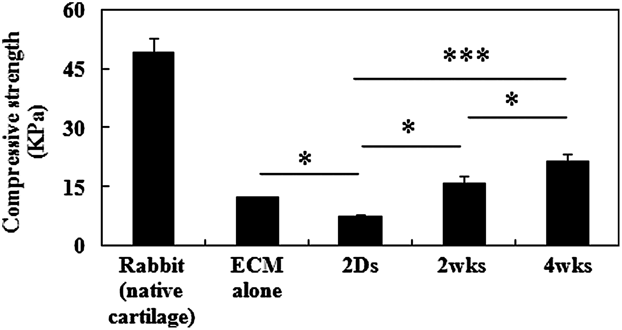
The compressive strength of the ECM scaffold alone and implants were measured at each time of culture in vitro as described in the Materials and Methods section. The compressive strength at the break point of 10% stain was significantly increased with culture time, reaching about 44% of the normal cartilage at 4 weeks. *p<0.05, ***p<0.001.
In vivo study
Gross examination of cartilage defects
Gross appearance of cartilage defects immediately after implantation of engineered cartilage is shown in Figure 4. Implants were stably inserted into the defect sites. At 1 month after surgery, repaired defects in groups 3 and 4 were smooth and glistening in appearance, and exhibited continuity with the surrounding host cartilage tissue. In contrast, the defect was not well repaired in group 1 (control), and was only partially filled with fibrous tissue in group 2. At 3 months, the repaired tissues in defects exhibited a white and glistening appearance in all groups. However, the surface of repaired tissue in groups 2, 3, and 4 was smooth and hard by a forceps test, whereas the surface of repaired tissue in group 1 was slightly rough with many fissures.

Gross findings of the cartilage defect with implants. Presented are gross images of the engineered cartilages
Histological evaluation
At 1 month, construct of group 2 in the defect area seemed to be resolved and became a mixture of fibrous and osseous structure. No GAG stained by Safranin-O, even though some abutment from neighboring cartilage was observed. The defect was partially filled with fibrous tissue in groups 1 and 2, but was not well integrated with surrounding host cartilage and bone (Fig. 5A, B, E, and F, arrows). In contrast, the construct of groups 3 and 4 became more mature. The ECM scaffold was completely degraded and replaced with hyaline material, which stained metachromatically by Safranin-O. This was more evident in group 4. Defects were repaired with hyaline cartilage-like tissue in both group 3, which was incompletely filled with rugged surfaces, and group 4, which was completely filled with smooth surfaces (Fig. 5C, D, G, and H). Successful integration of repaired tissues with the surrounding cartilage or the subchondral bone was not observed in any of the groups at 1 month (Fig. 5D and H, arrows). At 3 months, all defects were repaired with cartilaginous tissue, with some differences in quality. The control group recovered its osseous defect completely, but repaired cartilage had poor structure with rugged surface. The repaired host tissue interface consisted mostly of fibrous tissue with low amount of GAG (Fig. 5I and M, arrows). In group 2, cartilaginous tissue remained in the osseous defect region, on which well-developed articular cartilage tissue was restored. The interface consisted of immature tissue with high density of proliferating cells (Fig. 5J and N, arrows). In group 3, cartilagenous tissue still remained in the osseous defect region. They showed more uniform structure than that of group 2, but still contained many elongated cells found in fibrous tissue and did not stain intensively with Safranin-O in the cartilage defect region (Fig. 5K and O, arrows). Repaired cartilage in groups 1, 2, and 3 showed poor production of GAG; in addition, lacunae were barely evident, and cellular arrangement was not restored. Also, at this time, repaired tissue in group 1 exhibited a rough surface, and the subchondral bone was partially ossified in groups 1, 2, and 3 (Fig. 5I–K and M–O). In contrast, group 4 showed hyaline cartilage tissue with a mature matrix and a columnar organization of chondrocytes in the repaired tissues at 3 months. They consisted mainly of mature cartilaginous tissue containing round cells and GAG rich in neoECM that is well integrated with the host tissue (Fig. 5L and P, arrows). Moreover, the subchondral bone was well ossified in this group at this time (Fig. 5L and P). No immune or adverse response was observed in all experimental groups.
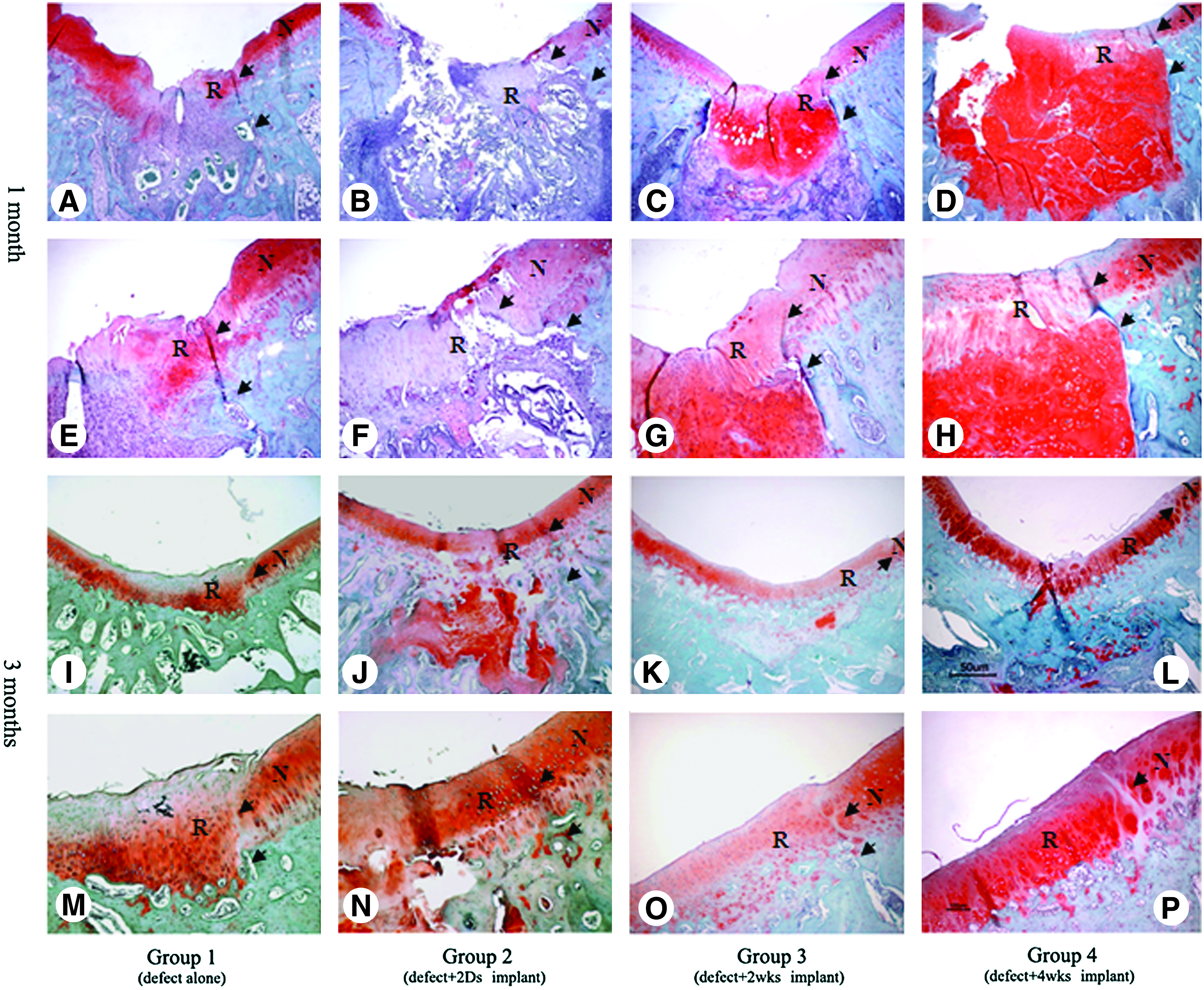
Safranin-O staining of repaired cartilages from each group at 1 and 3 months postimplantation.
Total International Cartilage Repair Society (ICRS) histological score increased significantly along with time in all groups (Fig. 6). At 1 month, the ICRS score was higher in groups 3 and 4 than in groups 1 and 2, and was highest in group 4 at 3 months.

The International Cartilage Repair Society (ICRS) score was determined from the results of the Safranin-O staining according to the rating system shown in Table 1. The highest ICRS score was measured in group 4 at 3 months. The statistical differences were compared among the groups at each time point. *p<0.05, **p<0.01.
Expression of type II collagen
At 1 month after implantation, expression of type II collagen was observed intracellular area in all groups, but more prominent in the whole defect area in groups 3 and 4. At 3 months, the expression of type II collagen was observed at the pericellular region in repaired tissues of groups 2, 3, and 4, but not in group 1 (Fig. 7, arrows). Expression of type II collagen was enriched in a zonal fashion particularly in group 4 at 3 months (Fig. 7H; dark brown color).

Immunohistochemical staining of type II collagen synthesized in the repaired tissues at 1 and 3 months after implantation (×200). Arrows indicate pericellular region. (
Discussion
This study showed that the more mature the construct was in vitro (i.e., cultured up to 4 weeks), the better remodeled and integrated was the regenerated cartilage after implantation. Quality of repaired tissues at 3 months after implantation of engineered cartilage cultured in vitro for 4 weeks was similar to that of the surrounding native cartilage. This is quite contrary to previous reports. It has generally been recognized that immature engineered cartilage is likely to integrate itself well with the surrounding tissues, owing to high cellular activity at the interface20,21 but could be easily broken down by the mechanical load. In contrast, mature cartilage might be more resistant to the mechanical load, making it more suitable for a weight-bearing joint such as the knee. However, it is less likely to integrate into surrounding cartilage, as shown in autologous osteochondral grafts, necessitating optimization of in vitro culture conditions. Obradovic et al. reported in their in vitro study that immature constructs using engineered cartilage with bovine articular chondrocytes had poorer mechanical properties, but integrated better than either more mature constructs or cartilage explants through increased cell proliferation and progressive formation of cartilaginous tissue, in contrast to integration of more mature constructs or native cartilage, which involved only secretion of ECM components. 21 Silverman et al. underlined the cellular activity for adhesion and integration of engineered cartilage to native cartilage. They used fibrin glue polymer mixed with fresh swine articular chondrocytes, which was sandwiched between two 6-mm-diameter discs of fresh articular cartilage. The experimental group showed a layer of neocartilage between the two native cartilage discs that filled all irregularities along the surface of the cartilage discs, whereas control samples (fibrin glue without chondrocyte) showed no evidence of neocartilage formation. Mechanical properties of the experimental constructs were significantly greater than those of the control group. Their study demonstrated that young premature tissue-engineered cartilage does adhere to adjacent native cartilage in a nude mouse model. 22 However, there are few reports that degree of maturity of engineered cartilage, representing chemical composition and histological stability can affect the regeneration of cartilage.
In this study, 4-week-cultivated construct unexpectedly showed complete remodeling as well as good integration with surrounding host cartilage. We assumed that it could be attributed to higher mechanical strength of 4-week-cultivated constructs than that of other groups. We learned from autologous osteochondral transplantation that good fixation is the primary determinant of success. Stable fixation of the donor fragment not only provides good integration with recipient cartilage, but also plays an important role in preserving the histological properties of cartilage. 23 After culturing for 4 weeks in vitro, the physical strength of the engineered cartilage reached 44% that of the native cartilage of rabbit, and could bear body weight, even immediately after surgery, which could thus readily support not only regeneration and remodeling of engineered cartilage, but also integration with neighboring cartilage. Also, we cannot exclude the possibility that recipient chondrocytes at the cut margin of cartilage and bone marrow cells that dislodged when the hole was made played some role. The engineered constructs even after 4 weeks of cultivation did not reach the normal rabbit cartilage in their amounts of collagen and GAG (data not shown). We speculate that this caused the loose entrapment of cells in ECM of cartilage, thus enabled free movement of cells between the construct and host cartilage, which was crucial to adhesion of donor cartilage as reported by Silverman and Obradovic. 22 Another influencing factor could be blood clot from the bone marrow. When the osteochondral defect was created in this study, some blood clot containing mesenchymal stem cells (MSCs) could be formed at interface between more mature implants cultured for 4 weeks in vitro and surrounding cartilage. Unlike the report by Silverman and Obradovic, our study was undertaken in an in vivo model, so that mechanical stress and natural biological environments must have enhanced cellular activity for achievement of better integration.
Interestingly, the histology showed the presence of newly mineralized bone-like tissue in the subchondral bone of the implanted area. This seemingly unusual finding was not unexpected because our previous clinical experience showed that autologous chondrocytes implanted into deep cartilage defects below the subchondral cortex developed into bone and completely filled the defect within 1 year, based on follow-up MRI and biopsy results (unpublished observations). Although the mechanism of ossification at the cartilage donor site remains to be determined, it is likely that endochondral ossification occurred in the engineered cartilage. According to Groot, 24 even fully differentiated chondrocytes can transdifferentiate into osteoblasts; thus, chondrocytes in engineered cartilage itself could differentiate into bone. Chondrocytes have recently been shown to possess osteoinductive ability, stimulating osteogenic differentiation of cocultured MSCs. 25 Therefore, implanted chondrocytes could have promoted bone formation by inducing osteogenic differentiation of bone marrow stem cells. We found that immature cartilage in vitro took more time to become ossified than mature cartilage. Group 4 at 1 month of implantation showed many hypertrophied chondrocytes in the osseous defect area. This may mean that mature cartilage goes further in endochondral ossification linage. Regardless of the mechanism, engineered cartilage became successfully anchored into the defect area, with 4-week-cultivated engineered cartilage yielding the most effective transition from cartilage to bone.
In any experimental implantation model, the intrinsic capacity of the animal to heal cartilage defects may complicate interpretation of results. However, because the rabbit model has been used extensively in cartilage implantation studies and offers low inter-animal variance due to extensive inbreeding, it is the most representative and justifiable experimental animal model for use in cartilage research. 26 Rudert have reported that success in repair of a 3-mm-diameter defect in the trochlea does not offer any benefit, since, according to the literature, perfect healing has never been observed, even with common 3-mm-diameter defects. 26 Although spontaneous regeneration of cartilage was observed to some extent in the defect-only group, the quality of the regenerated cartilage was poor and contained a considerable amount of fibrotic tissue on the surface, even 3 months after implantation. Formation of this type of fibrocartilage might be attributable to bone marrow stem cells and growth factors that originated from subchondral bone. 27 This process might be a particularly important contributor to repair of cartilage defects in the rabbit model because thickness of hyaline articular cartilage is approximately 300–400 μm in mature rabbits. 28 Therefore, larger animals, such as goats and pigs, might make better models for evaluation of the activity of scaffolding material for use in cartilage tissue engineering.
One major limitation of this study was that in vitro cultivation did not extend beyond 4 weeks. Longer cultivation might yield harder constructs containing larger amounts of ECM. However, cultivation for more than 4 weeks does not produce further changes in the chemical or physical properties of rabbit chondrocytes. Optimal culture time will need to be determined empirically for other combinations of scaffolding material and cell sources. Another limitation is that the young rabbit cells were used, which will limit the clinical relevance of our result. Future study of ours will evaluate the ECM scaffold using human chondrocytes under the current experimental condition. Biochemical and compression test for the implanted tissues in vivo will be also carried out.
Conclusion
We demonstrate that differences in remodeling outcome using implanted engineered cartilage depend on the degree of cartilage maturity in vitro. We found that the more matured the engineered cartilage was, the better repaired the osteochondral defect was, although we anticipate that further studies designed to optimize the cultivation period may be informative and perform the biomechanical and compression testing of regenerative cartilage in vivo. To the best of our knowledge, this study is the first to report on an effect of in vitro maturity of tissue-engineered cartilage on remodeling of repaired cartilage in an animal osteochondral defect model.
Footnotes
Acknowledgments
This study was supported by the Foundation of the Ministry of Knowledge Economy, Republic of Korea (Grant 10024099), and supported by a grant of the Korea Healthcare Technology R&D Project, Ministry for Health and Welfare, Republic of Korea (A091120).
Disclosure Statement
No competing financial interests exist.