
Abstract
The organ- or tissue-printing approach, based on layered deposition of cell-laden hydrogels, is a new technique in regenerative medicine suitable to investigate whether mimicking the anatomical organization of cells, matrix, and bioactive molecules is necessary for obtaining or improving functional engineered tissues. Currently, data on performance of multicellular printed constructs in vivo are limited. In this study we illustrate the ability of the system to print intricate porous constructs containing two different cell types—endothelial progenitors and multipotent stromal cells—and show that these grafts retain heterogeneous cell organization after subcutaneous implantation in immunodeficient mice. We demonstrate that cell differentiation leading to the expected tissue formation occurs at the site of the deposited progenitor cell type. While perfused blood vessels are formed in the endothelial progenitor cell–laden part of the constructs, bone formation is taking place in the multipotent stromal cell–laden part of the printed grafts.
Introduction
Organ- or tissue-printing is a new technique in regenerative medicine that can help to investigate whether imposed cell organization is actually necessary for obtaining fully functional newly formed tissues, as it enables defined placement of different cell types in a construct. Using 3D fiber deposition, layers of cell-laden hydrogel strands are deposited according to a rapid prototyping design. 6 The resulting 3D scaffolds show a highly reproducible architecture (size, shape, porosity, interconnectivity, pore-geometry, and orientation), whereas a great variation in material composition is possible. The porosity of the implants is easily tailored, which is important for mechanical and conductive properties of the construct. Pores enhance nutrient supply and waste product removal by allowing ingrowth of blood vessels and supporting homogeneous tissue formation. Further, the 3D fiber deposition method can be used for rapid formation of constructs with defined organization of multiple cell types. Printable hydrogel matrices, including thermosensitive gelatinous protein mixtures such as Matrigel and seaweed-derived ion-sensitive alginates, are well-suited materials for cell encapsulation and support viability and differentiation of embedded cells. 7 Previously, 3DF was used to print osteogenic progenitors in alginate scaffolds. 8 Multipotent stromal cells (MSCs) survive the deposition process and retain the ability to differentiate toward osteogenic lineage after printing. Deposition of multiple cell populations has also been achieved by other organ-printing techniques, including ink jet printing, laser deposition, and dispensing tools.9–11 However, limited evidence is available on the functionality of the designed cellular structures, due to short follow-up times after printing.
Currently, no data are present on performance of multicellular printed constructs in vivo. It would be interesting to know whether in vivo heterogeneous cell organization introduced by printing is retained and is being translated to heterogeneous tissue formation. In this study we follow the retention of defined cell arrangement and heterogeneous tissue formation in printed grafts in vivo. We printed intricate, multicellular constructs using MSCs and endothelial progenitor cells (EPCs), and subsequently analyzed the nature of tissue formed after subcutaneous implantation in immunodeficient mice. Cells were fluorescently labeled to assess the dispersion of the transplanted cell populations in vivo and to determine whether the resulting tissue formation corresponded to the printed cell type.
Materials and Methods
Cells
Goat MSCs (gMSCs) obtained from iliac bone marrow aspirates of adult Dutch milk goats were isolated by adhesion to tissue culture plastic. 12 Briefly, aspirates were plated at a density of 5×10⁵ cells/cm2 and cultured in expansion medium consisting of αMEM (Invitrogen) supplemented with 10% FBS (Lonza), 2 mM L-glutamine (Glutamax; Invitrogen), 100 U/mL penicillin, and 100 μg/mL streptomycin (Invitrogen). Medium was refreshed twice a week and cells were used for further subculturing or cryopreservation. Cell cultures were maintained in a humidified incubator at 5% CO2 and 37°C. gMSCs passage 2–6 were used in the study.
Goat EPCs (gEPCs) were isolated from peripheral blood. The peripheral blood samples were harvested from adult Dutch milk goats, which had not received prior stimuli to mobilize EPCs from the bone marrow. Mononuclear cells (MNCs) were isolated by density gradient centrifugation with Ficoll solution (Ficoll Paque™ Plus; GE Healthcare Biosciences AB). Cells from individual goats were plated in 25 cm2 culture flasks coated with fibronectin (2.5 μg/mL in phosphate-buffered saline [PBS], 1 h; Harbor Bioproducts) in EBM-2 (Cambrex), supplemented with singlequots (hEGF, VEGF, hFGF-B, R3-IGF-1, ascorbic acid, heparin, and gentamicin-amphotericin-B) and 20% fetal calf serum (FCS; Cambrex), and cultured at 37°C and 5% CO2 in a humidified chamber. After 4 days of culture, nonadherent cells were removed by washing with PBS, a fresh medium was applied, and cells were cultured for at least 6 weeks, replating them twice a week. Colonies from a single donor were cultured together. After colony outgrowth, the cells were detached using 0.25% trypsin (Invitrogen) and stored in liquid nitrogen until further use.
Fluorescent labeling of cells
Before printing, cells were fluorescently labeled to facilitate distribution-analysis of the two cell types. For this, gEPCs were incubated with PKH26 (red; Sigma-Aldrich) and gMSCs with CFSE (green; Molecular Probes). The presence of fluorescent cells was analyzed directly after printing and after 1 and 2 weeks in in vitro samples and in frozen sections of in vivo samples after 2 weeks. For this, a fluorescence microscope (Olympus BX51 microscope, Olympus DP70 camera) was equipped with an epifluorescence setup (Leica DM IRBE), excitation/emission setting of 488/530 nm to detect green-fluorescent gMSCs, or 530/580 nm to detect red gEPCs.
Cell-laden hydrogel preparations
BD Matrigel™ Basement membrane matrix (#354234; BD Biosciences) was used for encapsulation of EPCs at 5×10⁶ cells/mL gel. For encapsulation of MSCs we used alginate or growth factor-depleted Matrigel (#354230; BD Biosciences) (both gels contained 5×10⁶ cells/mL gel), supplemented with osteoinductive biphasic calcium phosphate (BCP) microparticles (10% [w/v], see below for details).
Matrigel preparations were processed at 4°C to keep the matrix fluid, and formed a gel at room temperature during printing. High-viscosity alginate powder (International Specialty Products) was autoclaved and subsequently mixed (10% [w/v]) overnight at 37°C with osteogenic medium (αMEM [Invitrogen] supplemented with 10% FBS [Lonza], 0.1 mM ascorbic acid 2-phosphate [AsAP; Sigma-Aldrich], 2 mM L-glutamine [Glutamax; Invitrogen], 100 U/mL penicillin, 100 μg/mL streptomycin [Invitrogen], 10−8 M dexamethasone [Sigma-Aldrich], and 10 mM β-glycerophosphate [Sigma-Aldrich]). For gel formation, after printing alginate was incubated for 15 min with 102 mM CaCl2 supplemented with 10 mM HEPES pH 7.4 (Invitrogen). BCP microparticles (Progentix Orthobiology BV) were sintered at 1150°C and ranged from 106 to 212 μm in size. To assess contribution of BCP to bone formation in vivo, MSC-laden gel samples (alginate and growth factor depleted Matrigel) without BCP served as controls.
3D fiber deposition presets and printing of heterogeneous scaffolds
The Bioscaffolder pneumatic dispensing system (SYS+ENG) was used for 3D printing of hydrogel scaffolds. This system was previously employed for extrusion of (cell-laden) hydrogels and is described in more detail elsewhere.8,13 Briefly, the Bioscaffolder is a three-axis dispensing machine, which builds up 3D constructs by coordinating the motion of a pneumatic syringe dispenser. The dispenser deposits extrudate consisting of empty or cell-laden hydrogel on a stationary platform. Composite models of the scaffolds are loaded via CAD/CAM software, which translates this information for the layer-by-layer fiber deposition by the machine.
To illustrate the ability of 3DF to print complex multicellular constructs, fluorescently labeled EPC and MSCs were mixed with a translucent thermosensitive hydrogel (Lutrol® F127 25% [w/v] in medium; BASF) at 106 cells/mL and printed with the Bioscaffolder at two different configurations of deposited fibers (strand orientation of 90° relative to the underlying layer for MSCs and circular for EPCs). Each cell type was placed in a separate syringe loaded into the machine and a four-layer construct was printed by automatically exchanging the syringes between each layer, resulting in heterogeneous scaffolds with red- and green-labeled cell populations. Images were taken using an epifluorescence microscope (Leica) directly after the deposition.
Rectangular (10×20×1 mm) 10-layer scaffolds were made with the CAD/CAM software for the study of cell dispersion in vitro and for design of in vivo implants, which consisted of two parts (10×10 mm) directly adjacent to each other (experimental groups are described in Table 1). The speed of deposition was set at 300 mm/min and the pneumatic pressure that was applied to the dispensing syringe was set at 0.175 MPa to yield uniform, continuous extrusion of fibers. An inner nozzle diameter of 420 μm was used. The Bioscaffolder interchanged the cooled cartridges (4°C) with the loaded syringes every two layers, in the case of heterogeneous scaffolds. The alginate-containing scaffolds were subsequently crosslinked in CaCl2 solution as indicated. Upon printing the constructs were cultured in vitro overnight and implanted the next day.
MG, matrigel; A, alginate; E, endothelial progenitor cells; M, multipotent stromal cells; B, BCP, biphasic calcium phosphate; MSC, multipotent stromal cell.
In vivo implantation
Tissue development in heterogeneous grafts was studied in composite constructs 2 and 6 weeks after implantation. Implanted constructs included Matrigel/Matrigel grafts (EPCs in Matrigel; MSCs in growth factor depleted Matrigel/BCP) and Matrigel/alginate grafts (EPCs in Matrigel; MSCs in alginate/BCP). Control samples included printed gel constructs (alginate or growth factor depleted Matrigel) without BCP and printed scaffolds without cells (all n=3 per time point). Female nude mice (NMRI-Foxnu), 6 weeks old, were anesthetized with 1.5% isoflurane, after which the implants were placed in separate subcutaneous dorsal pockets. The incisions were closed using a Vicryl 5-0 suture. The animals were postoperatively treated with the analgesic buprenorphine (0.05 mg/kg, sc; Temgesic, Schering-Plough) and housed together at the Central Laboratory Animal Institute, Utrecht University. Experiments were conducted with the permission of the local Ethics Committee for Animal Experimentation and in compliance with the Institutional Guidelines on the use of laboratory animals.
Sample processing
Two weeks after implantation, part of the implants was retrieved for analysis of cell distribution in printed grafts. For this, the samples were embedded in TissueTek and processed to frozen sections (5 μm). Another part of the samples was fixed overnight in 4% buffered formalin and processed for paraffin histology.
Implants retrieved after 6 weeks were fixed in 4% buffered formalin, decalcified in Luthra's solution (0.35 M HCl, 2.65 M formic acid in distilled water) for 24 h, and processed for paraffin sections.
Cell dispersion
The constructs were assessed with a fluorescence microscope to analyze cell distribution in vitro and after in vivo implantation, and included samples cultured in vitro for 2 weeks (n=3) and grafts implanted in vivo for 2 weeks (n=3), respectively.
For analysis of in vitro samples, three groups were used: Matrigel/Matrigel scaffolds (EPCs in Matrigel; MSCs in growth factor depleted Matrigel), Matrigel/alginate constructs (EPCs in Matrigel; MSCs in alginate), and Matrigel/Matrigel constructs supplemented with BCP particles (EPCs in Matrigel; MSCs in growth factor depleted Matrigel/BCP). For analysis of cell distribution in vivo, two groups were compared: Matrigel/Matrigel scaffolds (EPCs in Matrigel; MSCs in growth factor depleted Matrigel) and Matrigel/alginate constructs (EPCs in Matrigel; MSCs in alginate).
Evaluation of tissue formation
To analyze tissue formation, 5-μm-thick paraffin sections were cut, and general histological staining was performed with hematoxylin/eosin (HE) and Goldner's trichrome.
To detect blood vessels, the sections were stained for endothelial marker von Willebrand factor (vWF) and α-smooth muscle actin (α-SMA). vWF detection was performed on rehydrated sections, which were preincubated in 3% H2O2 for 10 min and 10% (v/v) normal goat serum in PBS for 20 min, and subsequently incubated with rabbit anti-human vWF antibody (15 μg/mL; DAKO) for 1 h. As secondary antibody we used powervision goat anti-rabbit HRP for 1 h. To detect α-SMA, rehydrated sections were incubated with alkaline-phosphate conjugated mouse primary antibody against α-SMA (1:100 in 5% BSA/PBS; Sigma-Aldrich A5691, clone 1A4) for 1 h and the presence of ALP was detected by incubation with Fuchsin Substrate-Chromogen system (DAKO) for 30 min, and counterstained with Mayer's hematoxylin (Merck).
To demonstrate the presence of osteogenic marker collagen type I, immunohistochemical analysis was performed on rehydrated sections that were preincubated in 0.3% H2O2 for 10 min and 5% BSA/PBS for 30 min, and subsequently incubated with rabbit polyclonal antibody against collagen type I (3.3 μg/mL in 5% BSA/PBS; Abcam 34710) overnight at 4°C. As secondary antibody we used goat anti-rabbit HRP (2.5 μg/mL in 5% BSA/PBS; DAKO) for 1 h.
To illustrate cartilage formation, Safranin-O staining of proteoglycans and collagen type II immunolocalization were conducted. For detection of collagen type II, rehydrated sections were preincubated in 0.3% H2O2 for 10 min and 5% BSA/PBS for 30 min, followed by antigen retrieval with 1 mg/mL pronase and 10 mg/mL hyaluronidase in PBS-0.1% Tween, each for 30 min at 37°C. The sections were subsequently incubated with rabbit polyclonal antibody against collagen type II (10 μg/mL in 5% BSA/PBS; Abcam 53047) overnight at 4°C. As secondary antibody we used goat anti-rabbit HRP (2.5 μg/mL in 5% BSA/PBS; DAKO) for 1 h. The immunohistochemical stainings were developed with 3,3′-diaminobenzidine (DAB) and counterstained with Mayer's hematoxylin (Merck).
Results
Printing of heterogeneous implants and analysis in vitro
To illustrate the possibilities of heterogeneous cell printing, EPCs and MSCs were each combined with a thermosensitive hydrogel and printed by the Bioscaffolder to yield structured scaffolds with distinct cell populations at predetermined locations (Fig. 1). Cells were homogeneously encapsulated throughout the gel, when placed in predefined strands within one construct (Fig. 1D).

Printing heterogeneous constructs for vascularized bone tissue engineering.
Cell distribution in dual heterogeneous grafts (Matrigel/Matrigel grafts (EPCs in Matrigel; MSCs in Matrigel±BCP) and Matrigel/alginate grafts (EPCs in Matrigel; MSCs in alginate; n=3 per group) was retained in vitro 2 weeks after printing in the three groups (Fig. 2). The MSC-laden parts of the constructs containing BCP particles were opaque, and limited the analysis with fluorescence microscopy (Fig. 2A). Some of EPCs, restricted to one part of the heterogeneous graft, formed networks (Fig. 2D), whereas MSCs remained homogeneously dispersed in the other (Fig. 2C). Cell distribution between alginate/Matrigel and Matrigel/Matrigel constructs differed. A distinct demarcation line was visible between green-labeled MSCs and red-labeled EPCs in alginate/Matrigel samples (Fig. 2B), whereas a transition zone with both cell types was observed in Matrigel/Matrigel constructs (Fig. 2A, C).
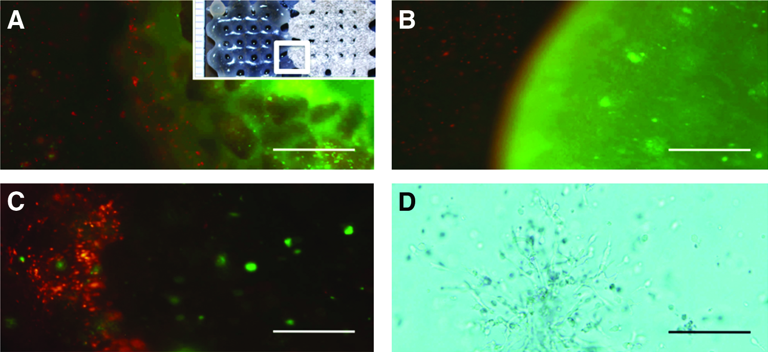
Cell dispersion in heterogeneous printed grafts in vitro.
Tissue formation in heterogeneous grafts in vivo
Cell distribution
After 2 weeks in vivo, fluorescently labeled cell populations were observed in frozen sections of the printed heterogeneous scaffolds (Fig. 3). In Matrigel/alginate constructs (Fig. 3A), the cells were mostly restricted to the part of the scaffold they were printed in, whereas in Matrigel/Matrigel grafts (Fig. 3B), a small fraction of the cells was observed to migrate into the transition zone between the two compartments.

Cell dispersion in heterogeneous printed grafts in vivo.
Tissue formation
Upon retrieval, the constructs were well integrated and vascularized by the host tissue, both at 2 and 6 weeks. Heterogeneous ECM formation in printed Matrigel constructs was observed both macroscopically (Fig. 4A) and upon histological evaluation (Fig. 4B, C) and the overview is presented in Table 2.
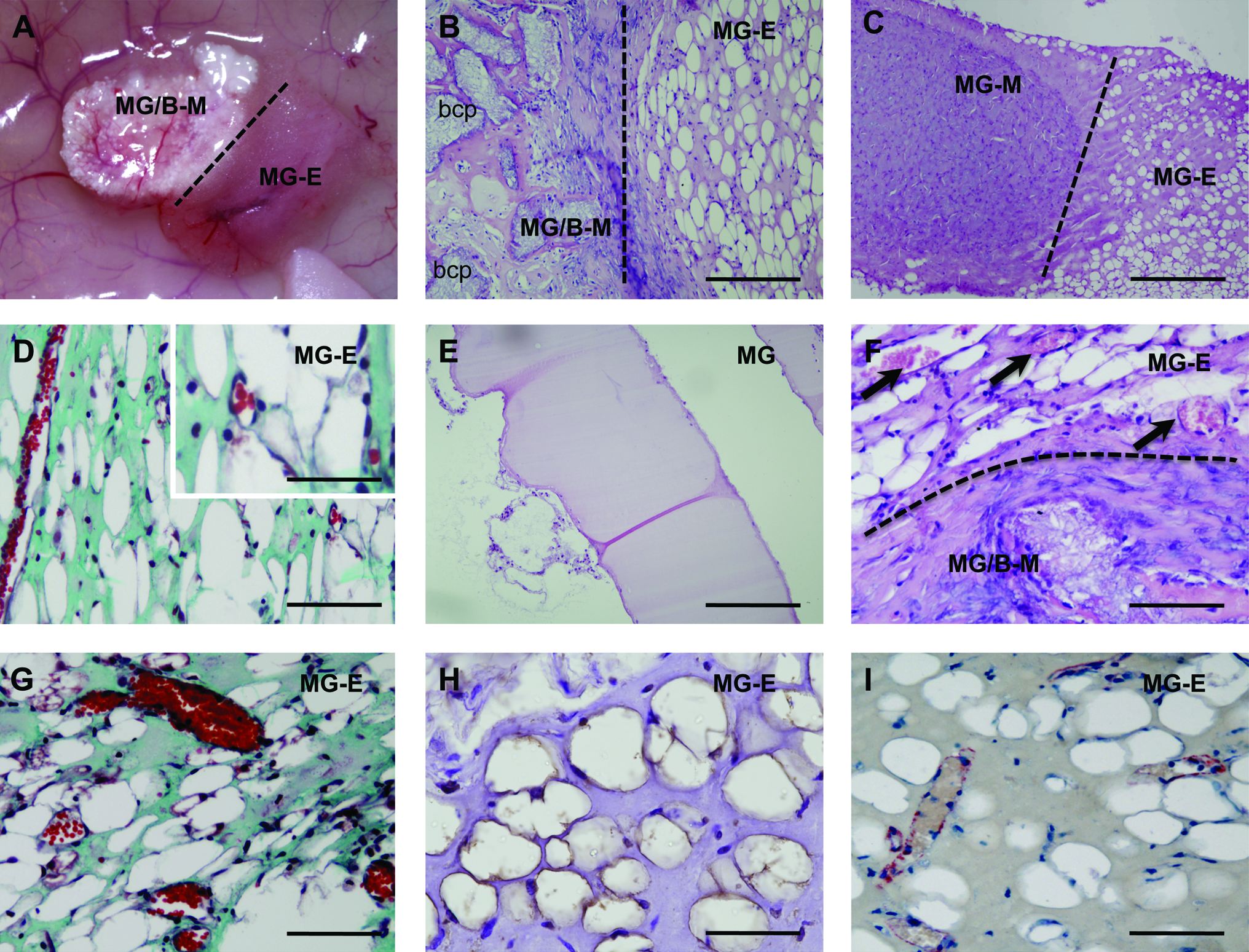
Heterogeneous tissue formation in vivo.
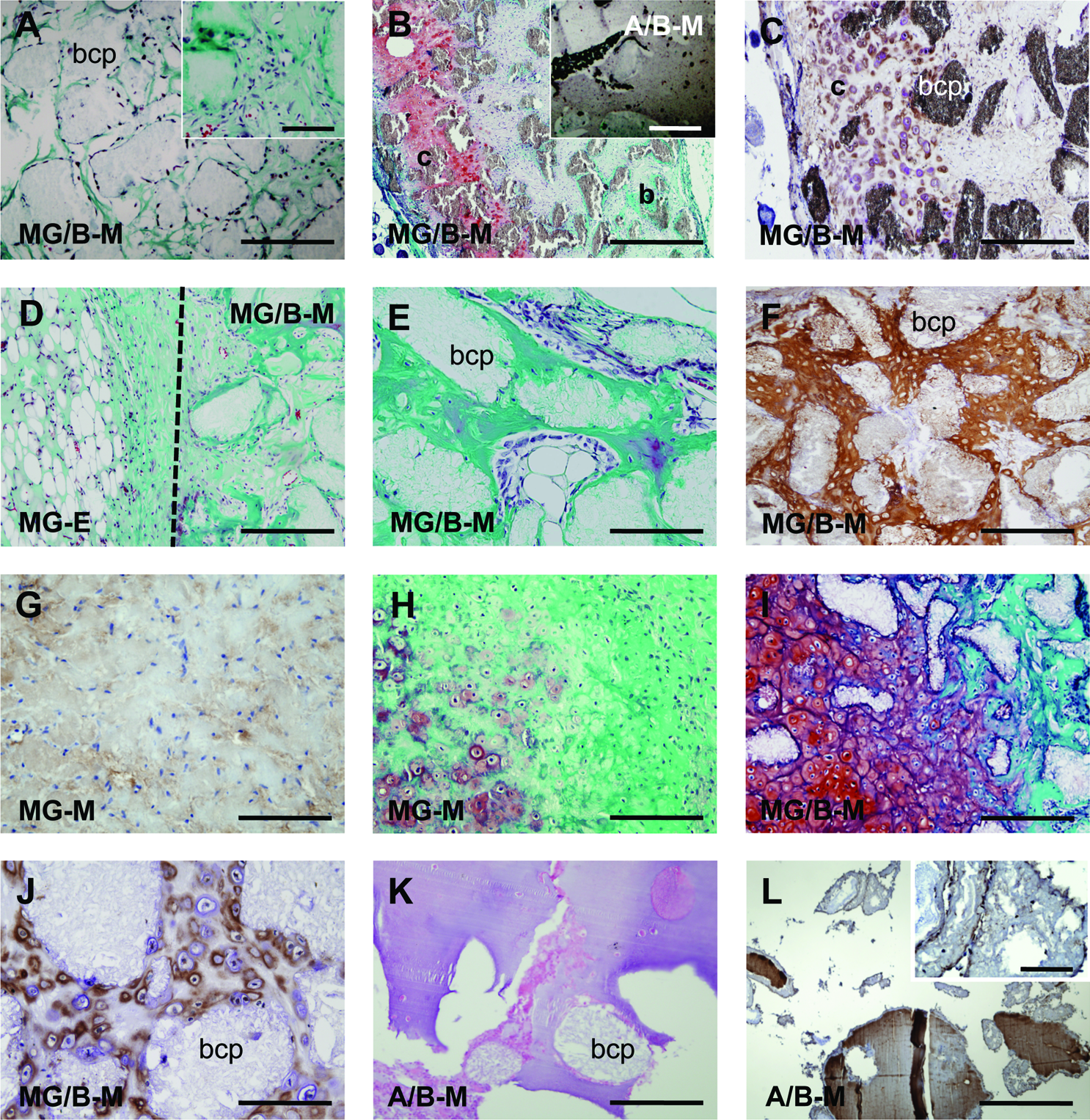
Two weeks after implantation of heterogeneous Matrigel grafts, blood vessels started to form in EPC-laden part of the construct (Fig. 4D). Some of the newly formed tubular structures were empty, and some were perfused with erythrocytes. No tubules were seen in control Matrigel without seeded cells (Fig. 4E), indicating that inclusion of EPCs is crucial for tubule formation. Six weeks after implantation, erythrocyte-filled tubules were observed throughout the EPC-laden part (Fig. 4F, G). Immunostaining for endothelial-specific marker vWF and α-SMA underscored the formation of stabilized blood vessels structures (Fig. 4H, I).
Early bone apposition and cartilage formation took place in the MSC/BCP-laden Matrigel part 2 weeks after implantation (Fig. 5A–C). Here, osteoblasts lining the BCP particles were frequently seen (Fig. 5A, inset). While cartilage formed in the vicinity of skin, early bone development started in deeper regions of the constructs, adjacent to the muscle layer (Fig. 5B, C). Very limited extracellular matrix formation was seen in the MSC-laden alginate part of the constructs (Fig. 5B, inset), and no collagen type I staining was detected (data not shown).
Bone tissue formation in MSC-laden part of the grafts in vivo.
After 6 weeks of implantation, the MSC-laden part of the construct with Matrigel and added BCP exhibited evident bone formation as confirmed by Goldner's trichrome staining and collagen type I immunolocalization (Fig. 5D–F). In the MSC/Matrigel-part of the construct without BCP, very limited collagen type I formation occurred next to some cartilage development (Fig. 5G, H), indicative of the osteoinductive role of the microparticles. Strikingly, again cartilage was detected in the MSC-laden part of the samples, as evidenced by Safranin-O and collagen type II stainings (Fig. 5I, J). The MSC-laden part of the construct with alginate/BCP demonstrated regions of newly formed matrix (Fig. 5K), which stained positive for osteogenic marker collagen type I (Fig. 5L). No collagen type I formation was seen in control MSC-laden alginate without added BCP (data not shown), indicating that inclusion of osteoinductive particles plays a role in the observed early bone formation in this hydrogel.
Discussion
In this study we validate the development of tissue equivalents printed with 3DF. Dual, heterogeneous constructs comprising two parts with distinct cell types (EPCs and MSCs) are printed with a 3DF machine and exhibit heterogeneous ECM formation corresponding to the deposited cell type after in vivo implantation.
In literature, the interactions between endothelial cells and osteogenic progenitors are well described, 14 and require further research into the ideal combinations and ratio of cells that would yield functional grafts. 15 In this respect, the Bioscaffolder printing technique represents a unique tool to study 3D interactions between the two cells types and provides us with a model to analyze the relevance of anatomical structuring in TE. As an early step toward this goal, in this study we demonstrate that the two cell types can be combined during the deposition process.
Various patterning and organ printing techniques enable formation of multicellular, heterogeneous constructs. Among others, combined photo- and electropatterning technique, 16 optofluidic maskless lithography 17 and robotic dispensing 11 were recently used to build layered grafts to modulate cell's biosynthetic activity, to precisely study cellular interactions, and for defined arrangement of multiple cell populations in skin tissue engineering, respectively. However, all of the conducted studies were performed in vitro and commonly provide little evidence on retention of cell organization introduced by printing, limited by their short-term observation periods. 11
In our experiments we demonstrate retention of printed cell organization 2 weeks after printing, both in vitro and after implantation in vivo. We observed that Matrigel/Matrigel composite scaffolds promote cell migration at the interface zone, compared to Matrigel/alginate constructs. This difference can be explained by low interactive properties of alginate, which as a rule entraps the cells and does not support their migration. The majority of the cells, however, are retained at its original printed location. In a recent in vivo study, which addressed short-term retention of stratified chondrocyte populations implanted in a patellofemoral defect in minipigs for 1 week, the stratified organization was lost after implantation. 18 Possibly, this could be explained by the mechanic pressure on the implants in the knee, which was much higher than at the ectopic location in our experiments. The organized deposition of cells and the subsequent development of viable, 3D hydrogel structures is an important achievement of modern TE technologies, but it is unknown whether these constructs will lead to more functional tissues in vitro and in vivo. It was previously demonstrated that patterning and dispensing techniques can build viable cell-laden structures, that singular cell populations retain their functionality for several weeks in vitro,8,19,20 and that functionality of embedded cells can be further modulated by specific cell arrangement. 16 In this study we demonstrate for the first time that multiple printed cell types also retain their functionality after in vivo implantation, and produce extracellular matrix according to the deposited cell type. Blood vessel formation takes place in the EPC-laden part of the printed constructs, whereas bone formation occurs in the MSC-laden part, starting 2 weeks after implantation.
In the EPC-laden Matrigel part of the printed construct, we observed formation of erythrocyte-filled tubules. EPCs isolated from peripheral blood are a potent source of endothelial cells (ECs). Under comparable conditions, EPCs perform better than ECs with respect to blood vessel network formation. 21 EPCs are highly proliferative, exhibit a stable endothelial phenotype, 22 support neovascularization of the ischaemic hindlimb,23–25 regenerate infarcted myocardium, 26 and support bone formation upon coimplantation with MSCs.27,28 In this study, addition of EPCs was essential for the formation of potent, erythrocyte perfused vessels, whereas no vessel ingrowth was seen in acellular Matrigel. The newly formed vessels stained positive for endothelial-specific marker vWF and were stabilized by smooth muscle cells. We cannot claim that the seeded EPCs actually form the blood vessels, as the exact origin of the cells forming the new blood vessel networks remains to be elucidated. It is likely that vWF-positive endothelial lining is in part derived from the seeded progenitors, whereas α-SMA positive vessel-stabilizing cells are either host-derived pericytes or MSCs. Limited migration of cells in the hydrogel matrix makes the contribution of coprinted MSCs to blood vessel stabilization less likely.
Bone tissue started to form around the BCP particles in Matrigel scaffolds 2 weeks after implantation and progressed to abundant bone formed at 6 weeks, a potent outcome considering that the concentration of encapsulated MSC used for implantation in this study was lower than in other bone TE studies.29,30 Inclusion of osteoinductive BCP particles has proven to be of importance for bone formation in this context. This finding is supported by other reports illustrating potent bone formation when calciumphosphate precipitates are added to the hydrogel. 31 The positive effect of microparticles on bone formation has been attributed to a number of factors, of which serum protein adsorption to the surface of particles is important. 32 Dissolution of calcium phosphates is responsible for calcium and phosphorus release into the microenvironment of the cells, which creates a favorable milieu for new bone formation, supported by the rough surface of the particles.
While a large amount of bone formed in the printed MSC-laden Matrigel part of the scaffold, as evidenced by Goldner's trichrome and collagen type I staining, limited bone formation was observed in alginate. A small degree of extracellular matrix development, as shown by Goldner's trichrome staining, next to some positive staining for collagen type I in alginate samples at 6 weeks, is indicative of osteogenesis taking place in this gel. Limited interaction of the alginate matrix with cells, restricting the migration of the cells through the matrix, and a relatively high concentration of this slowly resolving polysaccharide in the printed gel may be responsible for above findings. The degree of this delay in bone formation depends on the hydrogel type used and its degradation speed. 33
We also observed formation of substantial amounts of cartilage in the MSC-laden part of the Matrigel grafts. The development of bone by endochondral ossification is a common mechanism displayed by transplanted MSCs at ectopic locations in the murine model. 34 Cartilage formation appeared to be initiated at the skin-side of the constructs, whereas bone formation seemed to occur in deeper regions. The development of oxygen gradients that modulate the differentiation of MSCs35,36 after implantation may be responsible for this phenomenon.
While millimeter-sized bone tissue implants are successful in animal models, formation of large-scale tissues is limited due to death and restricted osteogenic differentiation of transplanted cells. Currently, the formation of printed centimeter-scaled tissue equivalents like we presented here lies a considerable distance away from the actual printing of bone tissue. However, in the future, 3DF may result in development of larger, clinically relevant-sized grafts. In vivo implantation of heterogeneous printed grafts at an orthotopic location will provide insight in the quality and quantity of tissue formation within the printed grafts. An important issue to investigate is whether the imposed cell organization is actually necessary for obtaining fully functional newly formed tissues upon in vivo implantation. Another essential matter to consider is the cell density in printed grafts, which is likely to have a profound effect on tissue formation12,37,38 due to a strong positive effect that direct cellular interactions can have on ECM development. Controlled delivery of biological factors in printed constructs is expected to promote further functionalization of complex tissue grafts.
Conclusions and Future Directions
In summary, we demonstrate here for the first time the retention of spatially organized, functional osteo- and endothelial progenitor cells in printed grafts after in vivo implantation. Heterogeneous extracellular matrix formation in printed grafts occurred analogous to the deposited cell type. In the next step, it would be attractive to design more intricate printed bone structures with blood vessel channels and test these implants in vivo in a large animal model, also at orthotopic locations, and to investigate the necessity of imposed organization.
Footnotes
Acknowledgments
We thank H. Yuan from Progentix Orthobiology BV (Bilthoven, The Netherlands) for kindly providing the BCP particles. We acknowledge the financial support from the Netherlands Organisation for Scientific Research to NF (NWO; grant number: 017.001.181) and the Smart Mix Program of the Netherlands Ministry of Economic Affairs and the Netherlands Ministry of Education, Culture and Science.
Disclosure Statement
No competing financial interests exist.