
Abstract
A sufficient cell source and minimal invasiveness in obtaining human adipose stromal cells (hASCs) hold great promise for their utilization in wound repair. However, little is known about how cell-residing microenvironments regulate the cellular response. In this study we explored the effects of polycaprolactone (PCL)/collagen nanofibers with distinct spatial arrangements (aligned and random) on phenotypic expression of hASCs in vitro. Elongated cell morphology, higher proliferation, and faster migration rate were observed for hASCs cultured on the aligned nanofibers, showing that hASCs could detect the nanofiber spatial arrangement and then distinctively respond. This study on the expression of extracellular matrix (ECM) related genes in hASCs revealed higher synthesis capacity for critical ECM molecules including tropoelastin, collagen I, and matrix metalloproteinase (MMP)-1 on the aligned nanofibers. Integrins α5, β1, β3, β6, and transforming growth factor (TGF)-β1 were differentially regulated by PCL/collagen nanofiber arrangements. Our results indicate that fiber orientation-induced phenotypic change of hASCs may be regulated by integrins and TGF-β signaling synergistically. These findings demonstrate the potential application of hASCs and aligned PCL/collagen nanofibers for accelerated wound repair.
Introduction
In this study, collagen-containing electrospun nanofibers with two types of spatial arrangements (aligned=fibers oriented in the same direction; random=fibers collected without specific orientation) were fabricated and used to culture human adipose stromal cells (hASCs). The morphology of hASCs on aligned on random fibers was examined by immunofluoresent staining of cytoskeletal proteins. The proliferation and migration of hASCs in association with fiber orientation were studied. Genes of various integrins and those involved in ECM synthesis and degradation were investigated to determine the phenotypic expression. It was found that fiber orientation greatly affected the cell morphology, proliferation, and migration by differentially regulating the expression of integrins and the formation of focal adhesion. The synthesis of collagen I, tropoelastin, and matrix metalloproteinase (MMP)-1 by hASCs was also influenced by fiber orientation, in which transforming growth factor (TGF)-β1 might be involved as suggested by the upregulation of TGF-β1 gene.
Materials and Methods
Materials
Poly(epsilon-caprolactone) (PCL, MW=80,000), MCDB201 medium, MCDB131 medium, ITS-X, bovine serum albumin (BSA), linoleic acid-BSA, L-ascorbate-2-phosphate, β-mercaptoethanol, recombinant human epithelial growth factor (rhEGF), recombinant human platelet derived growth factor (rhPDGF)-BB, and dexamethasone were purchased from Sigma-Aldrich (St. Louis, MO). Collagen type I was purchased from Elastin Products Company (Owensville, MO). 1,1,1,3,3,3-hexafluoro-2-propanol (HFIP) was obtained from Oakwood Products Inc. (West Columbia, SC). Fetal bovine serum (FBS) was purchased from American Type Culture Collection (ATCC, Manassas, VA). All other reagents and solutions were obtained from Invitrogen (Carlsbad, CA) except as indicated.
Electrospinning and characterization
Nanofibers were similarly prepared using electrospinning technique as described previously.17,23 Briefly, collagen type I solution (8% w/v), and PCL solution (8% w/v) dissolved in HFIP were mixed thoroughly at a volume ratio of 1:3. The solution was transferred to a 3 mL syringe attached with a blunt-tip capillary (inner diameter=0.9mm) and electrospun at 10 kV. Steady flow of the solution from the capillary spinneret was achieved using a syringe pump from KD Scientific (Holliston, MA), operating at a flow rate from 5 μL/min to 15 μL/min. To fabricate random nanofiber scaffolds, the collecting surface consisted of a stationary aluminum foil plate, and to collect the nanofibers with desired alignment, a customized rotating mandrel (φ=10 cm) was used. Nanofibrous scaffolds were collected on square glass cover slips. All fibers were fabricated under a sterile condition. To characterize the electrospun nanofiber using a scanning electron microscope (SEM), fibers were collected on silicon wafer and sputter coated with gold. The coated fibers were examined with a LEO 982 FEG SEM. To determine the diameter of nanofibers, images of five randomly selected areas were captured and analyzed by analysis software (NIS-elements BR 3.10) from Nikon. The orientation of electrospun nanofibers was determined by using the ImageJ program (NIH) along with the Oval Profile Plot plugin. Briefly, representative SEM images of the electrospun fibers were cropped into a square covering as much of the image as possible (the images used were 905×905 pixels in size). These cropped images were then processed with a fast Fourier transform (FFT) and then a circular selection of the image was made maximizing the amount selected (edges of the circle were tangent with the edges of the image). This selection then underwent a radial summation using the Oval Profile Plot plugin with 360 points such that the degree interval was 1°. The raw data were normalized to 0 and data from all sets were rescaled to an arbitrary range between 0 and 0.3. The final results were presented by plotting this arbitrary scale versus degrees, where the height of peaks represents a greater degree of alignment at a given angle.
Cell seeding and culture
Human adipose stromal cells (hASCs) kindly provided by Dr. Moscatello from Coriell Institute for Medical Research (Camden, NJ) were used in this study. Cells were cultured in the proliferation medium, which consisted of Dulbecco's modified Eagle medium (DMEM, low glucose), MCDB201 and MCDB131 at a ratio of 3:1:1, 0.5% FBS, 1× ITSX, 0.2% BSA, 1× linoleic acid-BSA, 100 μM L-ascorbate-2-phosphate, 100 μM β-mercaptoethanol, 10 ng/mL rhEGF, 5 ng/mL rhPDGF-BB, 1nM dexamethasone, 1% penicillin, and 1% streptomycin. Medium was refreshed every 3–4 days. Cell splitting was performed when the monolayer cells reached 70–80% confluence. Cells between the second to fifth passages were used in this study. To seed hASCs onto electrospun fibers, cells were trypsinized, centrifuged, and resuspended in the proliferation medium at a final concentration of 1×10⁵/mL. hASCs were then seeded onto various nanofibrous scaffolds (aligned and random) at a density of 5×10³ cells/cm2 and allowed to attach for 60 min before adding more medium to completely immerse the culture. The culture was kept in a humidified incubator at 37°C with 5% carbon dioxide for designated times. Monolayer culture of hASCs on glass cover slips served as controls.
Immunofluorescent staining
The morphology and adhesion of hASCs onto nanofibrous scaffolds were evaluated by immunofluorescently staining the cytoskeletal proteins and vinculin. Briefly, after fixation in 4% paraformaldehyde for 20 minutes the culture was permeabilized with 0.5% Triton X-100 (Fisher Scientific, Fair Lawn, NJ), and then incubated with phalloidin-tetramethyl rhodamine isothiocyanate (TRITC) (Sigma, 1:500), anti-α-tubulin (DSHB, 1:25), anti-vinculin conjugated with fluorescein isothiocyanate (FITC) (Sigma, 1:50), anti-vimentin (Biovision, Mountain View, CA, 1:20) or anti-αSMA (α-smooth muscle actin) conjugated with Cy3 (Sigma, 1:200) for 60 min at room temperature in dark. For those primary antibodies without fluorescent labels, the cells were further incubated with goat anti-mouse IgG-FITC conjugate secondary antibody (Caltag, PAB4971, 1:400) or goat anti-rabbit IgG-Alexa Fluor 594 conjugate secondary antibody (Molecular Probes, A11037, 1:500). Cell nuclei were stained with 4′,6-diamidino-2-phenylindole (DAPI) (Sigma, 1:1000). Excessive fluorescent conjugate was removed by washing with phosphate buffered solution (PBS). DAPI was used to stain cell nucleus. The staining was examined under a Nikon Eclipse 80i epifluorescent microscope (Nikon, Japan).
Cell proliferation
Cell proliferation on electrospun nanofibers (n=4) was determined by DNA assay using a CyQUANT® Cell Proliferation Assay Kit (Molecular Probes, Inc., Eugene, OR) as described previously. 24 Briefly, the samples harvested at different times were digested with proteinase K solution (Sigma). Two hundred microliters of CyQUANT® GR dye/cell-lysis buffer was added to each sample and incubated at room temperature for 2–5 minutes. The fluorescence intensity was measured with excitation at 480 nm and emission at 520 nm using a Synergy™ HT Multi-Detection Microplate Reader (BioTek Instruments Inc., Winooski, VT).
Cell migration
A collagen gel drop assay was developed and used to analyze cell mobility on nanofibrous scaffolds. In this assay, a small drop of collagen gel suspended with a high density of hASCs was placed on top of the nanofibrous scaffolds and then the cell migratory behavior out of gel was evaluated by optical microscopy. Briefly, hASCs was carefully resuspended in neutralized type I collagen solution (6 mg/mL in 0.01 M HCl) at a final concentration of 2×10⁶ cells/mL and then droplets of 10 μL cell/gel suspension were added onto the scaffold surface. Upon solidification of collagen gel at 37°C for 30 min, cell/gel droplets were completely covered with culture medium. After incubation for 3 and 7 days, samples were fixed in 4% paraformaldehyde, subsequently stained with methylene blue, and then examined under a stereo microscope (Nikon SMZ1500). Cell migration on different scaffolds was determined by measuring the longest distance that the cells migrated away from the boundary of gel drop.
Gene expression
Gene expression (n=4 per group) was measured using reverse transcriptase polymerase chain reaction (RT-PCR) at 1, 3, and 7 days. Total RNA was isolated using the RNeasy Mini Kit (Qiagen, Valencia, CA). The isolated RNA was reverse transcribed into complementary DNA (cDNA) using the SuperScript First-Strand Synthesis System (Promega, Madison, WI), and the cDNA product was then amplified using recombinant Taq DNA polymerase (Promega). Expression of integrins α2, α5, αV, β1, β3, β6, MMP-1, MMP-2, MMP-9, TGF-β1, tropoelastin, type I collagen, type III collagen, vimentin, and α-SMA (ACTA-2) were determined over time. β-actin served as housekeeping gene. Primer sequences for various genes are listed in Table 1. All genes were amplified for 30 cycles in a thermocycler (Eppendorf Mastercycler gradient, Brinkmann, Westbury, NY). Semiquantitative analysis of gene expression was performed, and band intensity was normalized to that of β-actin.
MMP, matrix metalloproteinase; ACTA2, alpha actin 2.
Statistical analysis
Each experiment was repeated at leastthree3 times on different days and data were expressed as the mean±standard deviation. All the cytotoxicity and cell attachment measurements were collected in triplicate for each group. Unpaired student t-test was used to evaluate the significance between experimental groups. A value of p<0.05 was considered statistically significant.
Results
Nanofiber meshes with various fiber orientations
By collecting the nanofibers either on a grounded rotating mandrel or on a grounded stationary flat surface, nanofiber meshes with fibers oriented either in one direction (i.e., aligned) or in all x-y directions (i.e., random) were obtained. SEM examination clearly showed various spatial arrangements of collected fibers (Fig. 1a vs. 1b). To better characterize the orientation, the SEM images of obtained nanofibers were analyzed with a fast Fourier transform (FFT) and the analysis results were shown in Figure1 (c–e). More than 90% electrospun nanofibers aligned in one direction in the aligned scaffolds, and no preferred orientation could be identified in the random scaffolds. Based on the SEM images, the average fiber diameter in the aligned scaffolds seemed slightly smaller than that of the random ones (Table 2), but there was no significant difference. SEM examination at a high magnification revealed that both aligned and random 3:1 PCL/collagen nanofibers had a smooth surface (data not shown).

Representative scanning electron microscope images of random
Attachment and morphology of hASCs on nanofiber meshes
The inclusion of collagen in PCL electrospun nanofibers favored the adhesion and spreading of hASCs. As the cell morphology was defined by the arrangement of intracellular cytoskeletal proteins, immunofluorescent staining for F-actin and α-tubulin was performed on the hASCs cultured on PCL/collagen nanofibers for 3 days. Both F-actin and α-tubulin staining showed that hASCs on random nanofiber surfaces had a polygonal morphology without preferred orientation, while those on aligned nanofiber surfaces elongated along with the nanofiber orientation, showing a typical spindle-like morphology (Fig. 2). The intracellular cytoskeletal protein fibers also oriented in the same direction as the electrospun nanofibers on the aligned PCL/collagen nanofiber surfaces and were more compacted as compared to those on the random nanofiber surfaces. The morphology remained similar for the entire experiment time (up to 7 days). To better quantify the morphology difference of hASCs on aligned and random fibers, fluorescent images of five randomly selected fields were analyzed using the NIH ImageJ analysis software. As shown in Table 3, the average cell area on aligned PCL/collagen nanofibers was consistently lower than that on the random nanofibers for both days 1 and 7 despite no statistical significance (p>0.05). The average cell area increased approximately one fold after cultured for 7 days compared to that from day 1. The long/short axis ratio of hASCs, an indicator of cell polarity, was also calculated (Table 3). Clearly, the cells on aligned fibers remained constantly elongated with the long/short axis ratio as high as 17, more than three times of the random ones. The formation of cell/fiber focal adhesion was evaluated by immunofluorescent staining of vinculin, a protein involved in the connection of focal adhesion point to cytoskeletal actin. It was found that vinculin of hASCs cultured on the aligned fibers showed long spikes with the same orientation as the fibers (Fig. 3b). In contrast, on random fibers vinculin were short strokes without preferred orientation (Fig. 3a).

Representative fluorescent images of human adipose stromal cells (hASC) cultured for 3 days on random

Representative fluorescent images of the distribution of vinculin in hASCs cultured on random
Statistically significant, p<0.001.
Cell proliferation and migration
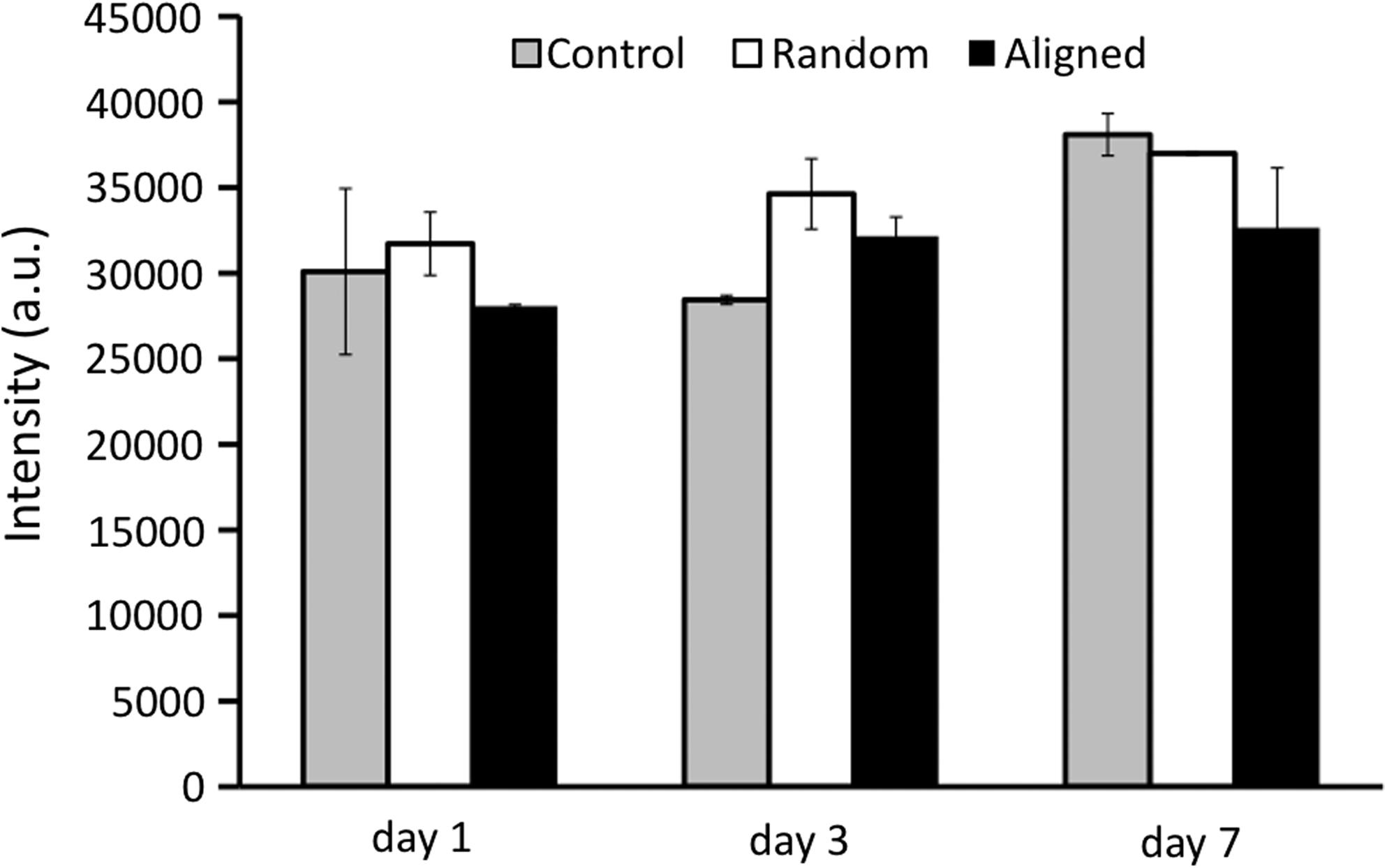
Cell proliferation on aligned and random nanofiber meshes was evaluated by DNA assay. Continuous cell proliferation was observed for hASCs on both aligned and random nanofibers. However, cells on aligned nanofibers proliferated faster than those on random fibers (Fig. 4), and this trend remained for all the times investigated. The migration of hASCs on the aligned and random fiber surfaces was determined by a collagen gel drop assay. Obviously, cells migrated out of the original droplet after 3 days on both aligned and random fiber surfaces. Interestingly, an isotropic hASC migration was observed on the random nanofibers, while an anisotropic migration was observed on the aligned nanofibers (i.e., accelerated migration along the fiber alignment in contrast to an inhibited migration perpendicular to fiber alignment) (Fig. 5). Besides, hASCs migrated in a longer distance on the aligned nanofiber meshes than they did on the random fibers. The migration distance away from collagen gel was measured on the samples stained with methylene blue (n=3 per time point). The results showed that hASC migration along the aligned fibers was more than two times faster than that on the random fibers, while the migration perpendicular to fiber alignment was less than half of that on the random one (Fig. 6).
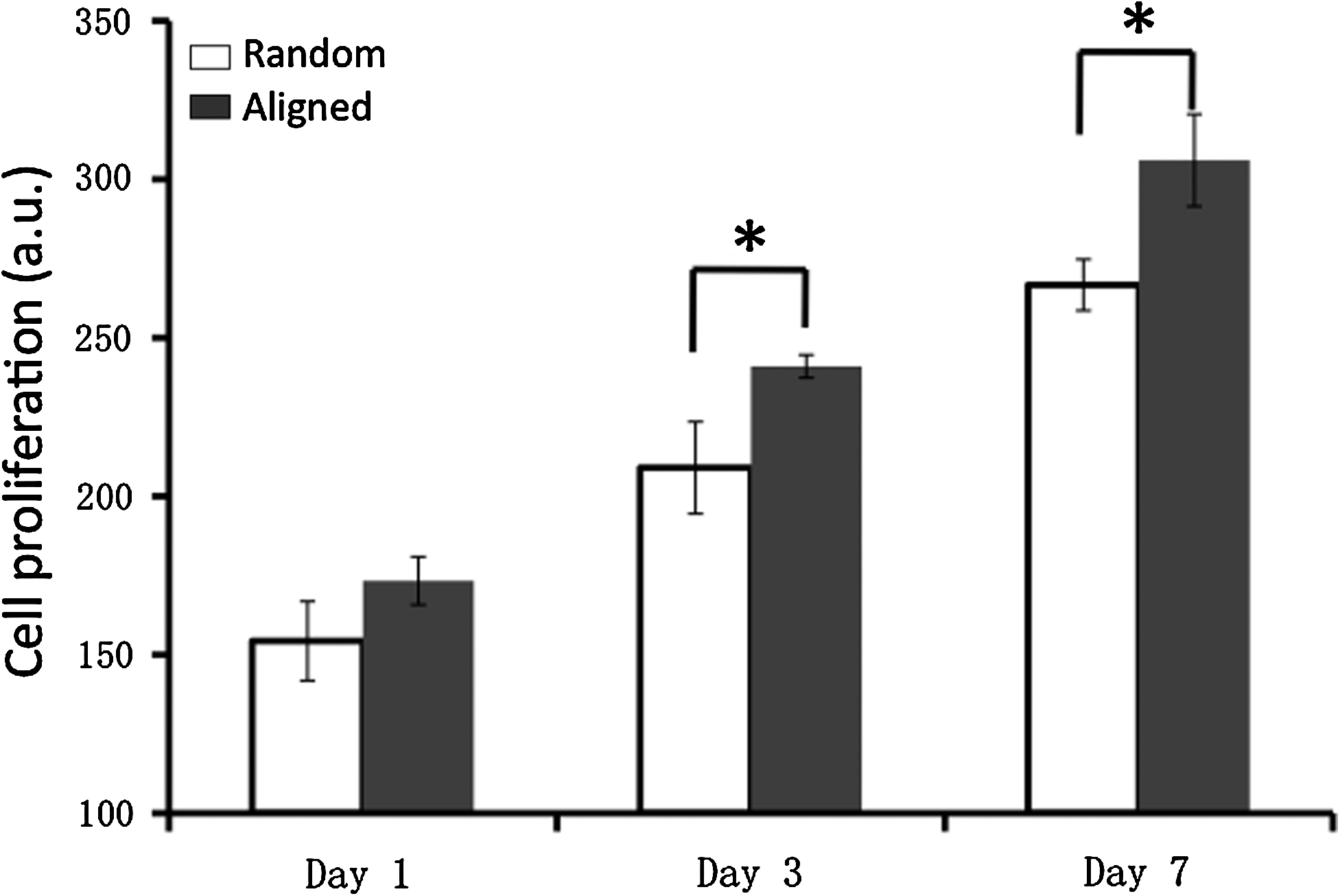
Proliferation of human adipose stromal cells on nanofiber meshes. Cell amount was determined by DNA assay. *Statistically significant, p<0.05.
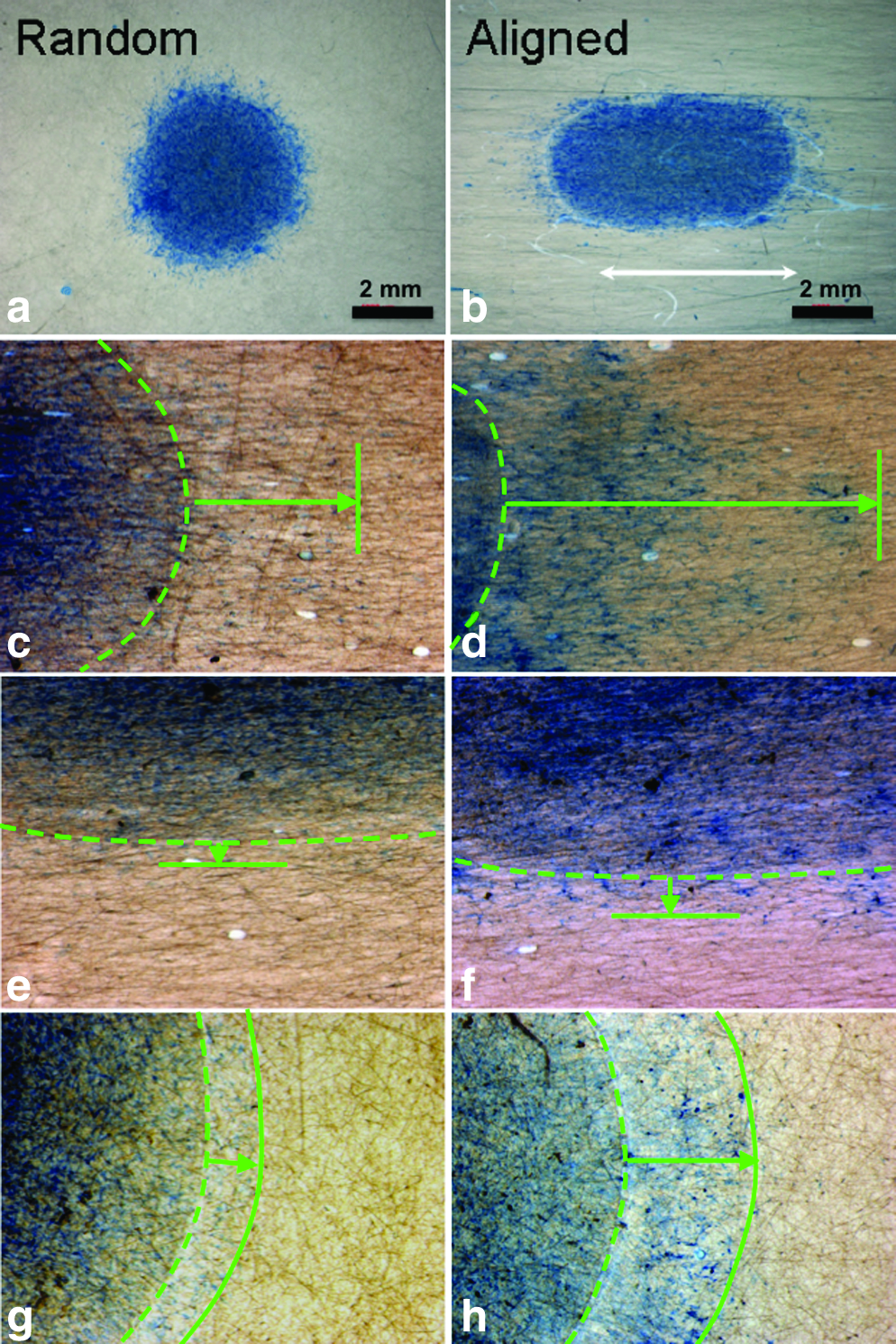
Optical images of hASCs migrated out of the collagen gel borderline (green broken line) on random
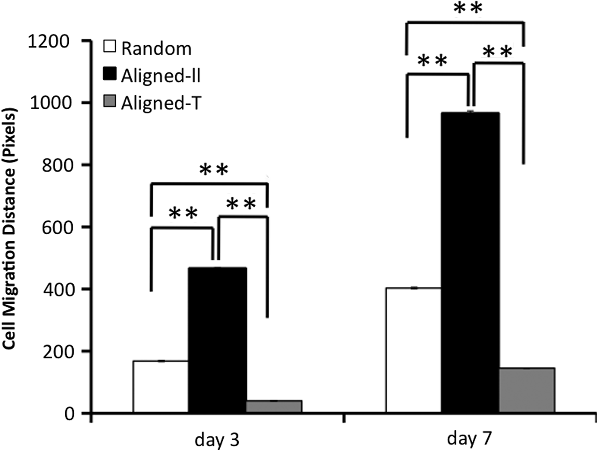
Quantitative measurement of hASC migration on nanofiber meshes. Aligned-ll: migration parallel to fiber alignment; Aligned-⊥; migration perpendicular to fiber alignment. **Statistically significant, p<0.01.
Expression of integrins
To determine whether integrins were involved in the fiber-orientation induced cell phenotypic change, gene expression for integrins (α2, α5, αv, β1, β3, and β6) in hASCs was analyzed using RT-PCR (Fig. 7). The results showed that gene expression of α2 and αv was not affected by various fiber and glass cover slip (controls) surfaces (data not shown). Western blot was also performed to determine the protein level of α2 and αv integrins on the cell membranes. Again, no noticeable difference was identified (data not shown). However, integrins α5, β1, β3, and β6 were significantly regulated by fiber orientation. A significant upregulation of β6 integrin was observed after 3 days. The fiber orientation upregulated the gene expression of integrins β1 and β3 as early as day 1 and remained for the rest of the experimental times (Fig. 7a, b). Somehow, the fiber orientation down-regulated integrin α5 expression for all the times investigated.
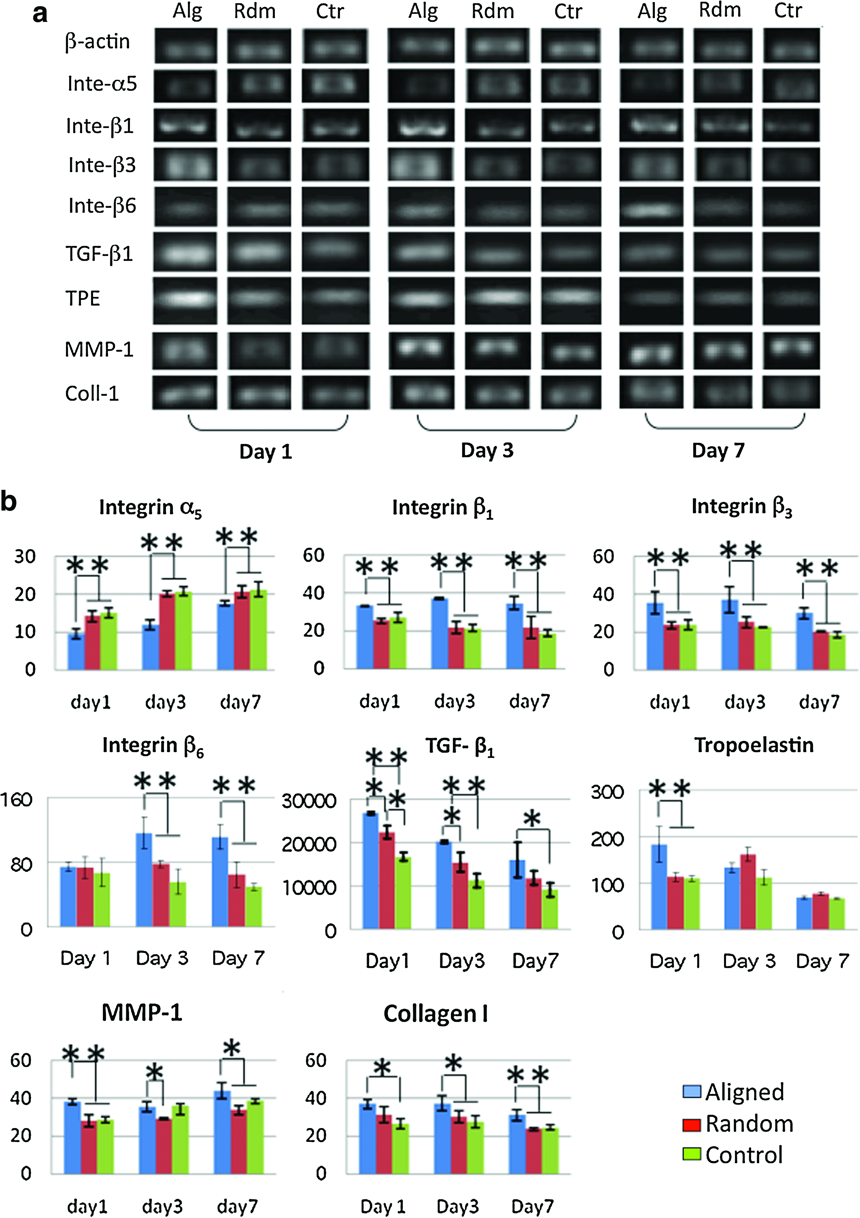
Gene expression of hASCs cultured on random (Rdm) and aligned (Alg) nanofiber meshes and glass coverslips (Ctr).
Expression of ECM related genes
As part of the phenotypic expression of hASCs, the capability of regulating ECM turnover is a crucial measure. In this regard, the gene expression of major ECM proteins related to wound healing (i.e., collagen type I [Coll I], collagen type III [Coll III] and tropoelastin [TPE]) were particularly investigated. Interestingly, higher gene expression of Coll I was observed for the hASCs cultured on aligned nanofibers compared to those on random nanofibers on days 3 and 7 (Fig. 7a, b), while no difference was observed in the expression of Coll III (Supplementary Fig. S1; Supplementary Data are available online at www.liebertonline.com/tea). Significantly higher expression of TPE was found on the aligned nanofibers, but only on day 1 (Fig. 7a, b). ECM degrading matrix metalloproteinase (MMP-1, MMP-2, MMP-9) were also investigated. Gene expression of both MMP-2 and MMP-9 was not significantly different between the aligned and random nanofibers (data not shown). Gelatin zymography further confirmed the gene result with no significant difference in MMP-2 enzymatic activity between the two groups (Fig. 8). In contrast, noticeably high MMP-1 was consistently found in the hASCs cultured on aligned nanofibers (Fig. 7a, b). Gene expression for TGF-β1, an important ECM regulator, was also studied. It was found that TGF-β1 was highly upregulated by aligned nanofibers (Fig. 7a, b).
Semiquantification of matrix metalloproteinase (MMP)-2 activity as measured by zymography.
Fibroblastic differentiation of hASCs on nanofibers
To determine the fibroblastic differentiation of hASCs on nanofibers, gene expression for vimentin, a reliable fibroblast marker, was evaluated using RT-PCR. Semiquantitative analysis showed that gene expression for vimentin was similarly detected on hASCs cultured on aligned or random nanofibers (Fig. 9a). Immunofluorescent staining for vimentin further confirmed the gene expression result. hASCs had a strong fluorescent staining for vimentin on both nanofiber meshes despite a distinct variation in spatial arrangement (spindle [aligned] vs. polygonal [random]; Fig. 9b), suggesting a fibroblastic phenotype. This observation remained consistent for all the time points up to 7 days (data not shown). α-SMA, involved in wound contraction, normally appeared in the wound myofibroblasts. To assess whether nanofiber orientation regulated the expression of α-SMA in hASCs, cells cultured on nanofiber meshes were immunofluorescently stained for α-SMA. No obvious myofibroblastic differentiation was detected in the hASCs either on aligned or random nanofibers (Fig. 9b). The expression level for α-SMA gene, (i.e., ACTA-2) was checked using RT-PCR. A low level expression was detected and it was comparable between aligned and random culture groups (Fig. 9a).
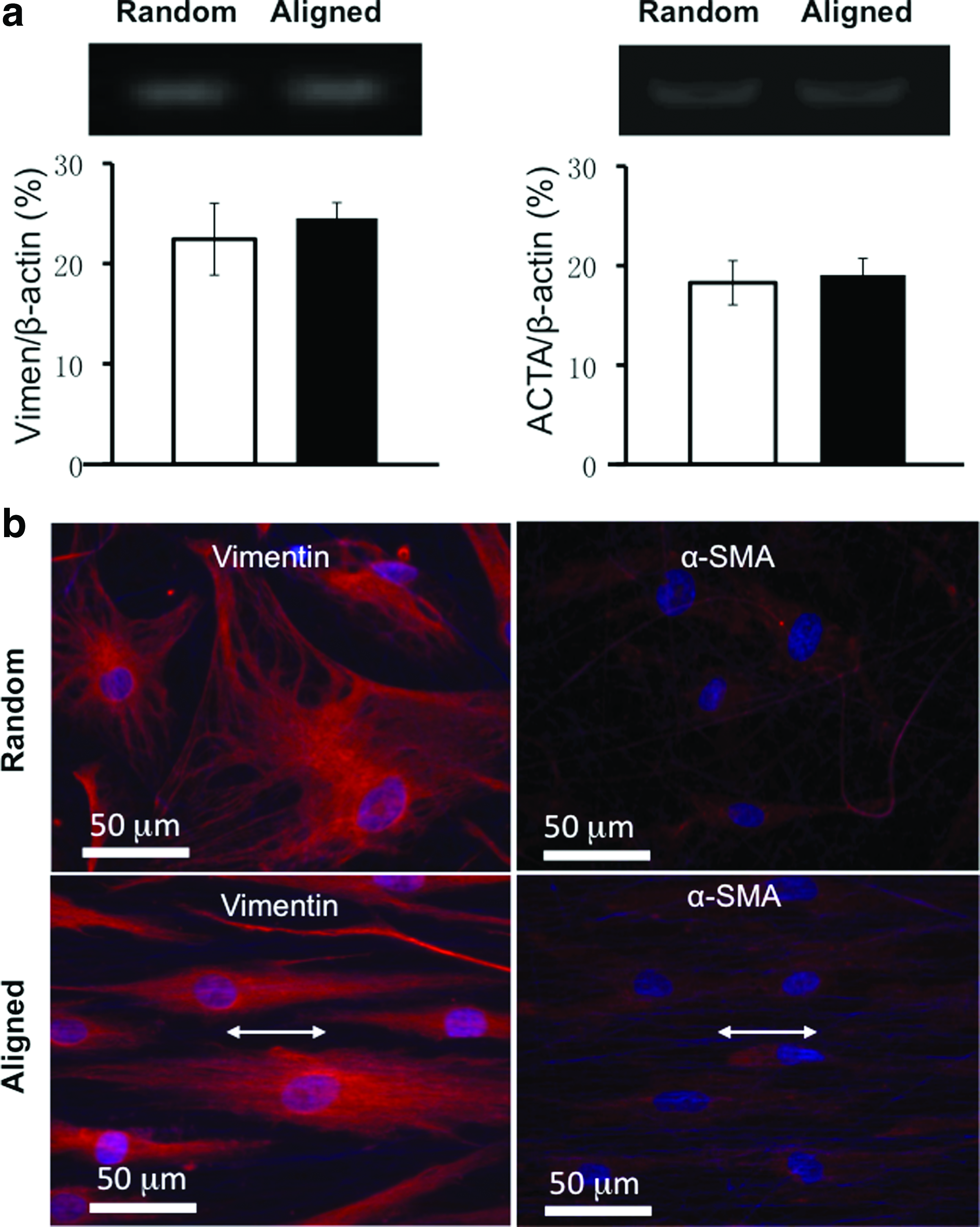
Fibroblastic differentiation of hASCs on nanofiber meshes.
Discussion
The advantages of electrospun nanofibers in promoting cell adhesion and maintaining cell phenotype, which mainly comes from the morphological similarity to native ECM fibrils, have been well recognized. 19 As a result, extensive efforts have been made to explore the possible utility of such nanofibers for regenerating a variety of tissues including skin.25–31 It is known that native ECM not only provides a substrate for cells to attach, but also regulates cellular function via cell/matrix interactions. Inclusion of ECM molecules (e.g., type I collagen) into electrospun nanofibers can better recapitulate the bioactivity of ECM 32 and therefore lead to cell/nanofiber interactions similar to the cell/ECM interactions in tissues. With the presence of type I collagen in electrospun nanofibers, hASCs can rapidly attach and spread on PCL/collagen nanofibers (Fig. 2). Moreover, fiber arrangements (aligned vs. random; Fig. 1) have a significant impact on the morphology of attached hASCs: cytoskeletal proteins (F-actin and α-tubulin) oriented along with the PCL/collagen nanofibers in the aligned group, but no preferred orientation in the random group (Fig. 2). Meanwhile, the hASCs on aligned PCL/collagen nanofibers are more elongated (long/short axis ratio: 11–17 [aligned] vs. 3 [random], p<0.001) with a lower mean cell area (Table 3). Although higher Young's modulus was measured on aligned PCL/collagen fibers in the orientation in other study compared to that of random ones (8.98±3.72 MPa vs. 2.83±0.60 MPa), the mechanical contribution to fiber-induced hASC response can be neglected in this study due to the use of rigid glass coverslips to collect nanofibers for cell culture, and no fiber detachment from coverslip surface has been observed during the culture. In addition, comparable fiber diameter has been measured between aligned and random nanofibers (Table 2); therefore, it is reasonable to hypothesize that the striking variation in cell morphology and other behaviors mainly comes from fiber arrangement. This fiber organization-induced cell morphology change has been reported for several types of cells, including cardiomyocytes, crucial ligament fibroblasts, and dermal fibroblasts.22,33,34 Although the exact mechanism is not fully understood, studies have shown that various integrins such as α2β1 are involved in fiber-induced cell adhesion.35,36 It is believed that integrins, particularly those involved in focal adhesion formation, are important in regulating cell morphology. 37 Indeed, the size and spatial distribution of focal adhesion plaques in hASCs seems well correlated with cell morphology and are controlled by fiber arrangement, as observed from the immunofluorescent staining of vinculin. Compared to short and random focal adhesion plaques formed on random PCL/collagen nanofibers (Fig. 3a), long focal adhesion plaques were observed for hASCs cultured on the aligned fibers and they also oriented in the fiber alignment (Fig. 3b). The distinct vinculin distribution would inevitably lead to corresponding cytoskeletal arrangement as shown in Figure 2.
In this study, we also found that aligned PCL/collagen nanofiber meshes promoted the proliferation and migration of hASCs. Approximately 15% increase in cell number was consistently observed in the aligned group compared to random one (p<0.05) (Fig. 4), contradicting to the results observed with ligament fibroblasts, 34 in which no significant difference in cell proliferation was noted between aligned and random nanofibers. We believe that the mitosis of fully differentiated cells like ligament fibroblasts will not be affected by the fiber organization, however, pluoripotent cells like hASCs may have various mitotic responses as a result of the commitment to different cell lineages induced by substrate topography.38,39 Accelerated migration of hASCs was also observed on the aligned nanofibers, however, only in the direction parallel to fiber alignment, but not in the perpendicular direction (Figs. 5, 6). This fiber orientation regulated cell migration was similarly reported with other cells. 40 Cell migration is controlled by a highly orchestrated set of events involving the polymerization and depolymerization of actin filaments and the exertion of force through actinomyosin mediated contraction.41,42 The role of integrins in cell migration has been well recognized for both their involvement in binding to ECM and in regulating the turnover of intracellular cytoskeletal proteins. 43 Gene expression for several integrin genes was particularly studied in this study and indeed a notable difference was observed between aligned and random nanofibers. Significantly higher expression of integrins β1 and β3 were consistently observed in the aligned group (Fig. 7). It is known that both integrin β1 and β343,44 are involved in cell migration, which suggests that accelerated hASC migration on aligned fibers may be the result of elevated integrins β1 and β3. Our ongoing efforts will further determine whether integrin β1 plays the determinant role by using a gene knockout model. In the gene expression study, significantly lower α5 was observed in the aligned group. Previous studies have shown that integrin α5β1 negatively regulates cell proliferation. 45 The down-regulated α5 integrins may partially explain the promoted cell proliferation on the aligned group.
Further studies on several ECM-related genes showed that tropoelastin, collagen I, and MMP-1 were all upregulated by the aligned PCL/collagen nanofibers (Fig. 7). Active ECM synthesis and degradation is crucial to wound healing, especially in the case of a significant loss of tissue. Elevated expression of ECM-related genes implies the activation of ECM turnover, which may favor the migration and proliferation of fibroblast and keratinocytes and therefore facilitate wound healing. Our previous study demonstrated that increased deposition of collagen by hASCs in the skin grafts could significantly promote wound closure with accelerated re-epithelialization. 16 TGF-β1 signaling, known for its essential role in regulating ECM turnover and the expression of integrin subunits that facilitate keratinocyte and fibroblast migration, and in inducing myofibroblastic differentiation that is involved in wound contraction,46–50 can come from either new synthesized TGF-β1 or those of activated latent ones.51,52 Although no TGF-β1 was supplemented to the culture in this study, upregulation of TGF-β1 gene was observed for hASCs cultured on both aligned and random nanofibers, and a significantly higher TGF-β1 expression was particularly observed in the aligned group (Fig. 7). Cumulative evidence indicates that integrins β6 can activate latent TGF-β through possible binding to latent association protein (LAP) to release the active TGF-β.53–55 Indeed, elevated gene expression for integrin β6 was observed for hASCs cultured on aligned nanofibers (Fig. 7). Thus, aligned nanofibers may not only promotes the synthesis of new TGF-β1 (most likely latent one) by hASCs, but also elevates the synthesis of integrin β6 to activate the latent TGF-β1 for initiating TGF-β1 signaling, which greatly regulates the production of new ECM molecules (collagen I and tropoelastin 52 and MMP-1 56 ). However, the increased TGF-β1 did not induce hASCs to further differentiate into myofibroblastic phenotype (Fig. 9b), which instead still retained a high expression of fibroblastic marker protein of vimentin (Fig. 9a). Clearly, the enhanced migration and proliferation of hASCs as well as promoted ECM turnover by aligned PCL/collagen nanofibers would be beneficial to regenerate large wounds in consideration of both abundant hASCs and the biocompatible and biodegradable nanofibers. Our ongoing efforts that focus on the in vivo evaluation of the effect of hASCs and PCL/collagen nanofibers on wound healing will provide critical evidence to the in vitro findings reported in this study.
Taken together, it is clear that hASCs can detect the spatial arrangement of nanofibers by exhibiting various cell morphologies initially upon their attachment and then distinctively respond by showing various cell proliferations, migration, and synthesis of new ECM molecules. The fiber orientation-induced phenotypic change of hASCs is most likely regulated by integrins and TGF-β signaling synergistically.
Conclusions
This study for the first time systematically explored the effects of PCL/collagen nanofibers with distinct spatial fiber arrangements (aligned and random) on cellular response of hASCs in vitro. Our results clearly show the behavioral difference of hASCs on nanofibers in response to various spatial arrangements. hASCs grown on aligned scaffolds have elongated morphology, higher proliferation and migration rate, and higher synthesis of ECM molecules, all of which occur during wound healing process. Moreover, differential expression of integrins and TGF- β1 on various nanofiber scaffolds suggests their involvement in regulating the distinct cell behavior. These findings demonstrate the potential application of hASCs and aligned PCL/collagen nanofibers for accelerated wound repair.
Footnotes
Acknowledgments
The project described was supported by Grant Number 1R21 AR056416 from NIAMS. Xiaoling Fu is also supported by an Innovation and Entrepreneurship Doctoral Fellowship from Stevens Institute of Technology. The authors would like to thank Dr. Chengyang Huang for his scientific review and important discussion of this manuscript.
Disclosure Statement
No competing financial interests exist.
References
Supplementary Material
Please find the following supplemental material available below.
For Open Access articles published under a Creative Commons License, all supplemental material carries the same license as the article it is associated with.
For non-Open Access articles published, all supplemental material carries a non-exclusive license, and permission requests for re-use of supplemental material or any part of supplemental material shall be sent directly to the copyright owner as specified in the copyright notice associated with the article.