
Abstract
Mesenchymal stem cells (MSCs) can give rise to osteoblasts and have therefore been suggested as a cell source for bone engineering. Here we hypothesized that MSC osteoblastic differentiation and maturation can be supported by three-dimensional cultures in collagen hydrogels (hydrogel culture) to ultimately give rise to mechanically robust bone-like tissue. We first compared the osteoblastic differentiation efficiency of MSCs using osteoinductive supplements (β-glycerophosphate, vitamin C, and dexamethasone) in a hydrogel culture and in a two-dimensional culture (2D culture) by assessing surrogate parameters for osteoblastic differentiation, including osteocalcin (OC) secretion and calcium (Ca) deposition. We next constructed ring-shaped bone-like tissues using MSCs in the hydrogel cultures, and assessed their mechanical (strain–strain analysis), biochemical/molecular (OC secretion, Ca deposition, and Runx2/osterix mRNA levels), and morphological (von Kossa staining) properties. OC secretions and Ca depositions were significantly higher in the hydrogel cultures than those in the 2D cultures, suggesting better osteoblastic differentiation and maturation in the hydrogel cultures. Collagen hydrogel-based ring-shaped bone-like tissues conditioned with osteoinductive supplements developed enhanced biomechanical properties, including high tissue stiffness and ultimate burst strength, superior molecular/biochemical properties, and morphological signs typically found in mineralized bone. These results may be exploited not only to generate bioartificial bone, but also to elucidate the basic mechanisms of bone physiology.
Introduction
Tissue engineering as a means to repair defective organs is developing rapidly. 29 It would ideally provide completely autologous tissue from the patient's own cells. Moreover, bioartificial tissue would have the capacity to grow and thus offer physiological support to diseased organs throughout the whole lifespan of a recipient patient. Accordingly, we have previously provided initial evidence for the principle utility of heart cell/collagen hydrogel cultures in myocardial restoration.30–34
Tissue engineering of bone from stem cells has been attempted by many groups with considerable success.22–28 However, the utility of pure collagen hydrogel cultures without porous scaffolds, according to our recently established myocardial tissue engineering technology, has so far, at least to our knowledge, not been used in bone engineering. Moreover, a report that measured the changes in mechanical properties of the tissue constructed from osteoblasts differentiated from stem cells was difficult to find. 23 A potential advantage of this technology is that the mechanical properties of the resulting tissue can easily be assessed under defined conditions using classical force transducers in standard organ baths.
The use of collagen hydrogels, as well as MSCs, does principally open the door for clinical applications, given the wide biomedical utilization of collagen such as for hemostats and the availability of already clinically approved protocols for MSC isolation and autologous application in cell-based therapies.
Here we provide early data on the combined use of MSCs, osteogenic induction, and collagen hydrogels for in vitro bone engineering in a rat model.
Materials and Methods
Materials
Eagle's minimal essential medium (EMEM) was obtained from Nacalai Tesque Inc., and fetal bovine serum (FBS) was purchased from JRH Biosciences. A mixture of antibiotics (100 U/mL penicillin, 100 μg/mL streptomycin, and 0.25 μg/mL amphotericin B; MSC medium), Trypsin-EDTA solution (0.25% trypsin, 0.53 mM EDTA-4Na), β-glycerophosphate (β-GP), ascorbic acid 2-phosphate magnesium salt (termed Vit. C), and dexamethasone (Dex) were obtained from Sigma-Aldrich Co. Acetic acid-solubilized collagen type I (rat tail) was obtained from Becton-Dickinson. Wistar rats were purchased from Kiwa Laboratory Animals Co., Ltd. Experimental animals received humane care, and experiments were performed in compliance with the National Institute of Health Guide for the Care and Use of Laboratory Animals (Revised 1996) and in accordance to the guidelines on animal experimentation by the Nara Medical University School of Medicine.
Preparation of rat MSCs and cell culture
Rat MSCs were cultured according to previously reported methods.3,7,14,15,35,36 Briefly, fresh bone marrow plugs were obtained by flushing out the femoral shafts of 7-week-old Wistar rats with 10 mL of culture medium expelled gently from a syringe through a 21-gauge needle. The released cells were collected in culture dishes containing standard medium (MSC medium) comprising of EMEM supplemented with 15% FBS and a mixture of antibiotics, and maintained in a humidified cell culture incubator of 95% air and 5% CO2 at 37°C. After 10 to 14 days of primary culture, adherent fibroblastic cells were released using trypsin-EDTA, centrifuged at 1,000 rpm for 5 min at room temperature, and resuspended in MSC medium. These cells, defined as MSCs, were not passaged before use. Osteoinductive supplements (10 mM β-GP, 82 μg/mL Vit. C, and 10 nM Dex) were added to the MSC medium for inducing osteogenic differentiation as indicated and reported previously.3–5,15
Hydrogel and 2D cultures
A reconstitution mixture (200 μL) containing MSCs (2×10⁴ cells) and 0.16 mg neutralized soluble collagen type I was dropped onto a 24-well plate as a hydrogel culture, and incubated for 2 h at 37°C in a humidified cell culture incubator to facilitate hardening. Subsequently, MSC medium was added slowly, taking care not to disturb the hydrogel/cell mixtures. For 2D cultures, the same number of MSCs was inoculated onto collagen-coated 24-well plates, which were coated with collagen type I according to the manufacturer's instruction. Hydrogel and 2D cultures were performed in 1 mL culture medium.
Mold construction
We placed seven Teflon cylinders (diameter 12 mm, height 25 mm) in a 6-cm glass culture dish, subsequently filled with silicone. We removed the Teflon cylinders after the silicone hardened (10–14 days) to create a cylinder-shaped recess. A polypropylene mandrel (diameter 5 mm) was placed in the recess to construct a ring-shaped casting mold with an outer diameter of 12 mm and an inner diameter of 5 mm (Fig. 1A-a, B). Casting molds were sterilized by autoclaving before use.
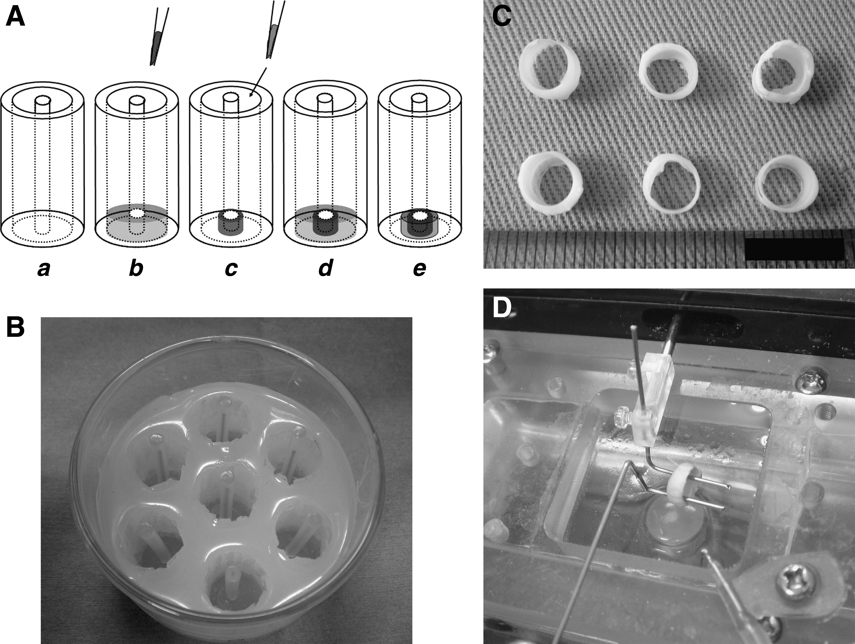
Construction of ring-shaped tissue. Principal methods of constructing ring-shaped tissue
Construction of ring-shaped bone-like tissue
The principal technique is shown in Figure 1A and initially required casting of a reconstitution mixture (750 μL) containing MSCs (7.5×10⁴ cells) and 0.6 mg neutralized soluble collagen type I into the mold (Fig. 1A-b). Directly after casting, the reconstitution mixture was incubated for 2 h at 37°C in a humidified cell culture incubator to facilitate hardening. Subsequently, MSC medium was added slowly, taking care not to disturb the hydrogel/cell mixture. After 2 days in culture, tissue constructs condensed around the mandrel (Fig. 1A-c). This process was repeated thrice to generate 3-layer ring-shaped tissue (Fig. 1A-d, e). Ring-shaped tissues were transferred to a 24-well plate with 2 mL of MSC medium with (Ring+) or without (Ring−) osteoinductive supplements and continued to culture for 8 weeks.
Biochemical analyses
Secreted osteocalcin (OC), a surrogate parameter for osteoblastic differentiation, 15 was measured in the hydrogel culture, 2D culture, or ring-shaped tissue culture conditioned medium using the rat OC ELISA system (DS Pharm Biomedicals).
Calcium (Ca) deposition in the mineralized matrix in the hydrogel culture, 2D culture, or ring-shaped tissue was extracted by shaking in 0.5 mL of 20% formic acid for 24 h at 4°C. The total amount of Ca in the formic acid extracts was measured using a Calcium E-test Wako kit (Wako Pure Chemicals).
Cell numbers in the hydrogel or 2D cultures were evaluated using a Cell Counting Kit-8 (WST-8; Dojindo). 4
Total RNA was isolated from cells cultured in the hydrogel or 2D cultures using an Isogen RNA Extraction kit (Nippon Gene) and Lysing Matrix D (MP Biomedicals). The extracted total RNA was converted into complementary DNA (cDNA) with a High Capacity cDNA Reverse Transcription Kit (ABI). mRNA levels of two typical osteogenic transcription factors, Cbfa1 type 2 (also known as Runx2 type2) and osterix, were quantified using a TaqMan Gene Expression Assay kit (Rn01512296_m1, ABI [Runx2]) and a custom-made primer/probe set for rat osterix (forward primer: 5′-AGCCCTGGGAAAAGGAGG-3′; reverse primer: 5′-GACCATTGGTGCTTGAGAAGG-3′; fluorogenic [Fam/MGB] probe: 5′-CCATACACTGACCTTTC-3′), respectively. 15 Quantitative PCR was performed using the TaqMan® Universal PCR Master Mix in a StepOnePlus Real-Time PCR System (ABI). The expression levels of Runx2 and osterix were normalized to GAPDH (Rn99999916_s1, ABI).
Mechanical properties testing
Ring-shaped tissues (Fig. 1C) were removed from the casting molds, mounted on a static holder on one side and an isometric force transducer on the other side in phosphate-buffered saline (PBS) in thermostated (37°C) organ baths (Fig. 1D; Unique Medical). They were then subjected to stress–strain analyses until tissue failure to assess ultimate burst strength. Stress (g) at defined steps of strain (0.5 mm) was plotted to generate stress–strain curves (Fig. 4A). Tissue stiffness was expressed as the modulus of stress–strain (g/mm), which was calculated from the slope of the approximately linear part of the stress–strain curves (Fig. 4A, arrow).
Morphologic evaluation
Ring-shaped tissues were washed with PBS and fixed in 10% neutral buffered formalin. Transverse sections of the paraffin-embedded samples were stained with von Kossa to assess mineralization of the extracellular matrix.
Statistical analysis
Data are presented as mean±standard error of the mean (SEM), and were analyzed using paired and unpaired two-tailed Student's t-tests or ANOVA followed by a Bonferroni's multicomparison test. Values of p<0.05 indicated significant differences between groups.
Results
Osteoblastic differentiation in the hydrogel cultures
OC secretions (n=3) and Ca depositions (n=3) were measured at 4 weeks of MSCs culture under 8 different culture conditions (MSC medium with no additional substances, only β-GP, only Vit. C, only Dex, β-GP and Vit. C, β-GP and Dex, Vit. C and Dex, or all three substances; equivalent respective concentrations per condition) in the hydrogel cultures. OC secretions and Ca depositions were highest under the osteoinductive supplements (MSC medium with β-GP/Vit. C/Dex, Fig. 2A, B), indicating advanced osteogenic differentiation of the MSCs.
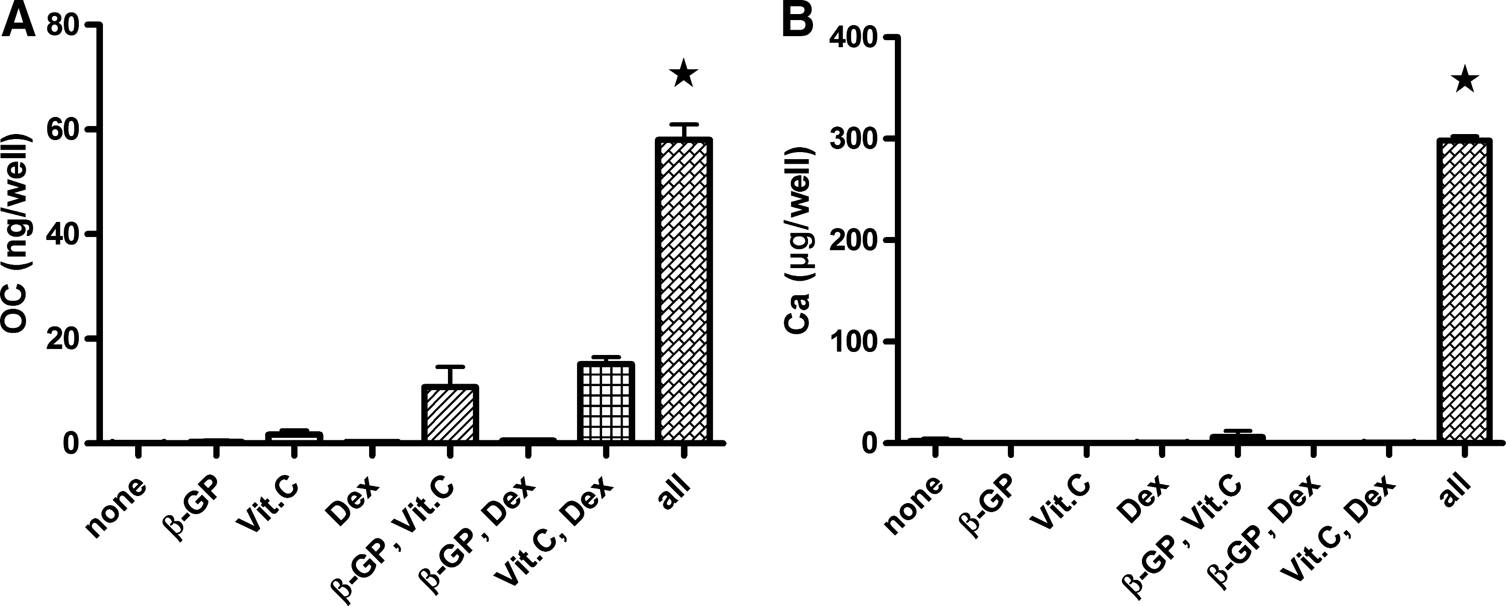
Osteoblastic differentiation in the hydrogel cultures. MSCs were cultured in eight different culture conditions (as indicated on the abscissa) in the hydrogel cultures. Osteocalcin (OC) secretions
Osteoblastic differentiation efficiency in the hydrogel and 2D cultures
OC secretions (n=4) and Ca depositions (n=4) were measured at 1, 2, 3, and 4 weeks of MSC culture with or without osteoinductive supplements in the hydrogel (3D+/3D−) or 2D cultures (2D+/2D−). WST-8 cleavage (reflects number of cells, n=4) was measured in parallel to calculate differentiation efficiency (OC secretions or Ca depositions per cell). WST-8 cleavages of 2D+/2D− and 3D− continued to increase until 4 weeks of culture; in contrast, those of 3D+ decreased from 3 weeks of culture. In addition, those of 3D+ were significantly lower compared with those of 2D+ at 3 and 4 weeks of culture (Fig. 3A). OC secretions per cell of 3D+ were significantly higher than those of 2D+ at all time points (Fig. 3B). Ca depositions per cell of 3D+ were also significantly higher than those of 2D+ from 2 weeks of culture (Fig. 3C). In Figure 3B and C, OC secretion and Ca deposition appear increased from baseline (3D−, 2D−) only in 3D+. Those in 2D+ do not appear increased from baseline. However, OC secretion from 2 weeks and Ca deposition from 3 weeks in 2D+ significantly increased from those in 2D− according to the results of the unpaired t-test (OC: 2D+, 3.2±1.0 vs. 2D−, 0.01±0.01 at 2 weeks; 2D+, 4.5±0.5 vs. 2D−, 0.09±0.01 at 3 weeks; and 2D+, 7.6±1.9 vs. 2D−, 0.30±0.09 at 4 weeks; p<0.05. Ca: 2D+, 7.2±1.9 vs. 2D−, 0.5±0.3 at 3 weeks; and 2D+, 17.1±5.5 vs. 2D−, 1.1±0.3 at 4 weeks; p<0.05). In 3D− and 2D−, almost no OC secretions or Ca depositions were detected.

Osteoblastic differentiation efficiency in the hydrogel and two-dimensional (2D) cultures. MSCs were cultured in the hydrogel or 2D cultures in MSC medium with (3D+/2D+) or without (3D−/2D−) osteoinductive supplements. WST-8 cleavages (reflects number of cells), OC secretions, Ca depositions, and Runx2 and osterix gene expressions were measured at 1, 2, 3, and 4 weeks of culture. WST-8 cleavage p<0.0001, 
 p<0.01 3D+ versus 2D+, ★p<0.0001, ★★p<0.01 3D+ versus all others (ANOVA with Bonferroni's multicomparison test).
Runx2 (n=4) and osterix (n=4) gene expression was also measured at 1, 2, 3, and 4 weeks of MSC culture to further evaluate osteoblast differentiation and maturation. Indeed, both parameters were elevated in 3D+ at most investigated time points as compared with the other groups (Fig. 3D, E).
Mechanical properties of ring-shaped tissues
Ring-shaped tissues reached a stable geometry after 2 weeks in culture and maintained that structure for at least 8 weeks whether cultured with or without osteoinductive supplements (Fig. 1C).
Stress–strain behaviors of 3-layer ring-shaped tissues cultured in the presence (Ring+) or absence (Ring−) of osteoinductive supplements were tested at 2, 4, and 8 weeks of culture to assess biomechanical properties (Fig. 4A). Tissue stiffness (modulus of stress–strain, Fig. 4B) and ultimate burst strength (Fig. 4C) increased under osteoinductive conditions (n=4) over time. Stress–strain behaviors of Ring− did not change over time (Fig. 4A); therefore, tissue stiffness and ultimate burst strength also were unchanged (Fig. 4B, C). In agreement with these findings, OC secretions (Fig. 5A) and Ca depositions (Fig. 5B) increased markedly in Ring+ compared with Ring− in which almost no OC secretions or Ca depositions were detected.
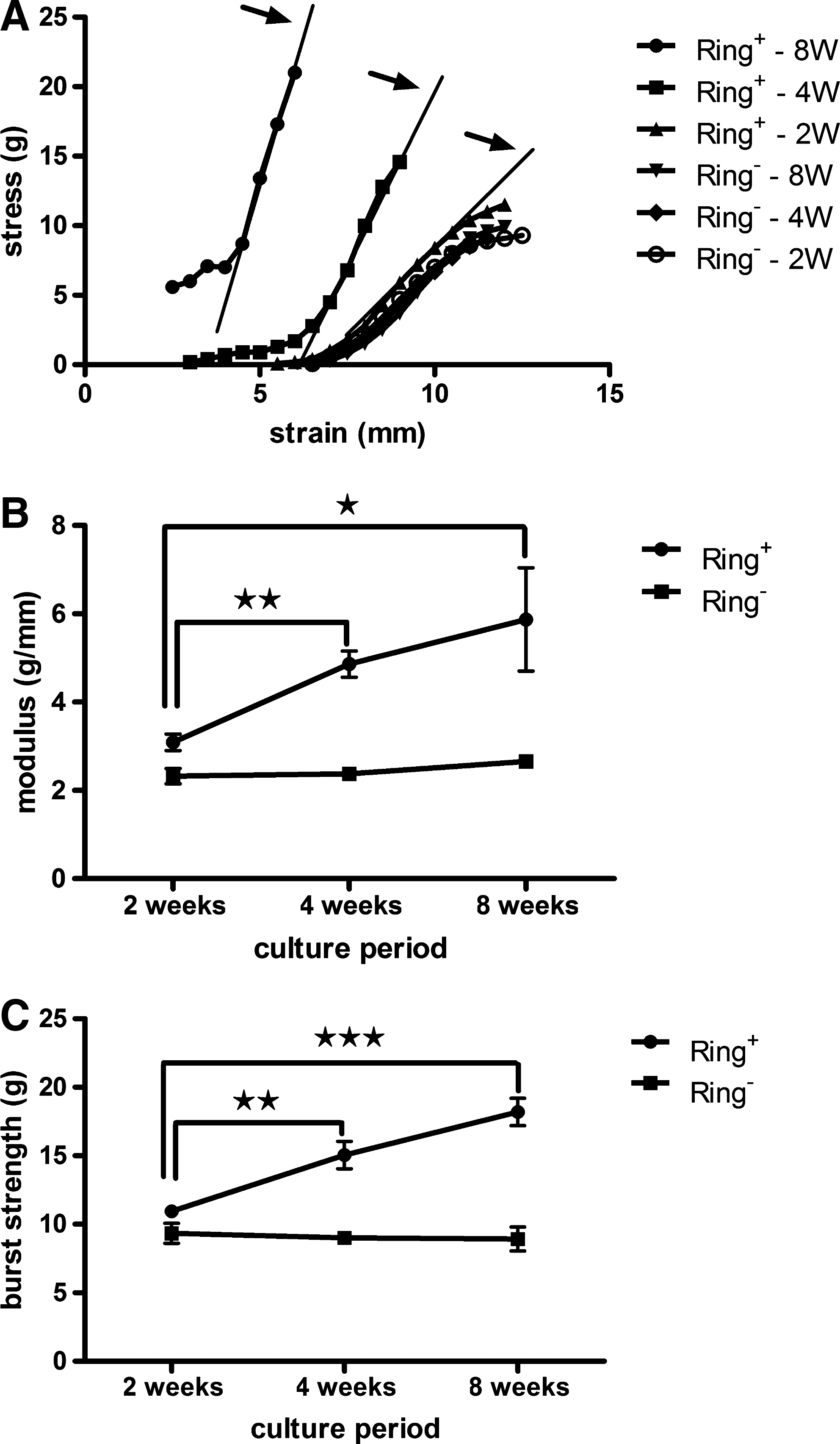
Mechanical properties of ring-shaped tissues. Ring-shaped tissues were cultured with MSC medium with (Ring+) or without (Ring−) osteoinductive supplements. Mechanical properties were measured at 2, 4, and 8 weeks of culture. Stress at defined steps of strain (0.5 mm) was plotted to generate stress–strain curves. Representative stress–strain curves are shown
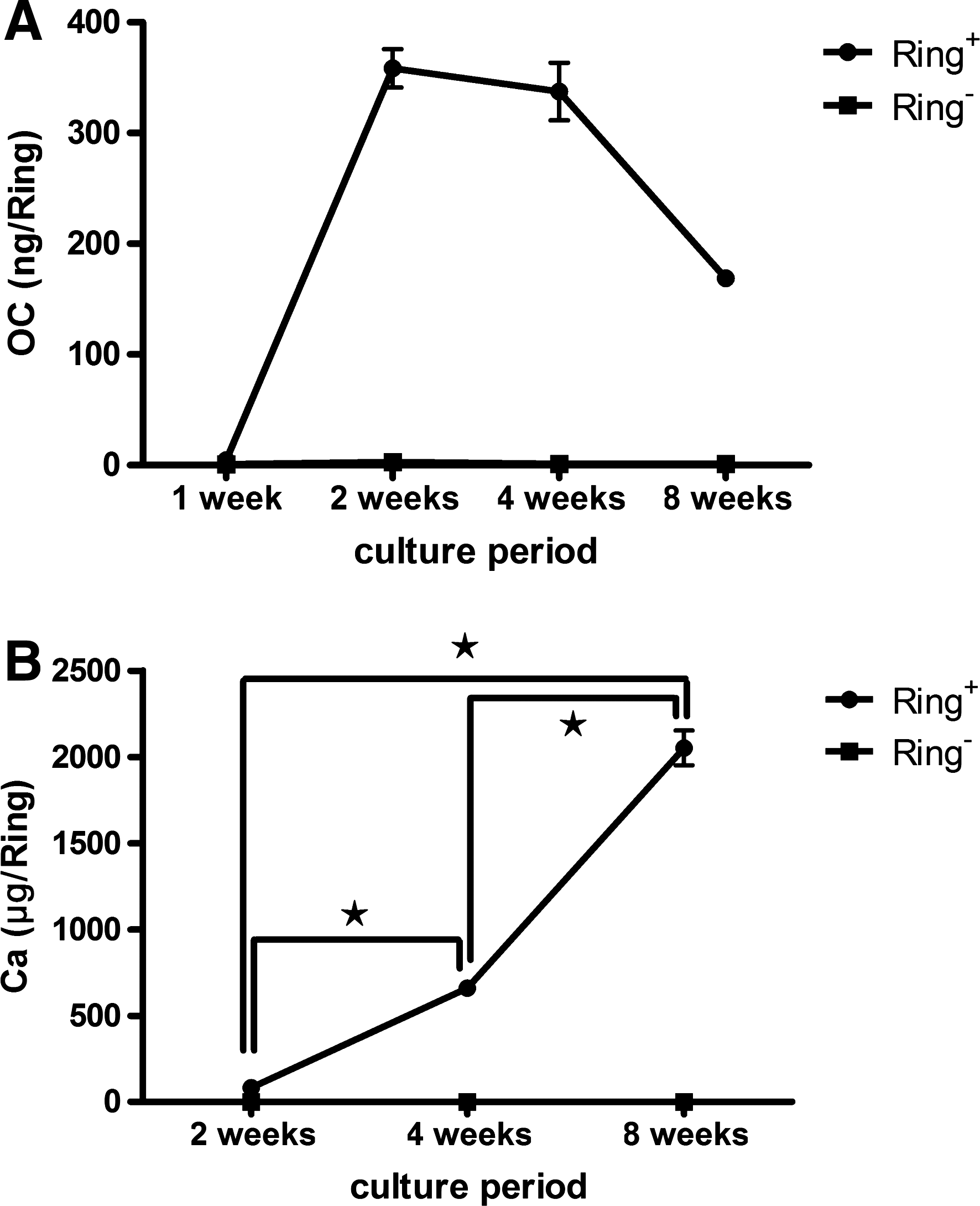
Osteoblastic differentiation in ring-shaped tissues. OC secretions
Histological study of ring-shaped tissues
To confirm whether mechanical strength of three-layer ring-shaped tissues reflected osteogenic development, we performed histochemical staining to assess the mineralization of extracellular matrices. The extracellular matrix in Ring+ tissues stained negative for von Kossa (calcification; Fig. 6A) at 2 weeks of osteogenic culture, but they stained positive at 4 and 8 weeks of culture (Fig. 6B, C). In contrast, Ring− tissues were negative for stain even at 8 weeks of culture (Fig. 6D).
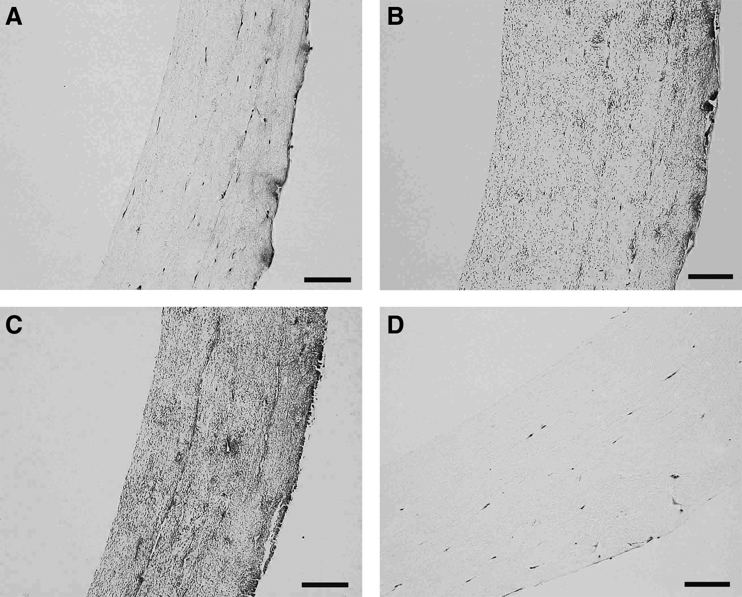
Histological study of ring-shaped tissues. von Kossa staining for calcification (black) of Ring+
Discussion
Ideally, tissue engineering would provide completely autologous tissue from the patient's own cells. Many kinds of stem cells, such as MSCs, embryonic stem (ES) cells, or induced pluripotent stem cells, are presently discussed and partially already tested as candidates in tissue engineering for clinical applications. The use of stem cells, in particular, requires control of stem cell differentiation into the desired phenotype. A large body of literature on stem cell differentiation into osteoblasts exists.1–3,6–13,20,22–28 However, most studies have analyzed the osteogenic differentiation of stem cells in 2D cultures.1–3,6–10 We identify this hydrogel culture system as a caveat and propose a novel tissue engineering model to overcome this limitation. Recently, the advantages of 3D cultures in the induction of differentiation from stem cells to osteoblasts have been reported.11,12 Hanging-drop cultures, as an alternative 3D culture format for studies of stem cell aggregation and tissue formation, have also been exploited.37–39 While ES cells can differentiate under floating conditions, MSCs need to attach onto a substrate for proliferation and differentiation. In light of our own expertise in collagen hydrogel-based tissue engineering,30–34 we sought to adapt this technique to MSC-based bone tissue engineering using osteogenic induction.
Other groups and we have reported that MSCs can be osteogenically induced by the addition of β-GP, Vit. C, and Dex.3–5,15 In this study, we confirmed this finding and advanced it by applying pharmacological osteogenic induction to MSC-containing collagen hydrogels. Collagen I is an essential part of native bone, and it has been reported that collagen I facilitates osteogenic differentiation and enhances the development of engineered bone in vitro better than other hydrogels.6,24 Here, it is important to note that MSCs cultured within the collagen hydrogel showed a clearly higher degree of osteoblastic differentiation than MSCs cultured on a collagen substrate. We base this inference on the identification of a clear elevation of surrogate parameters for osteoblastic differentiation and maturation (i.e., OC secretion and Ca deposition). As shown in Figure 3A, cell numbers decreased significantly in 3D+ compared with 2D+ from 3 weeks of culture; therefore, the remarkable differences of OC secretion per cell and Ca deposition per cell were emphasized. These cell number decreases might have occurred because of the contribution of maturation and organization of the extracellular matrix surrounding osteoblasts to the shutdown of proliferation.8,9 The absolute quantities of OC secretion and Ca deposition were also significantly higher in 3D+ compared with 2D+ (OC, 3D+ 113±3 ng/well vs. 2D+ 19±3 ng/well; Ca, 3D+ 560±28 μg/well vs. 2D+ 59±17 μg/well at 4 weeks of culture; p<0.0001). Thus, we considered that OC secretion and Ca deposition significantly increased in 3D+ compared with 2D+.
MSCs differentiate into mature osteoblasts through preosteoblasts and immature osteoblasts. Runx2 affects all of these stages and is particularly important for controlling multipotent mesenchymal precursor cells toward an osteoblastic lineage. 40 It has been reported that Runx2 expressions had 2 peaks at days 8 to 16, indicating the beginning of differentiation, and at day 32, resulting in bone formation. 8 In contrast, osterix acts as a downstream gene of Runx2 in the osteoblast differentiation signal pathway and allows preosteoblasts to differentiate into immature osteoblasts.4,5,41–44 On the other hand, OC is expressed from mature osteoblasts to osteocytes, and Ca deposition is used as the hallmark of mature osteoblasts.15,41 In this study, Runx2 and osterix expressions, as well as OC secretions, were significantly greater in the hydrogel cultures compared with those in the 2D cultures at 1 week of culture in osteoinductive conditions. In contrast, Ca depositions were gradually increased over time from 2 weeks of culture. These results indicate that 3D culture in the collagen hydrogels accelerated Runx2, which steered MSCs to differentiate into osteoblasts. As a result, osterix was also activated, and finally OC secretions and Ca depositions were increased.
The advantages of 3D culture for the induction of stem cells have been reported.11,12 However, biological markers were used as the evidence of osteoblastic differentiation in most of these studies. A report that measured the changes in mechanical properties of the tissue constructed from osteoblasts differentiated from stem cells was difficult to find. 23 In that study, gradual increases of the tissue stiffness in collagen I hydrogel/beta-tricalcium phosphate scaffolds were demonstrated by biomechanical compression testing. We also believe that not only biological function but also mechanical property is important in some cases of stem cell differentiation, such as those associated with mechanical strength of artificial bone or contractile function of artificial muscle. As such, we have originally developed hydrogel cultures to measure contractile function of artificial heart muscle.30–34 Using the advantage of easy shape distortion of the hydrogel culture, we could measure the mechanical properties easily. In this study, collagen hydrogel-based ring-shaped bone-like tissues conditioned with osteoinductive supplements developed enhanced mechanical properties, including high tissue stiffness and ultimate burst strength, superior molecular/biochemical properties and morphological signs typically found in mineralized bone. These results indicate that mechanical properties increased by the differentiation of MSCs into osteoblasts. It is also reported that matrix elasticity can affect the differentiation of stem cells into osteoblasts, and osteoblasts could be highly differentiated on a hard matrix. 16 In the present study, the extracellular matrix became hard over time, and MSCs may have progressively differentiated into osteoblasts.
A similar technology to entrap various cell types, including fibroblasts, smooth muscle cells, epithelial cells, and cardiomyocytes, has been described.30–34,45–47 Hydrogel cultures might be used not only for inducing MSCs into osteoblasts, but also for other stem cell differentiation. Moreover, this method has the advantage of constructing any shape of tissue and providing layered structures. Constructing tissue layers from different types of cells enables us to construct more complex organs. For example, we have previously constructed a prototypical bioartificial trachea, which has a structure of ring-shaped tissues that were constructed from mixtures of rat MSCs and collagen and fused to the tissue-engineered tube that was constructed from mixtures of rat fibroblasts and collagen to function as a reinforcement. 48 We aim to finally obtain a bioartificial trachea that has the structural and functional properties of native trachea by constructing an epithelial tissue layer in the inner aspect, a smooth muscle tissue layer in the middle layer, and cartilage tissue in the outer layer from the patient's own cells.
Notably, hydrogels lack the initial mechanical strength needed for weight bearing, which is a serious disadvantage for implantation as artificial bone. In this study, MSCs were differentiated fully to provide mineral deposition, but ring-shaped bone-like tissue was transformed only around 5 g of stress. We do not think that it is clinically possible to use this bone-like tissue alone. Porous scaffolds, such as hydroxyapatite (HA) or β-tricalcium phosphate, combined with suspensions of cultured MSCs have been reported. The construction of bone-like tissue by wrapping HA with osteogenic cell sheets was recently reported; in addition, an osteogenic cell sheet contributed to the formation of new bone in a rat nonunion model.49,50 We also think that by wrapping these scaffolds with this bone-like tissue, we can construct bone-like tissue that might promote bone regeneration. In addition, osteoblast differentiation in hydrogel cultures might further elucidate the basic mechanisms of bone physiology and may be suitable for the study of the osteogenic and growth potentials of bone derived from MSCs.
In conclusion, hydrogel cultures exhibited an advantage compared with 2D cultures in the induction of MSC osteoblastic differentiation. The mechanical properties of the bone-like tissues constructed with MSCs in the hydrogel cultures were enhanced with the differentiation of MSCs into osteoblasts. These results may be exploited not only to generate bioartificial bone, but also to elucidate the basic mechanisms of bone physiology.
Footnotes
Acknowledgment
We thank Ms. Mamiko Yoshimura for excellent technical assistance.
Disclosure Statement
No competing financial interests exist.