
Abstract
Innovative strategies based on regenerative medicine, in particular tissue engineering of skeletal muscle, are promising for treatment of patients with skeletal muscle damage. However, the efficiency of satellite cell differentiation in vitro is suboptimal. MicroRNAs are involved in the regulation of cell proliferation and differentiation. We hypothesized that transient overexpression of microRNA-1 or microRNA-206 enhances the differentiation potential of human satellite cells by downregulation quiescent satellite cell regulators, thereby increasing myogenic regulator factors. To investigate this, we isolated and cultured human satellite cells from muscle biopsies. First, through immunofluorescent analysis and quantitative reverse transcription-polymerase chain reaction (qRT-PCR), we showed that in satellite cell cultures, low Pax7 expression is related to high MyoD expression on differentiation, and, subsequently, more extensive sarcomere formation, that is, muscle differentiation, was detected. Second, using qRT-PCR, we showed that microRNA-1 and microRNA-206 are robustly induced in differentiating satellite cells. Finally, a gain-of-function approach was used to investigate microRNA-1 and microRNA-206 potential in human satellite cells to improve differentiation potential. As a proof of concept, this was also investigated in a three-dimensional bioartificial muscle construct. After transfection with microRNA-1, the number of Pax7 expressing cells decreased compared with the microRNA-scrambled control. In differentiated satellite cell cultures transfected with either microRNA-1 or microRNA-206, the number of MyoD expressing cells increased, and α-sarcomeric actin and myosin expression increased compared with microRNA-scrambled control cultures. In addition, in a three-dimensional bioartificial muscle construct, an increase in MyoD expression occurred. Therefore, we conclude that microRNA-1 and microRNA-206 can improve human satellite cell differentiation. It represents a potential novel approach for tissue engineering of human skeletal muscle for the benefit of patients with facial paralysis.
Introduction
Muscle tissue has its own endogenous repair and maintenance system which is based on myogenic progenitor cells, that is, satellite cells. On stimuli, such as damage, satellite cells proliferate and differentiate, which contributes to the regeneration of damaged muscle.4–7 The regenerative capacities of human satellite cells derived from skeletal muscle appear to make them a suitable source for tissue engineering. 8 However, myogenesis, that is, efficient differentiation of human satellite cells toward adult skeletal muscle, remains a major hurdle in vitro. Moreover, with increasing age, the population of satellite cells per myofiber decreases.9,10 Furthermore, the myogenic capacity of satellite cells in vitro decreases.10–12 Therefore, tissue engineering of skeletal muscle from autologous satellite cells will be impaired for elderly patients. Due to donor variation, the efficiency of tissue engineering of skeletal muscle will vary between individual patients.13,14 Therefore, novel approaches to improve myogenesis are mandatory to augment tissue engineering of human skeletal muscle. The process of myogenesis is strongly regulated by epigenetic factors, in particular by microRNAs.15–19 Therefore, we hypothesized that microRNAs could contribute to improving tissue engineering of human skeletal muscle for future clinical application.
MicroRNAs are small, noncoding RNAs, 20–22 nucleotides in length, involved in post-transcriptional gene regulation through inhibition of protein translation or enhancing messenger RNA degradation. Thereby, they also offer great potential as a tool to modify cell fate and function. 20 MicroRNA-1 and microRNA-206 appear prominent in myogenesis through regulation of the pairedbox genes PAX3 and PAX7.21–23 These pairedbox genes are quiescent satellite cell regulators that are essential in myogenesis. They function upstream of both myogenic regulator genes MYOD and MYF5 to initiate proliferation and muscle differentiation.6,24–26 If Pax7 is either overexpressed in satellite cells, or if its expression is prolonged, MyoD expression is inhibited, and the onset of myogenesis delayed, which prevents muscle differentiation. 27 In satellite cells, microRNA-1 and microRNA-206 downregulate Pax7 such that MYOD is no longer inhibited, and muscle differentiation progresses. Another microRNA involved in myogenesis is microRNA-133 that promotes proliferation through repressing Serum Response Factor, which results in inhibited muscle differentiation. 15
We propose to modify the microRNA profile of satellite cells to facilitate tissue engineering of skeletal muscle, which is a novel concept in regenerative medicine. It offers an opportunity to transfect satellite cells with a pre-microRNA or anti-microRNA in order to modulate myogenesis. By transfecting murine satellite cells with anti-microRNA-133, the contractile force of a bioartificial muscle increased. 28 However, the potential of human satellite cells transfected with microRNA-1 or microRNA-206 in tissue engineering remains to be investigated.
This current study aims at investigating whether modulation of human satellite cells through microRNA-1 and microRNA-206 could contribute to the differentiation potential of human satellite cells in a three-dimensional bioartificial muscle model, thereby improving tissue engineering of human skeletal muscle.
Our hypothesis is that transient overexpression of microRNA-1 or microRNA-206 enhances the differentiation potential of human satellite cells by downregulation quiescent satellite cell regulators, thereby increasing myogenic regulator factors.
Materials and Methods
Satellite cell isolation and culture
Muscle biopsies were obtained from six healthy donors undergoing reconstructive surgery. The age of the donors was 49.5±8 years (30–60 years). The study protocol was approved by the institutional medical ethics committee, and patients gave their informed consent. Satellite cells were isolated with 0.04 mg/mL (0.16 Collagenase Wünsch units/mL) Liberase Blendzyme 3 (Roche Applied Science) as previously described. 29 Proliferation medium (PM) consisted of Dulbecco's modified Eagle's medium (DMEM; Invitrogen/Gibco), 20% fetal bovine serum (FBS; Invitrogen/Gibco), and 1% penicillin/streptomycin 50 μg/mL (Sigma-Aldrich).
Differentiation medium (DM) contained DMEM, 2% FBS, 1% penicillin/streptomycin, 1% insulin-transferrin-selenium-A (100×; Invitrogen), and 0.4 μg/mL dexamethason (Sigma-Aldrich). Medium was refreshed thrice per week. Cells were plated at 5.0×10³ cells/cm2 in culture flasks precoated with 1% gelatine/phosphate-buffered saline (PBS) for 30 min. When cells reached 70% confluence, they were enzymatically harvested using Accutase (Invitrogen) and passaged.
Passage number (Px) was defined as the xth sequential harvest of a subconfluent cell population. All experiments were performed using P8–15.
Transfection with microRNA-1 and microRNA-206
For gain-of-function studies, pre-microRNA molecules specific for the mature human microRNA-1 sequence, UGGAAUGUAAAGAAGUAUGUAU (pre-miR for hsa-miR-1, PM10617), and microRNA-206 sequence, UGGAAUGUAAG GAAGUGUGUGG (pre-miR for hsa-miR-206, PM10409; both Ambion/Applied Biosystems), were transfected with siPORT NeoFX transfection agent (Ambion) into satellite cells in accordance to the manufacturer's protocol. Briefly, the transfection agent was diluted in Opti-MEM I (Gibco) and after 10 min, was mixed with 50 nM of the microRNA, or with 50 nM scrambled microRNA control (a random, inert nucleic acid sequence). After incubating for 15 min, the mixture was dispensed into gelatin precoated tissue culture flasks. Cells were added to each flask at 1.0×10⁴ cells/cm2 and cultured in PM for 24 h. After 24 h, PM was refreshed, and cells were cultured for another 24–48 h until they reached 100% confluence.
Bioartificial muscle construct engineering
Bioartificial muscle construct were cultured as previously described. 30 Briefly, house-shaped pieces of Velcro were glued to the bottom of a six-well plate 12 mm apart, sterilized with ethanol (70%) and exposure to UV for 15 min. A gel mixture was prepared on ice by mixing 50% collagen type I (3.44 mg/mL; BD Biosciences), 39% PM, 3% 0.5 M NaOH (Sigma-Aldrich), and 8% growth factor reduced Matrigel® (BD Biosciences). Satellite cells transfected with microRNA-1, microRNA-206, or the scrambled control were harvested and mixed at a concentration of 4.5×10⁶ per mL gel. Then, 350 μL of gel mixture was pipetted in and between the Velcro attachment points. After 1 h gelation, 3 mL PM was added, which was replaced after 24 h with DM which was refreshed daily for 4 days at which point analysis was performed.
Immunofluorescent staining
Cells were cultured on Thermanox® coverslips, Lab-Tek chamber slides, or 96-well plates (all NUNC Brand Products) coated with 1% gelatine. At 100% confluence, cells were fixed or cultured for an additional 5 days in DM and subsequently fixed in 2% paraformaldehyde at room temperature for 10 min. A permeabilization step was performed with 0.5% Triton X-100 (Sigma-Aldrich) in PBS at room temperature for 10 min. Nonspecific binding-sites were blocked with 10% goat serum in PBS for 30 min. Cells were incubated with the primary antibody in PBS and 2% serum at room temperature for 60 min or at 4°C overnight. The primary antibody consisted of either (1) a myogenic marker, rabbit-anti-human desmin (1:100; Novus Biological), (2) a fibroblast marker, mouse anti-human MCA1399G (1:100; AbD Serotec), (3) a sarcomere component, mouse-anti-human α-sarcomeric actin IgM (1:200; clone Alpha Sr-1; Abcam) (4) a myogenic transcription factor, mouse-anti-human MyoD (1:100; Dako), (5) a sarcomere component, mouse-anti-human myosin (MF20; 1:500), and (6) a satellite cell marker, mouse-anti-human Pax7 (1:10; both Developmental Studies Hybridoma Bank). After three washes with 0.05% Tween in PBS, the cells were incubated with a secondary antibody cocktail at room temperature for 30 min. The secondary antibody cocktail constituted FITC-conjugated goat-anti-rabbit IgG (1:100; Southern Biotech), Alexa Fluor® 488 goat-anti-mouse IgM and Alexa Fluor 555 goat-anti-mouse IgG1 or IgG2b (all Invitrogen; 1:300) and 10% normal human serum in PBS/DAPI. Samples were mounted in Citifluor AP1 (Agar Scientific). For Odyssey® infrared imaging (LI-COR Biosciences), the secondary antibody was goat-anti-mouse IrDye800 (1:500 in PBS containing DRAQ5 [1:1000] and 10% normal human serum). Examination was performed by immunofluorescent microscopy using a Leica DMRXA microscope and Leica Software (Leica Microsystems), and further quantification was performed by either TissueFAXS using a Zeiss AxioObserver.Z1 microscope and TissueQuest Cell Analysis Software (TissueGnostics), or by Odyssey infrared imaging system.
Bioartificial muscle construct analysis
Constructs were washed in PBS and fixed in 10% formalin for 1 h. A permeabilization step was performed with 0.5% Triton X-100 (Sigma-Aldrich) in PBS at room temperature for 30 min. Nonspecific binding-sites were blocked with 1% horse serum in NET-gel twice for 20 min. Constructs were incubated with mouse-anti-human MyoD (1:100; Dako), rabbit-anti-human desmin (1:100; Novus Biological) and 10% serum in NET-gel at 4°C overnight.
After six washes with NET-gel, the constructs were incubated with Alexa Fluor 555 goat-anti-mouse IgG1 (1:300), FITC-conjugated goat-anti-rabbit IgG (1:100; Southern Biotech) and 10% normal human serum in PBS/DAPI at room temperature for 2 h. After three washes with NET-gel, the constructs were mounted between coverglasses with Mowiol. Confocal microscopy was performed using a Leica SP2 AOBS CLSM confocal microscope (Leica Microsystems).
Gene transcript analysis
Total RNA was isolated from ∼200,000 cells using the Rneasy Kit (Qiagen, Inc.), in accordance to the manufacturer's protocol. Briefly, a cell lysate was made and diluted with an equal volume of ethanol (70%). RNA was collected on an RNA binding filter by centrifugation. DNA was removed by incubation with a DNase I solution at 37°C for 15 min. The RNA-binding filter was washed twice and, subsequently, the RNA was eluted with 14 μL Elution Buffer. The RNA concentration and purity were determined by spectrophotometry (NanoDrop Technologies). For quantitative reverse transcription-polymerase chain reaction (qRT-PCR) analysis, total RNA was reverse transcribed using the First-Strand cDNA synthesis kit (Fermentas UAB). In summary, 1 μg of total RNA was diluted in a final reaction volume of 20 μL containing random hexamer primer (0.5 μg), RiboLock™ Ribonuclease Inhibitor (20 U), 1 mM dNTP mix, and incubated at 37°C for 1 h. The reverse-transcription reaction was terminated by heating the mixture to 70°C for 10 min, after which the samples were placed on ice. Quantitative RT-PCR analysis was performed in a final reaction volume of 10 μL, consisting of SYBR Green Supermix (Bio-Rad), 0.5 mM primer mix (Table 1), and 5 ng cDNA. For analysis of PKT9, Applied Biosystems “assay on demand” primer/probe sets were used to detect amplimers of PKT9 (Hs00702289_s1) and β-2-Microglobulin (β2M; Hs99999907_m1). Reactions were performed at 95°C for 15 s, 60°C for 30 s, and 72°C for 30 s, for 40 cycles. Analysis of the data was performed using Science Detection Software 2.2.2. To determine differences in expression, CT-values were normalized against GAPDH-expression using the ΔCT-method [ΔCT(gene)=CT(gene)−CT(GAPDH or β2M)]. Relative expression levels were calculated as 2−(ΔCT). All cDNA samples were amplified in triplicate.
MicroRNA analysis
Total RNA was isolated from ∼200,000 cells using the mirVana kit (Ambion), in accordance to the manufacturer's protocol. Briefly, a cell lysate was made and diluted with 1.25 volumes of ethanol (100%). Total RNA was collected on a RNA binding filter by centrifugation. The RNA concentration and purity were determined by spectrophotometry (NanoDrop Technologies). cDNA synthesis was performed using the microRNA Reverse Transcription Kit. In summary, 5 ng of total microRNA was diluted in a final reaction volume of 7.5 μL with 1.5 μL microRNA specific RT-primer mix (Table 2), and 3.5 μL RT-master mix, containing 1 mM dNTP mix, multiscribe RT enzyme, RT Buffer, RNase Inhibitor, and water. This was incubated at 16°C for 30 min, 42°C for 30 min, 85°C for 5 min, and subsequently mixed with 2 μL microRNA specific qRT-primer mix and 10.5 μL water. Quantitative RT-PCR analysis was performed with 5 ng cDNA-primer mix and 5 μL iTaq Supermix with ROX (Bio-Rad). Reactions were performed at 95°C for 15 s, 60°C for 60 s, for 45 cycles. Analysis of the data was performed using Science Detection Software 2.2.2. To determine differences in expression, CT-values were normalized against RNU6B-expression using the ΔCT-method [ΔCT(microRNA)=CT(microRNA)−CT(RNU6B)]. Relative expression levels were calculated as 2−(ΔCT). All cDNA samples were amplified in triplicate.
Statistics
All data are represented as means±SEM and were analyzed by Student's t-test or analysis of variance using Graph-Pad Prism Version 5 (GraphPad Software, Inc.).
Results
The balance between quiescent satellite cells and myotubes during myogenesis
Satellite cells were isolated and cultured from enzymatically dissociated muscle tissue. Initial passages comprised of heterogeneous cell populations, but at passage 8, satellite cells had nearly reached homogeneity (Fig. 1A). More than 95% of the cells expressed the satellite cell marker desmin. The remaining desmin negative cells were most likely residual fibroblasts (Fig. 1B). Differentiation of confluent satellite cell cultures was induced by switching to DM. Five days after switching to DM, a part of the satellite cells had differentiated, fused, and formed myotubes (Fig. 1C). Approximately 30% of the satellite cells remained mononucleated. These cells highly expressed Pax7. On the contrary, the nuclei of satellite cells that formed myotubes did not express Pax7. However, they expressed MyoD, and moreover, myotubes were highly positive for desmin, α-sarcomeric actin, and myosin (Fig. 2A–D).

Satellite cell culture, differentiation, and characterization. During passages 8–15, cultured cells display a homogeneous morphology, note the triangular-shaped cells that are typical for satellite cells, by differential interference contrast (DIC) microscopy
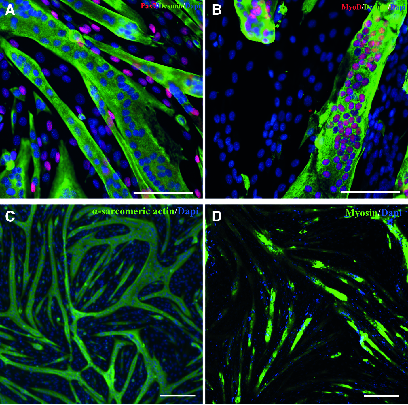
Differentiation of confluent satellite cell cultures was induced by switching to differentiation medium. Immunofluorescent staining of differentiated satellite cell cultures after 5 days showed myotubes that highly expressed desmin (green). The nuclei of quiescent satellite cells highly expressed Pax7 (red)
Since usually Pax7 is expressed by quiescent satellite cells, we hypothesized that satellite cell cultures which contain a relative low percentage of Pax7 expressing cells before differentiation are more prone to myotube formation. Indeed, undifferentiated satellite cell cultures that showed 10.3%±5.6% Pax7 expressing cells (Pax7low) showed 61.6%±4.9% more MyoD-positive cells than satellite cell cultures that showed 62.3%±11.9% Pax7 expressing cells (Pax7high) (Fig. 3A, B; p=<0.01). This was confirmed at the gene expression level, where undifferentiated satellite cell cultures that expressed relative low levels of PAX7 (2−(ΔCT)=0.003±0.08) showed 43.5%±9.5% higher MYOD expression compared with satellite cell cultures that expressed relative high levels of PAX7 (2−(ΔCT)=0.013±0.22) (Fig. 3C, D; p=0.02).
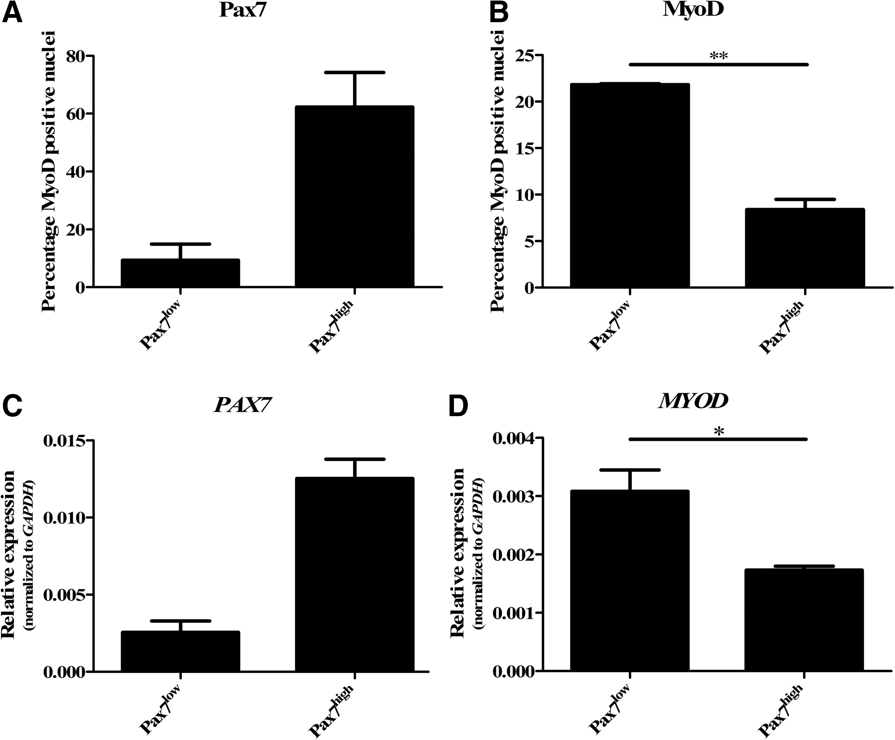
Immunofluorescent analyses by TissueFAXS showed that there are undifferentiated satellite cell cultures that contain a relative low percentage of Pax7 expressing cells (Pax7low), and cultures that contain a high percentage Pax7 expressing cells (Pax7high)
In these undifferentiated satellite cell cultures, we measured the protein expression of the sarcomere components α-sarcomeric actin and myosin using digitalized immunofluorescent imaging (i.e., Odyssey). It showed that both proteins had similar expression levels in Pax7low and Pax7high cultures. We confirmed this similar expression of α-sarcomeric actin (ACTA1) and myosin (MYL1; MYL3) at gene expression level (data not shown). However, after differentiation, α-sarcomeric actin expression was 17.7%±5.4% higher in Pax7low cultures compared with Pax7high cultures (Fig. 4A, B; p=0.03). Furthermore, also at the gene expression level, ACTA1 expression was 73.6%±7.4% higher in Pax7low cultures (Fig. 4C; p=0.01).
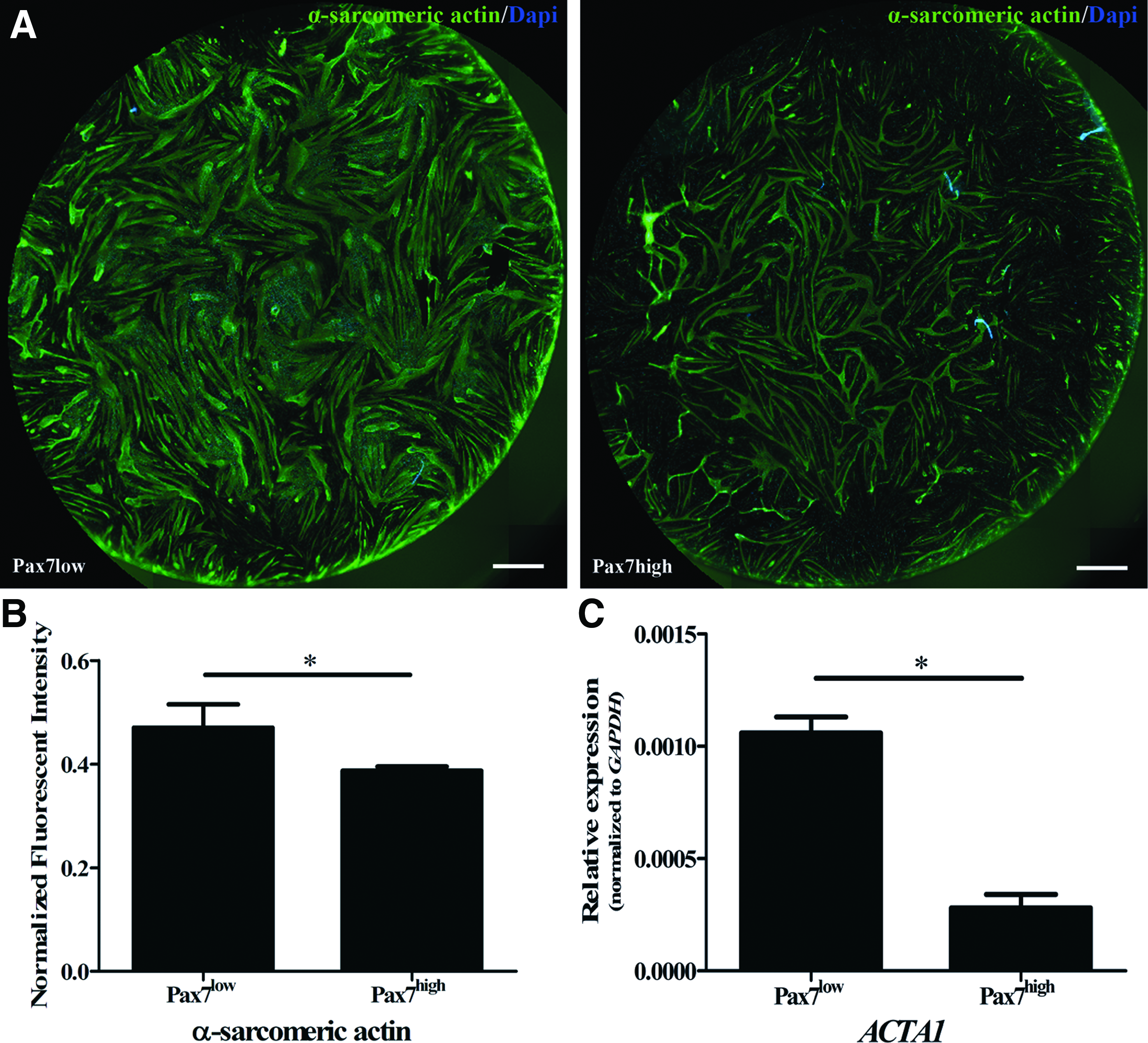
Differentiation of satellite cells in Pax7low and Pax7high cultures. Immunofluorescent analyses of α-sarcomeric actin (green) by Odyssey, 5 days after switching to differentiation medium, showed that in Pax7low cultures, α-sarcomeric actin expression was 17.7%±5.4% higher compared with Pax7high cultures (*p=0.03)
MicroRNA expression during satellite cell differentiation
We determined microRNA-1 and microRNA-206 expression in proliferation, confluent, and differentiated satellite cell cultures. Using qRT-PCR, we showed that both microRNAs were upregulated 10-fold when satellite cells reached confluence. During myotube formation, microRNA-1 is upregulated an additional 100-fold, and also microRNA-206 is upregulated an additional 10-fold. The levels at which microRNA-1 and microRNA-206 were expressed were comparable to levels we had found in normal human skeletal muscle biopsies (Fig. 5A, B).
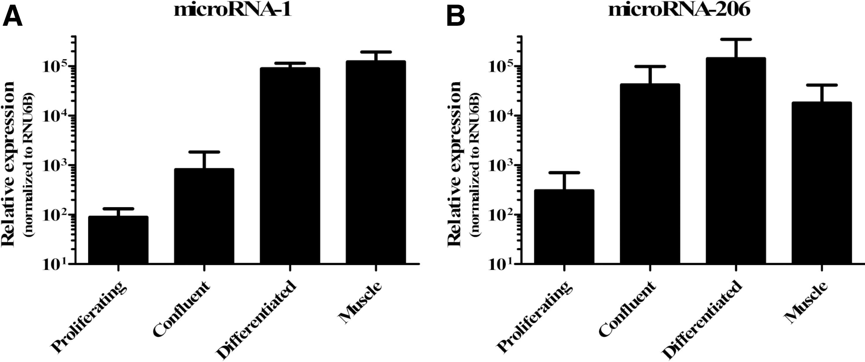
MicroRNA-1 and microRNA-206 expression in proliferating, confluent, and differentiated satellite cells. Quantitative microRNA expression analysis showed that both microRNAs were upregulated 10-fold in confluent satellite cells. Five days after switching to differentiation medium, microRNA-1 is upregulated 100-fold, and microRNA-206 is upregulated 10-fold. Levels at which microRNA-1 and microRNA-206 were expressed were comparable to the levels we had found in normal human skeletal muscle biopsies
Effective satellite cell transfection with microRNA-1
Satellite cells were transfected with microRNA-1 or scrambled microRNA for forty-8 h. We found that PKT9 (an experimental control gene) was downregulated by 79.5%±2.9% in satellite cell cultures transfected with microRNA-1, compared with satellite cell cultures transfected with the scrambled microRNA control. This confirmed an effective cell transfection. Furthermore, we observed only a 2.0%±0.2% cytotoxicity level in transfected satellite cells compared with non-transfected satellite cells.
MicroRNA-1 and microRNA-206 regulate satellite cell differentiation potential
Gain-of-function approach was used to investigate the potential of microRNA-1 and microRNA-206 in human satellite cell differentiation. Transfection of undifferentiated satellite cells for 48 h with microRNA-1 downregulated Pax7 protein expression by 43.3%±15.8% compared with the scrambled microRNA control (p=0.02). Transfection with microRNA-206 did not result in a significant decrease in Pax7 expression. After transfection for 48 h, satellite cells were confluent, and PM was switched to DM. Five days after switching to DM, part of the satellite cells differentiated. In these differentiated cultures, MyoD protein expression had increased by 82.0%±31.3% in satellite cells transfected with microRNA-1, compared with scrambled microRNA (p=0.03). MyoD expression was 59.2%±32.5% increased in satellite cells transfected with microRNA-206 (Fig. 6B; p=0.04).

The effect of microRNA-1 and microRNA-206 on satellite cells. Analysis by TissueFAXS showed a decrease of Pax7 expressing cells by 43.3%±15.8% in proliferating satellite cell cultures transfected with microRNA-1 for 48 h, compared with cultures transfected with the scrambled microRNA control (*p=0.02). There was no significant decrease in the percentage of Pax7 expressing cells in cultures transfected with microRNA-206
Finally, α-sarcomeric actin expression had increased by 51.1%±24.7% in satellite cells transfected with microRNA-1 (p=0.04), and by 47.9%±26.1% increased in satellite cells transfected with microRNA-206 (Fig. 6C; NS). Myosin expression had increased by 14.2%±6.2% in satellite cells transfected with microRNA-1 (p=0.03), and increased by 32.1%±8.3% in satellite cells transfected with microRNA-206 (Fig. 6D; p=<0.01). Cross-striations were observed in differentiated satellite cells transfected with microRNA-1, microRNA-206, or the scrambled microRNA. However, no significant difference between these groups could be detected.
MicroRNA-206 increases satellite cell differentiation in a three-dimensional bioartificial muscle construct
Satellite cells transfected with microRNA-1 or microRNA-206 were subsequently cultured in a three-dimensional bioartificial muscle construct. Analysis by confocal microscopy showed that 26.7%±0.6% of the nuclei in the microRNA-1 transfected muscle constructs were MyoD positive, and 31.7%±2.1% of the nuclei in the microRNA-206 transfected muscle constructs were MyoD positive. Compared with cultures transfected with the scrambled microRNA control, the percentage of MyoD expressing nuclei was increased by 19.4%±1.2% in cultures transfected with microRNA-1, and by 31.8%±4.5% in cultures transfected with microRNA-206 (Fig. 7; **p<0.01).

The effect of microRNA-1 and microRNA-206 on satellite cells in a three-dimensional bioartificial muscle construct. The percentage of MyoD expressing cells increased by 19.4%±1.2% in cultures transfected with microRNA-1, and by 31.8%±4.5% in cultures transfected with microRNA-206 compared with cultures transfected with the scrambled microRNA control (**p<0.01). (n=3; data are represented as means±SEM).
Discussion
The current study set out to investigate whether modulation through microRNA-1 and microRNA-206 of human satellite cells could positively influence myogenesis. One major finding is that transient overexpression of microRNA-1 or microRNA-206 in satellite cells enhances differentiation potential by downregulation of the satellite cell marker Pax7, thereby increasing the myogenic regulator factor MyoD and enhancing myogenic potential. In addition, in satellite cells cultured in a three-dimensional bioartificial muscle construct, this enhancement occurs. Moreover, sarcomere components such as α-sarcomeric actin and myosin become more abundant in microRNA transfected satellite cells.
Our finding that when Pax7 expression is low in proliferating satellite cells, MyoD expression is high, implies that these cells are geared up for myogenic differentiation, that is, myotube formation. This results in an increased upregulation of the sarcomere component α-sarcomeric actin, and more efficient myotube formation during differentiation. This differentiation process is caused by activation of the myogenic regulator factors MyoD and Myf5, after which Pax7, a transcription factor that inhibits differentiation and maintains quiescent satellite cell state, is downregulated.31,32
Our results show that microRNA-1 and microRNA-206 are highly upregulated during differentiation of human satellite cells. Through transient transfection of human satellite cells with microRNA-1 and microRNA-206, we show that microRNA-1 correlates with a downregulation of Pax7, resulting in an upregulation of MyoD and subsequently, a higher α-sarcomeric actin and myosin expression. The explanation for this increased differentiation is that microRNA-1 and microRNA-206 are transcribed simultaneously with MYOD, which is upregulated during differentiation.16,22 MicroRNA-1 and microRNA-206 bind to the 3’UTR of Pax3, thereby downregulating Pax3. Since Pax3 is responsible for preserving quiescent satellite cell state and preventing differentiation, downregulating Pax3 promotes differentiation of satellite cells. 22 Furthermore, microRNA-1 and microRNA-206 also bind to the 3′UTR of Pax7, downregulating Pax7 and even further promoting differentiation in satellite cells.21,23 Although we did not find a significant downregulation of Pax7 on transfection with microRNA-206, satellite cells did show a significant upregulation of MyoD, and also a higher α-sarcomeric actin and myosin expression. This discrepancy in microRNA-206 regulation of Pax7 might be caused by differences, such as rodent versus human or cell line versus primary cells,33,34 which makes translation to primary human cells difficult. Furthermore, heterogeneity with regard to function, behavior, and different subsets within the satellite cell population13,24,35–37 might be responsible for this discrepancy. The heterogeneity is caused by different capacities of satellite cells; they are capable of regenerating muscle fibers and meanwhile replenishing their own pool of progenitor cells.38,39 On the other hand, these qualities of simultaneous regeneration and self-renewal render satellite cells highly suitable for tissue engineering.
Furthermore, transient transfection with microRNA-206 alone may not be sufficient to downregulate Pax7 significantly, but it might be possible that a slight downregulation at an earlier time point is sufficient to influence downstream effects, such as upregulating MyoD, α-sarcomeric actin, and myosin. In addition, in recently published studies, microRNA-1 and microRNA-206 are coadministered to repress Pax7, 21 which may indicate that transfection with both microRNAs is necessary to have a significant effect.
Remarkably, we were unable to show a loss of function after transient transfection with anti-microRNA antisense molecules specific for the mature microRNA-1 and microRNA-206 sequence (anti-miR for hsa-miR-1, AM10617; anti-miR for hsa-miR-206, AM10409; Ambion). The expression of Pax7, MyoD, α-sarcomeric actin, or myosin did not change significantly in either proliferating satellite cell cultures or differentiated satellite cell cultures (data not shown). Most likely, the antisense molecules cannot be offered in sufficient amounts to reach the threshold necessary to exert a functional or measurable effect as a result of the high increase in endogenous microRNA-1 and microRNA-206 in differentiating human satellite cells, and the transient character of transfection in our study.
Our results demonstrate that microRNA-1 and microRNA-206 promote satellite cell differentiation through a downregulation of Pax7 and/or an upregulation of MyoD. However, transfection with either microRNA-1 or microRNA-206 solely was not sufficient to trigger myotube formation in proliferating or even confluent satellite cells. For that to occur, confluency and switching to DM was still required. Thus, microRNA-1 and microRNA-206 enhance muscle differentiation, but require other mediators to initiate myotube formation.
Several microRNAs have been identified that are involved in skeletal muscle proliferation and differentiation.19,40 Besides the pronounced role for microRNA-1 and microRNA-206, there is involvement of microRNA-27. Through downregulating Pax3, microRNA-27 forces satellite cells to start differentiation. 41 Furthermore, for tissue-engineering applications, transfection with microRNAs offers a novel tool for modulating efficient cell function. MicroRNA-133 is involved in maintaining the proliferation of satellite cells through repressing Serum Response Factor. The inhibition of microRNA-133 in murine satellite cells decreased their proliferation.15,34 In a three-dimensional model, that is, a bioartificial muscle,30,34 this inhibition of microRNA-133 improved expression of a differentiation marker Mef2 and moreover improved contractile force. 28 On culture in a three-dimensional bioartificial muscle construct, satellite cells transfected with microRNA-206 showed an increased MyoD expression. Therefore, we conclude that in a scaffold consisting of extracellular matrix, microRNA-206 improves the generation of myotubes. After thoroughly confirming the augmenting effect of microRNAs in two-dimensional cultures, we show that this also occurs in a three-dimensional set-up, paving the way for future functional in vitro and in vivo experiments.
We have shown that microRNA-1 and microRNA-206 improve differentiation of human satellite cells. However, in optimizing differentiation, it is key to prevent a complete depletion of the pool of quiescent satellite cells, known as reserve cells. 42 This population of satellite cells per myofiber naturally decreases with increasing age in vivo, as does their myogenic capacity.9,10,43 Therefore, to promote tissue engineering of skeletal muscle from autologous satellite cells, novel approaches such as microRNAs contribute to improving the tissue engineering of human skeletal muscle for clinical application. Furthermore, for in vivo implantation, preservation of a pool of progenitor cells and their regenerative capacity in the tissue engineered construct is important. Transient transfection is a good option, as it modulates satellite cells in entering the differentiation program, without being a complete knockdown of quiescent satellite cell preserving factors. It merely provides a kick start in the differentiation of satellite cells during the tissue engineering of skeletal muscle. Maintaining the balance between genetic regulatory transcription factors and epigenetic regulatory microRNAs is vital. Therefore, in future studies, we aim at identifying the players that are responsible for the balance between regeneration of skeletal muscle and quiescence of satellite cells.
In conclusion, we show that microRNA-1 and microRNA-206 improve human satellite cell differentiation potential. This represents a novel approach for the tissue engineering of human skeletal muscle for the benefit of patients with facial paralysis.
Footnotes
Acknowledgments
This study was funded by a research grant by the Graduate School W.J. Kolff Institute from the University Medical Center Groningen, University of Groningen, the Netherlands. The antibodies MF20 and Pax7 developed by resp. Fischman, D.A. and Kawakami, A. were obtained from the Developmental Studies Hybridoma Bank developed under the auspices of the NICHD and maintained by The University of Iowa, Department of Biology, Iowa City, IA 52242. The TissueFAXS, “the equivalent to flow cytometry for multiparameter quantitative analyses in tissues,” was acquired with an NWO-ZonMW Medium Investment Grant (40-00506-98-9021).
Disclosure Statement
No competing financial interests exist.