
Abstract
Many cell-based tissue-engineered cartilaginous constructs are mechanically softer than native tissue and have low content and abnormal proportions of extracellular matrix (ECM) constituents. We hypothesized that the load-bearing mechanical properties of cartilaginous constructs improve with the inclusion of collagen (COL) and proteoglycan (PG) during assembly. The objectives of this work were to determine (1) the effect of addition of PG, COL, or COL+PG on compressive properties of 2% agarose constructs and (2) the ability of mechanical compaction to concentrate matrix content and improve the compressive properties of such constructs. The inclusion of COL+PG improved the compressive properties of hydrogel constructs compared with PG or COL alone. Mechanical compaction increased the PG and COL concentrations in and compressive stiffness of the constructs. Chondrocytes included in the constructs maintained high viability after compaction. These results support the concepts that the assembly of cartilaginous constructs with COL+PG and application of mechanical compaction enhance the ECM content and compressive properties of engineered cartilaginous constructs.
Introduction
To treat damaged articular cartilage, several strategies, including tissue engineering approaches, have emerged aiming to repair cartilage and stimulate healing. 5 However, many cell-based tissue-engineered constructs for articular cartilage are mechanically soft and have an imbalance between its ECM components. In normal cartilage, the COL content is typically ∼2–10 times higher than sGAG content in cartilage ranging in maturity from immature fetal bovine to adult human cartilage, respectively (Table 1),6,7 whereas the typical ratio of COL:sGAG in engineered cartilage is 1:1 or less, even after prolonged culture.8–10 After culture, cartilaginous constructs may attain a PG content that approaches physiological levels, although its COL content is substantially below that of native cartilage.
COL, collagen; sGAG, sulfated glycosaminoglycan.
A variety of stimuli have been used in attempts to increase matrix content in and mechanical properties of cell-based, tissue-engineered cartilaginous constructs11,12 These factors include chemical stimuli such as morphogens, growth factors, and cytokines; mechanical stimuli such as dynamic compression, shear, tension, perfusion, and pressure; and other biophysical factors such as hypoxia and electric fields. These approaches typically take weeks, if not months, to achieve the concentrations that approach physiological levels.
More recently, efforts have been made to increase the concentration of ECM constituents by direct manipulation of the matrix. By biomimetic molecular reassembly and addition of assembled PG aggregates to a tissue-engineered construct, the desired PG concentration was rapidly achieved and maintained over a 3-day incubation period. 13 The addition and retention of assembled PG content increased the biomechanical properties of the engineered construct and reduced the time needed to produce a mature ECM. In another method, selective enzymatic degradation of PG in cartilage and engineered constructs resulted in increased COL concentration, COL:PG ratio, and tensile stiffness of the tissue.14–16 These approaches highlight the importance of the content of and balance between COL and PG in the mechanical function of tissue-engineered constructs. However, direct mechanical compaction to increase ECM concentration and mechanical function has not been studied previously. The ability to accelerate matrix accumulation by compaction of a larger, hyper-hydrated construct with appropriate COL:sGAG ratio into a targeted dimension may enable more rapid fabrication of grafts ready for implantation into a joint defect.
The fabrication of cartilaginous constructs that more closely approximates the COL:sGAG ratio and content of normal cartilage may allow for improved mechanical properties and more rapid functioning of such constructs in the mechanically demanding environment of the in vivo joint. Thus, the objectives of this work were to determine (1) the effect of addition of PG, COL, or COL+PG on compressive properties of hydrogel constructs, and (2) the feasibility of mechanical compaction to concentrate matrix content in tissue-engineered constructs and the effect of such compaction on the compressive properties of such constructs.
Materials and Methods
The effects of addition of PG, COL, or COL+PG and mechanical compaction on biochemical and compressive properties of tissue-engineered constructs were studied. Constructs with varying ECM components were assembled, measured for thickness and wet weight, and then mechanically compacted to 90% of their initial thickness while measuring their peak stress (σPEAK) and equilibrium stress (σEQ). After 1 day, the compacted constructs were analyzed for thickness and wet weight, tested under compression to obtain σPEAK and σEQ, and analyzed for sGAG and COL content. The mechanical properties were analyzed further by solely considering the ECM contribution to σEQ with subtraction of the agarose contribution, comparing the stress–compression curves of the initial and final compacted constructs, and comparing the PG contribution to the predicted πPG values.
Preparation of PG components and COL
PG aggregate was prepared for cartilaginous tissue assembly as describedpreviously. 12 AGC and LP were purified from bovine calf knee articular cartilage.17–19 Pharmaceutical-grade HA was obtained as Healon® (Abbott Medical Optics, Inc., Santa Ana, CA). The concentrated PG aggregate solution containing AGC + 1% HA + 5% LP was dialyzed (MWCO 8–10 kDa) against phosphate-buffered saline (PBS), pH 7.0 at 4°C for 18 h to remove the Guanidine-HCl and allow for aggregation, which was verified by S-1000 column chromatography. 20
COL was prepared from the articular cartilage of two calf knees, based on a modification of a previously described method.21,22 Cartilage, extending ∼2–5 mm from the articular surface and ∼10 g per knee, was chopped into ∼1-mm3 pieces and digested with 1 mg/mL trypsin from bovine pancreas in PBS, 0.9 mM CaCl2, and 0.5 mM MgCl2 (pH 7.1) overnight at room temperature with gentle mixing to remove PG. Then, the cartilage pieces were washed twice in PBS, followed by 2 mg/mL soybean trypsin inhibitor in PBS for 30 min, and then washed twice in PBS. The digested cartilage samples were cryomilled with dry ice, suspended in PBS, and then filtered through a 100-μm-pore-size nylon mesh filter (BD Falcon, San Jose, CA). The filtrate was analyzed for hydroxyproline 23 and sGAG, 24 respectively, and found to contain a high proportion (>95%) of COL relative to sGAG.
Formation of tissue-engineered constructs
Constructs were formed with 2% low-melting temperature agarose (SeaPlaque, Lonza, Rockland, ME) and variable amounts of PG and COL to achieve the final concentrations as follows (also in Table 2): (Group I) control with no PG or COL, (Group II) PG only (2.5 mg/mL AGC + 1% HA [by weight of AGC] + 5% LP), (Group III) COL only (10 mg/mL COL), and (Group IV) COL+PG. The concentrations of COL (10 mg/mL) and sGAG (2.5 mg/mL) were chosen since the COL:sGAG ratio of 4:1 was in the range of that for bovine and human adult articular cartilage (Table 1).1,6
HA, hyaluronan; LP, link protein; PG, proteoglycan.
The concentrated matrix solutions were warmed to 40°C and mixed well with a concentrated solubilized agarose solution, for a final agarose concentration of 2%. The PG-agarose solutions were gelled into ∼3-mm-thick sheets at 4°C for at least 1 h. Disk constructs (d=6.4 mm, h=∼3 mm; n=4–5/group) were punched from the PG-agarose sheets and placed -into PBS (pH 7.0), containing protease inhibitors (PIs; 0.5 mM EDTA, 1 mM phenylmethylsulfonyl fluoride, 5 mM benzamidine-HCl, and 5 mM N-ethylmaleimide)overnight at 4°C.
Mechanical compaction of the constructs
After the overnight incubation, the wet weights and thicknesses of the constructs were measured. For the mechanical compaction of the constructs, the constructs were placed in a radially confining chamber between two porous platens filled with PBS + PIs and compressed to a final compression of 90% of the initial thickness using a mechanical spectrometer. 13 The constructs were compressed to 30%, 60%, 75%, and 90% of the initial thickness during a 1600-s ramp compression, followed by 2400-s relaxation to equilibrium. At each compression level, σPEAK and σEQ were calculated from the measured load at the end of ramp compression and at the end of relaxation, divided by the area of the disk. After mechanical compaction, the constructs were incubated in PBS + PIs overnight at 4°C under free-swelling conditions to allow equilibration.
Biomechanical analysis of the compacted constructs
Following overnight incubation after mechanical compaction, the constructs were photographed and measured for wet weight and thickness. The constructs were tested in the same confined compression apparatus, with compression to 60%, 75%, and 90% of the initial thickness (before compaction) using 1600-s ramp compressions followed by 2400-s relaxation to equilibrium. These constructs were not tested at 30% compression of the initial thickness because the compacted thickness was less than 70% of the initial thickness. At each compression level, σPEAK and σEQ were determined and compared to corresponding values during the initial mechanical compaction and also to compacted constructs from other experimental groups.
Biochemical analysis of construct ECM components
After the final mechanical testing, the constructs were cut in half. One half of each construct was solubilized by proteinase K digestion at 60°C overnight and incubated at 70°C for 2 min. The constructs were analyzed for sGAG and COL content by dimethylmethylene blue 24 and hydroxyproline assays, 23 respectively.
The other construct halves were prepared for histology. The constructs were fixed in 4% paraformaldehyde for 3 h, snap-frozen with optimal cutting temperature compound (Sakura Finetek, Torrance, CA) in isopentane cooled with liquid nitrogen, and cryosectioned in the vertical orientation of the constructs at 30-μm thickness. The sections were assessed for sGAG with Alcian blue 25 and for COL type II by immunohistochemistry, 26 and then imaged using brightfield microscopy (Eclipse TE300; Nikon, Melville, NY).
Comparison of mechanical properties of initial and compacted constructs
The stress–compression relationship was plotted using the compression levels normalized to the measured thicknesses before compaction for initial constructs or mechanical testing for compacted constructs. At the last two compression levels of the compacted constructs, stresses were estimated from the stress–compression curves of the initial constructs. Then, the estimated stresses were compared to the measured σEQ for the compacted constructs from the same group.
Comparison of PG contribution to predicted πPG to compressive properties
To assess the matrix contribution to σEQ apart from the agarose contribution, assuming the contributions from each components were additive, the stress ascribed to PG or COL or COL+PG only at each compression level was calculated by subtracting σEQ of Groups II, III, and IV from σEQ of Group I, the agarose-only constructs, (σEQ – σEQ,I).
Using an FCD–πPG relationship 4 based on previous studies,27,28 πPG during compression was estimated for the constructs from Groups II and IV. πPG was calculated from FCDEF using experimentally obtained biochemical data, COL extrafibrillar (EF) water at each compression level, and the FCD–πPG relationship. The EF water varies with the COL content in this FCD–πPG relationship. Then, πPG was compared to the σEQ – σEQ,I of the corresponding group.
Initial test of effects of construct formation and compaction on chondrocyte viability
An initial test of compatibility of construct formation and compaction with viable cells was performed by addition of cells, in the form of alginate-recovered chondrocytes (ARC), 29 during assembly. Disk constructs (d=6.4 mm, h=∼3 mm) containing either no ECM (Group I, n=3 constructs) or COL+PG (Group IV, n=2 constructs) at 80% concentration (8 mg/mL COL and 2 mg/mL PG) with ARC at 1.7×10⁶ cells/mL in 2% agarose were formed. The ECM concentration was decreased to accommodate the additional volume of chondrocytes in the constructs. Groups I and IV were chosen as they represent the extreme cases of peak stress and equilibrium stress that the cells would have to withstand. The cell density was chosen to result in a ratio of cells to COL and PG content similar to native bovine adult cartilage.
Briefly, the ARC were prepared from bovine calf femoral condyle chondrocytes by culture in beads of 1.2% alginate for 8 days at 37°C in Dulbecco's modified Eagle's medium (DMEM)/F12 with additives (100 U/mL penicillin, 100 μg/mL streptomycin, 0.25 μg/mL fungizone, 0.1 mM MEM nonessential amino acids, 0.4 mM L-proline, and 2 mM L-glutamine), 10% fetal bovine serum, and 25 μg/mL ascorbate and then released with their cell-associated matrix from alginate using 55 mM sodium citrate.29,30 The constructs were cultured at 37°C in DMEM with additives, 10% FBS, and 25 μg/mL ascorbate for 1 day and compacted to 90%, as described above. The compacted constructs after 1-day culture, along with uncompacted constructs as controls, were assessed qualitatively for chondrocyte viability by live/dead staining (Life Technologies, Carlsbad, CA) and imaged by epifluorescence microscopy. The images were analyzed to quantify the proportion of viable cells at the edges and in the middle of the constructs with or without compaction (see Supplementary Data for details; Supplementary Data are available online at www.liebertonline.com/tea).
Statistical analysis
Data are presented as mean±standard error of the mean. Thickness, wet weight, sGAG/volume, COL/volume, σPEAK, σEQ, and σEQ – σEQ,I were analyzed by repeated measures analysis of variance (ANOVA) with compaction as a repeated factor and experimental groups as a main factor. To determine the effect of compaction within one experimental group, paired t-tests were performed (p<0.05). To determine the effect of experimental groups for either initial or compacted constructs, ANOVA was performed and was followed by post hoc Tukey test when significance was detected (p<0.05). Estimated σEQ for initial constructs at compression levels for compacted constructs and compacted σEQ were analyzed by unpaired t-tests (p<0.05). πPG and σEQ – σEQ,I for each group at one compression level were analyzed by ANOVA with post hoc Tukey test when significance was detected (p<0.05).
Results
The 2% agarose constructs containing PG, COL, or COL+PG (Groups II–IV) were formed, with the COL-containing constructs (Groups III and IV) appearing more white and opaque than the constructs without COL (Groups I and II) (Fig. 1). All constructs were able to withstand the mechanical compaction and the subsequent mechanical testing, maintaining macroscopic integrity (Fig. 1). Initially, the thickness and wet weights of the constructs were similar, with Group III constructs being slightly thicker (p<0.03; Fig. 2). Thicknesses and wet weights of the constructs were reduced to 39%–56% (p<0.001) and 41%–61% (p<0.001), respectively, of their initial values (Fig. 2). The ability of the constructs to maintain their compacted thickness was dependent on their initial formulation. Those constructs assembled with PG (Groups II and IV) swelled overnight more than the constructs assembled without PG (Groups I and III) to higher thicknesses and wet weights (p<0.01). The constructs with PG had similar thickness and wet weights (p>0.7), while the constructs without PG were also similarly compacted to each other (p>0.4).

Macroscopic images of constructs. The engineered cartilaginous constructs, atop spatulas as seen en face
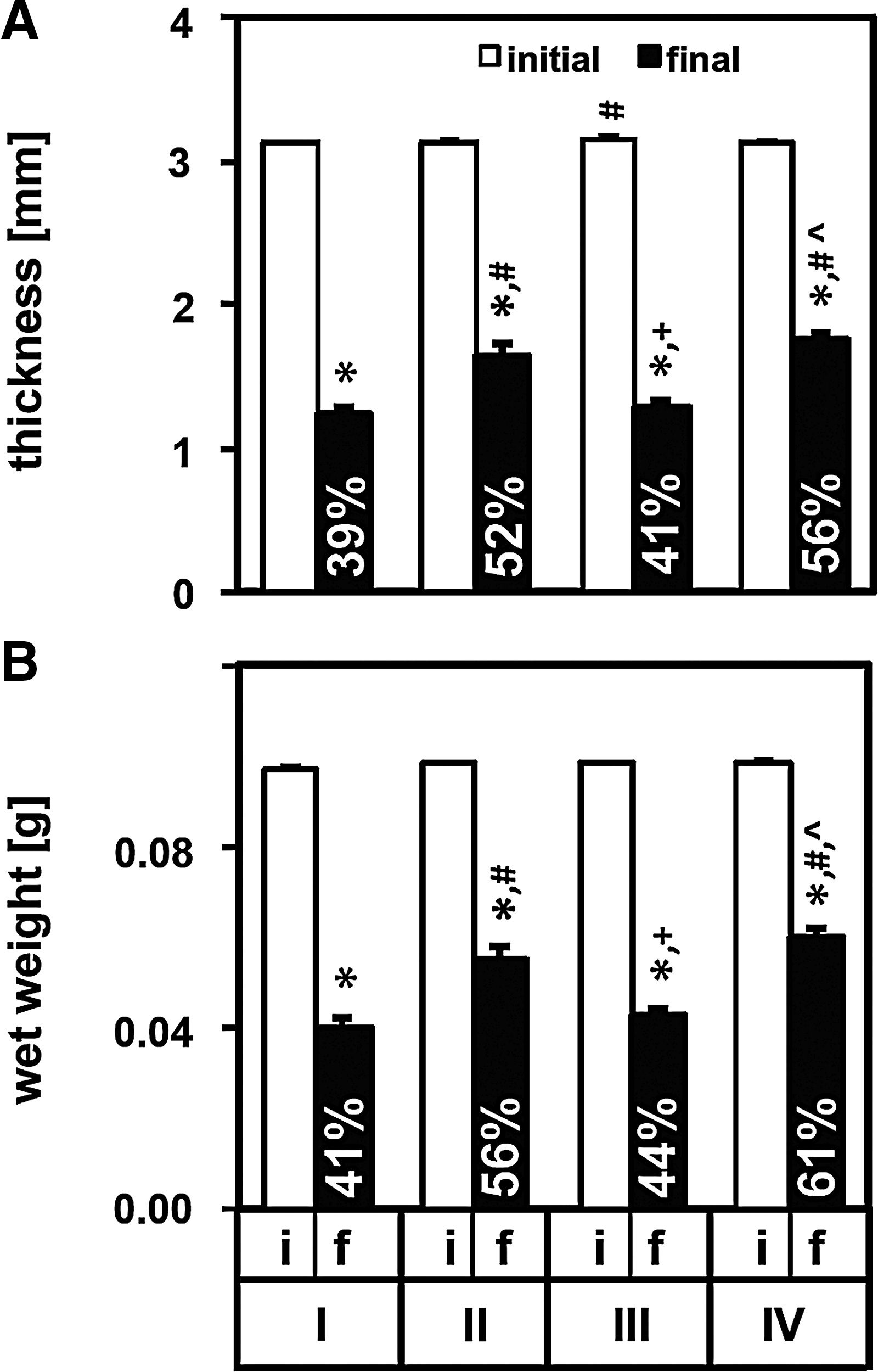
Dimension of constructs. Thickness
Mechanical compaction and initial formulation affected the final PG and COL concentrations of the constructs (Fig. 3). The constructs initially containing PG or COL (Groups II–IV) had similar content before and after the compaction and mechanical testing, maintaining most of their matrix content during the PBS + PIs incubations, mechanical compaction, and mechanical testing. When the matrix contents were normalized to the construct volume before and after compaction, the sGAG concentration increased by ∼1.8–1.9-fold in Group II and IV constructs to ∼4.6–4.8 mg/cm3 (p<0.001), while the COL concentration increased by ∼1.8-fold for Group IV constructs to 15.8 mg/cm3 (p<0.01) and ∼2.5-fold in Group III constructs to 24.6 mg/cm3 (p<0.001) after compaction as compared to before the compaction.

Biochemical content of constructs. The sGAG
Histological assessments for sGAG and type II COL distribution on cryosections of the constructs were consistent with the biochemical analysis (Fig. 4). The compacted constructs stained more intensely for both sGAG and type II COL than the corresponding constructs at day 0. The sGAG and type II COL staining for the compacted constructs were relatively even throughout the section when the constructs contained these ECM components. Sections from the compacted constructs with PG (Groups II and IV) stained strongly blue, indicative of sGAG throughout the section (Fig. 4A–H). Likewise, sections from the compacted constructs containing COL (Groups III and IV) immunostained intensely positive for type II COL (Fig. 4I–P).
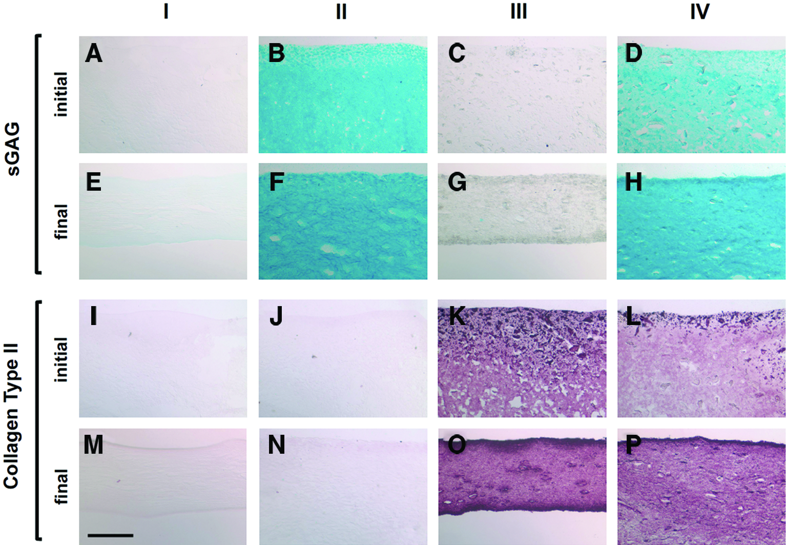
Staining of constructs for sGAG and COL type II. The constructs from Groups I (control)
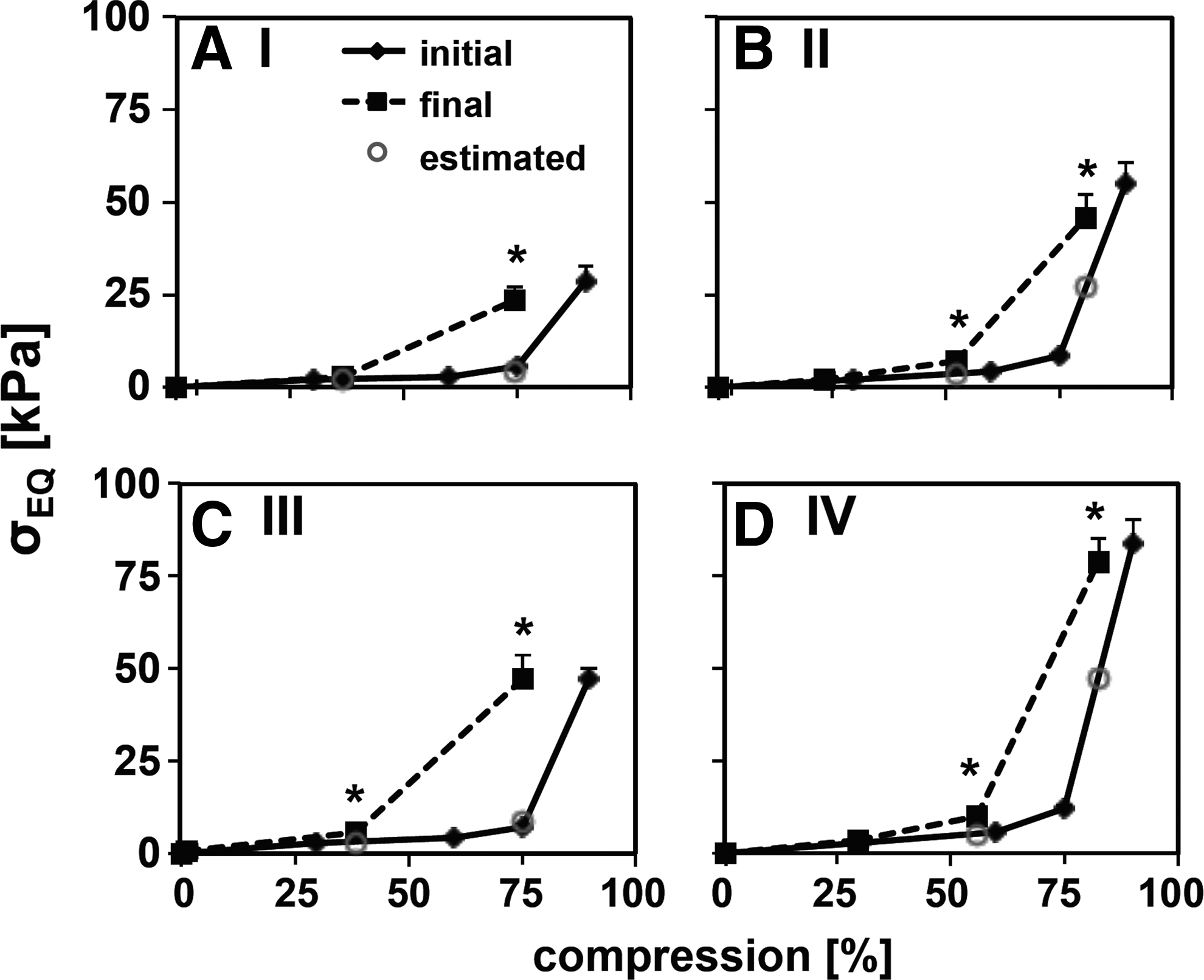
The initial mechanical properties of the constructs during the compaction were dependent on the construct formulation (Fig. 5A–C). σPEAK and σEQ increased with increasing compression levels for all constructs (Fig. 5A, B). The constructs containing PG, COL, or COL+PG (Groups II–IV) had higher σPEAK than the agarose-only constructs (Group I) (overall p<0.001). Also, the constructs with COL+PG (Group IV) had higher σPEAK and σEQ than that of the constructs containing PG or COL alone (Groups II and III) at 60%, 75%, and 90% compression (p<0.01). The σEQ – σEQ,I, the stress ascribed to the ECM components by subtracting the contribution from agarose (Group I), was higher for Group IV constructs than for Group II and III (p<0.01) (Fig. 5C).

Compressive properties of the constructs during and after compaction. Peak
The compacted constructs had similar σPEAK and σEQ values as during the initial compaction at 75% and 90% compression to the initial thickness (p>0.4) (Fig. 5D–F). σPEAK and σEQ at 75% and 90% compression were the lowest for Group I constructs and highest for Group IV constructs (p<0.001 vs. Group I, p<0.05 vs. Group II and III) (Fig. 5D, E). The σEQ – σEQ,I for Group IV constructs was higher than those from Groups II and III (p<0.05; Fig. 5F).
When compression levels were normalized to the thicknesses of the compacted constructs rather than the initial constructs, the computed compressive stiffnesses were higher (Fig. 6). The highest compression level (90% compression relative to initial thickness) was equivalent to 75%–83% compression relative to the thicknesses of the compacted constructs. At these compression levels, the compacted constructs had higher σEQ than the estimated σEQ for the initial constructs by 170%–560% (p<0.05).
Stress–compression relationship of the constructs during and after compaction. Stress–compression curves of constructs during compaction (initial) and after compaction (final) from Groups I
As calculated from the FCDEF–πPG relationship, the predicted PG contribution to σEQ was consistent with σEQ –σEQ,I values obtained experimentally (Fig. 7). At 90% compression of the initial thickness, the estimated πPG contribution to the σEQ were similar to the σEQ – σEQ,I for the initial and final constructs from Groups II (16.1, 26.8, and 22.3 kPa for πPG, initial, and final σEQ – σEQ,I, respectively) (p>0.3) and IV (55.8, 55.5, and 55.1 kPa) (p>0.99). The estimated πPG values at 75% compression of the initial thickness were not as similar (p<0.07) because the FCD values were very small (<0.05 mEq/g water) where the model is less accurate.
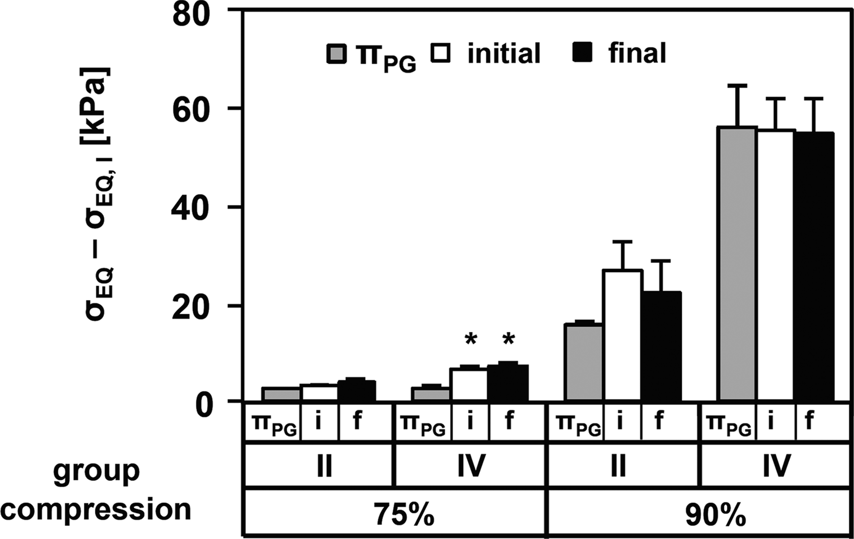
Prediction of PG contribution to compressive properties by πPG. Comparison of predicted πPG and experimentally obtained equilibrium stresses minus the agarose contribution (σEQ – σEQ,I) at 75% and 90% compressions of initial thickness for initial constructs (i) and final compacted constructs (f) from Groups II and IV (*p<0.05 vs. πPG).
In an initial test of cell compatibility, ARC in the constructs remained mostly viable before and after mechanical compaction to 90% of construct thickness (Fig. 8). Formation of constructs with ECM components was compatible with chondrocytes as 97% and 85% of the ARC were viable in uncompacted constructs from Group I and IV, respectively, with similar proportion of viable cells at the edges and middle of the constructs (Fig. 8A, B, E, F and Supplementary Fig. S2 in Supplementary Data). The ARC viability was slightly lower after compaction than in uncompacted constructs but still fairly high. For Group I constructs, 85% and 86% of the cells remained viable at the edges and the middle of the constructs, respectively (Fig. 8C, D and Supplementary Fig. S2). For constructs with COL+PG (Group IV), the cell viability was slightly lower at the edges (62%) than at the middle of the constructs (78%) (Fig. 8G, H and Supplementary Fig. S2).
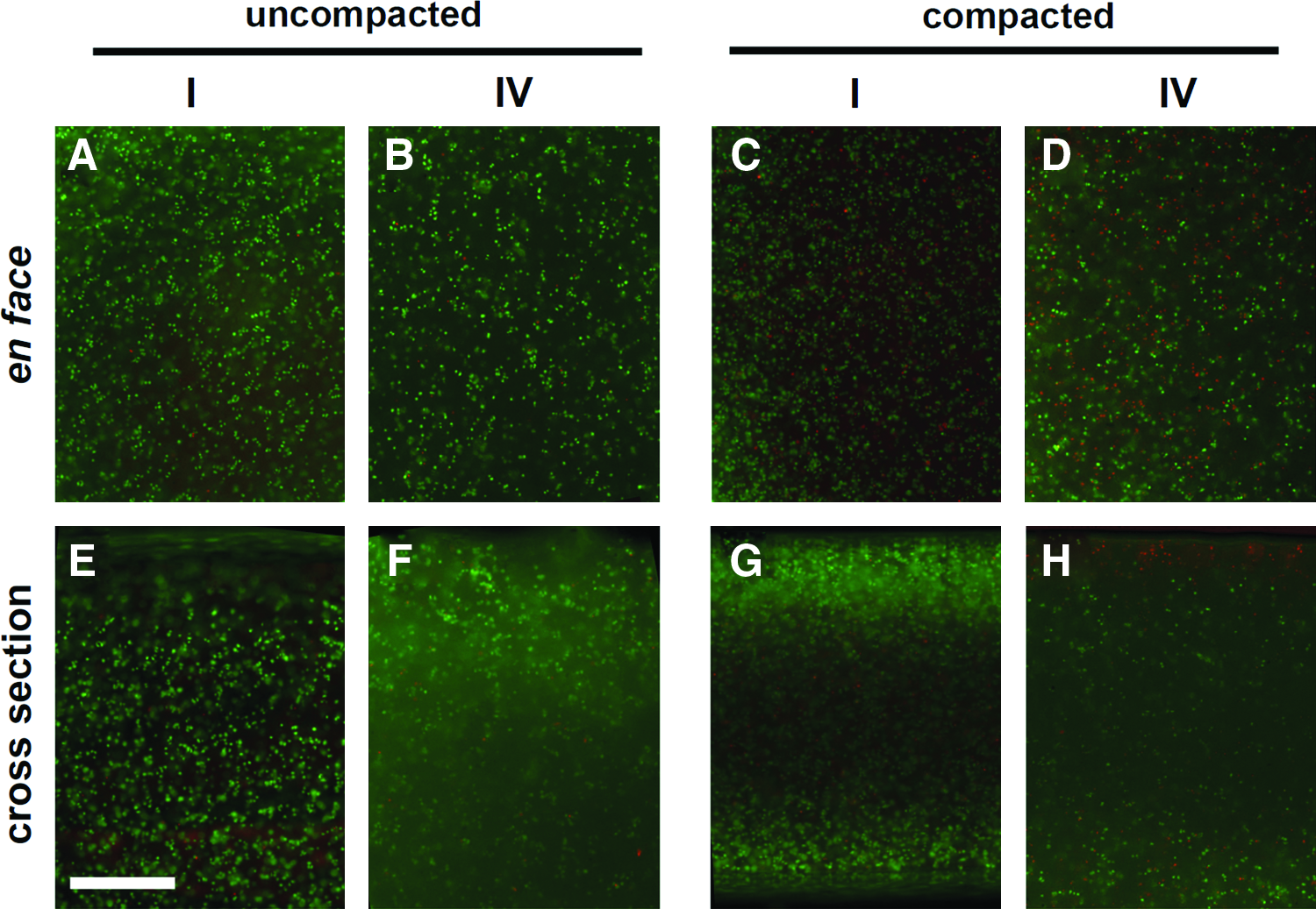
Live/dead staining of chondrocytes in compacted constructs. En face
Discussion
This study introduces a new approach, mechanical compaction, to rapidly increase the ECM concentration and associated mechanical properties of cartilaginous constructs. Mechanical compaction increased the PG and COL concentrations (Fig. 3) and the compressive stiffness (Fig. 6). The presence of COL+PG improved the compressive properties of hydrogel constructs compared with PG or COL alone (Fig. 5), highlighting the contribution of COL content, in combination with PG, to compressive properties of engineered constructs. At 90% compression of the initial thickness, the predicted πPG value from FCDEF from the PG content closely approximated the matrix contribution to the measured σEQ value of the constructs (Fig. 7). ARC remained mostly viable after compaction (Fig. 8), demonstrating the applicability of the method to cell-containing constructs. These results support the general concept that mechanical compaction provides a novel method to rapidly enhance the ECM content and compressive properties of engineered cartilaginous constructs.
The presence of both COL and PG in the appropriate proportions appears to play an important role in the compressive mechanical function of the engineered cartilaginous constructs. Constructs with COL+PG would be expected to have a higher effective FCD and πPG than constructs with only PG due to the space-filling effect of COL fibrils. 27 COL fibrils contain intrafibrillar (IF) water, which is inaccessible to the large PG molecules, effectively increasing the associated charge density of the PG. 31 Thus, the interaction between COL and PG is important to consider in relating the PG content to FCD and compressive properties; in particular, even with similar sGAG concentrations, the presence and level of COL in an engineered construct can alter the amount of PG contribution to the compressive properties.
The retention of most of the initial matrix content in the constructs after compaction and subsequent mechanical testing (see Supplementary Data and Supplementary Fig. S1) suggests that most of the compaction was due to fluid loss. The water content appeared to reach a steady state at the 1 day time point, since thickness and wet weight varied little among compacted constructs. In typical cell-based engineered constructs, the constructs tend to be highly hydrated with over 80% water content, 15 due to the limited restraining forces provided by the low COL content, and result in compressive modulus only in the order of tens of kPa.32,33 Recent efforts to improve construct mechanical properties by selective enzymatic degradation of PG in engineered constructs after weeks of culture have decreased the water content of the engineered tissue along with an increase in COL concentration.15,16 Others include cell-mediated compaction or contraction of the scaffold, which may increase the mechanical strength of the material but typically are associated with more fibroblastic phenotypes.34–36 Other approaches include functionalizing the scaffold or composing the scaffold wholly with the desired matrix proteins,12,37,38 which increase the initial matrix concentration of the construct. In the present study, an 80% increase in COL concentration was achieved rapidly in a few days by a matrix assembly and mechanical dehydration method.
During mechanical compaction, the gel network of the agarose constructs may have undergone restructuring since the constructs did not recover to their initial thicknesses. Initial thickness can be controlled well due to low πPG with low GAG concentration, 4 which could be balanced by the restraining force of agarose, consistent with our previous study. 13 The swelling of the PG-containing constructs (Groups II and IV) beyond the thickness of agarose only or COL-containing constructs (Groups I and III) after compaction indicates the presence of swelling pressure exerted by PG that was restrained with the increased thickness in the agarose gel network. A mechanism for providing higher restraining force with a smaller increase in construct thickness after compaction may be useful in further retention of the compacted state of the constructs and to further increase the matrix concentrations.
Compaction of the construct altered its geometry (i.e., thickness) and has the potential to be coupled with the application of more complex three-dimensional shape to the constructs (Fig. 9). The surface congruity of an implanted graft to the surrounding native cartilage has been shown to be important for the viability of such constructs 39 and likely for tissue-engineered constructs as well. There have been recent efforts to shape cartilaginous constructs using molding techniques.40–42 Using molds with anatomical contours, the compaction method has the potential to mechanically impose a shape to the constructs or further maintain and improve the shaping fidelity of already-shaped constructs, which may facilitate fabrication of grafts for large cartilage defects with complex surface contours.
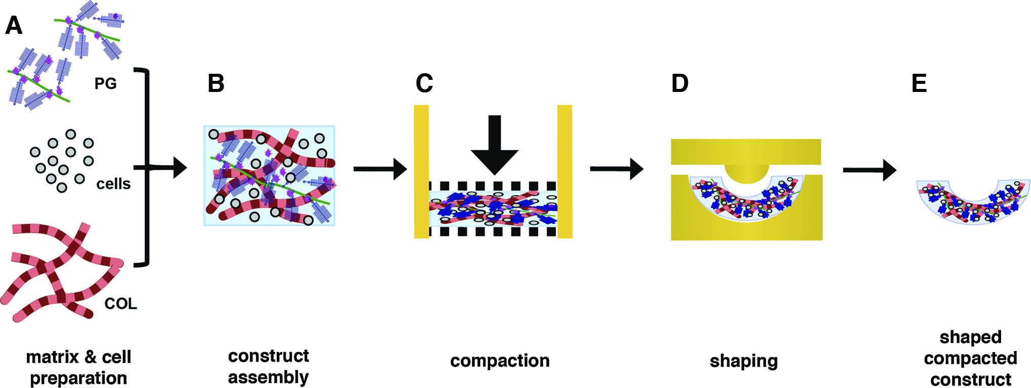
Schematic of application of matrix and cell preparation
Maintenance of high cell viability in compacted constructs in an initial test of cell compatibility demonstrates that the compaction method can be extended to cell-containing constructs. The cell-associated matrix on the ARC may have acted as a protective layer against high stresses and strains during compaction.43,44 Such compaction was likely to be lower in the middle than at the edges of the constructs, due to fluid exuding initially from the construct edges. Based on the measured thickness of compacted constructs without ARC, the final cell density is ∼3–4×10⁶ cells/mL, which is ∼1/4–1/2 of the cell density in native human adult cartilage. 7 As the present study involved short-term culture of constructs containing immature chondrocytes, substantial studies in vitro with longer-term culture and use of different cell types and densities, as well as in vivo studies, are needed to further elucidate the effect of compaction and addition of native ECM components in tissue-engineered constructs on cell viability, water content, and matrix deposition by the indwelling cells. With longer-term studies, cell survival and fates (viability, proliferation, differentiation, and migration) may become more challenging to assess.45–47 Additionally, subsequent dynamic loading and/or perfusion may counter diffusive limitations that may exist in compacted constructs. Nevertheless, the present studies establish short-term cell viability after mechanical compaction.
The compaction of cartilaginous constructs with the appropriate COL:sGAG ratio at a fraction of the targeted concentration may allow for rapidly achieving a more physiological matrix concentration. Working with lower concentrations of COL and PG allows for ECM manipulations at more manageable concentrations, as solutions with high PG concentrations are viscous and difficult to handle. The typical COL:sGAG ratios found in engineered constructs are 1:1 or less, which is far below those found in native tissue of 2–4:1 in bovine fetal and adult cartilage 6 and 3–10:1 in human adult cartilage7,48 (Table 1). Thus, preassembly of an engineered construct with a more physiological COL:sGAG ratio and compacting to increased matrix concentrations may lead to rapid fabrication of a more mechanically functional engineered construct for treatment of articular cartilage defects.
The compaction method described here has the potential to significantly reduce the time needed to fabricate mechanically functional cartilaginous constructs. Typical tissue engineering methods require weeks, if not months, of culture for the resident cells to produce enough ECM. Such constructs may not initially contain the appropriate balance of COL to PG or enough matrix molecules, the presence of which are known to foster chondrogenic phenotypes and cell compatibility.49–51 While compacted constructs presented here have lower matrix concentration than native cartilage, ∼1/2–1/4 compared to bovine fetal cartilage and ∼1/4–1/5 of bovine adult cartilage, the compaction method may be modified to include higher initial matrix concentrations and/or be repeated with longer duration to increase matrix concentrations toward physiological levels.
The methods presented here may provide a new assembly paradigm for cartilage tissue engineering (Fig. 9). The matrix macromolecules first can be preassembled into PG aggregates and fibrillar COL (Fig. 9A), and then mixed into an appropriate COL:sGAG ratio to form a hyper-hydrated construct (Fig. 9B). Such constructs can undergo mechanical dehydration by compaction to squeeze out excess fluid (Fig. 9C) and be shaped into an appropriate physiological contour, in sequence or simultaneously (Fig. 9D). This assembly approach may facilitate a more rapid engineering of mechanically functional cartilaginous constructs that are ready for implantation into a cartilage defect to re-establish joint function.
Footnotes
Acknowledgments
This work was supported by National Institutes of Health, National Science Foundation, and a grant to University of California–San Diego, in support of Prof. Robert Sah, from the Howard Hughes Medical Institute through the HHMI Professors Program. Additional support was received from an NSF Graduate Research Fellowship (EHH). The authors thank Mr. Johnny Du for careful reading of the article.
Disclosure Statement
No competing financial interests exist.
References
Supplementary Material
Please find the following supplemental material available below.
For Open Access articles published under a Creative Commons License, all supplemental material carries the same license as the article it is associated with.
For non-Open Access articles published, all supplemental material carries a non-exclusive license, and permission requests for re-use of supplemental material or any part of supplemental material shall be sent directly to the copyright owner as specified in the copyright notice associated with the article.