
Abstract
While silk-based microfibrous scaffolds possess excellent mechanical properties and have been used for ligament tissue-engineering applications, the microenvironment in these scaffolds is not biomimetic. We hypothesized that coating a hybrid silk scaffold with an extracellular matrix (ECM)-like network of self-assembling peptide nanofibers would provide a biomimetic three-dimensional nanofibrous microenvironment and enhance ligament tissue regeneration after bone marrow-derived mesenchymal stem cell (BMSC)-seeding. A novel scaffold possessing a triple structural hierarchy comprising macrofibrous knitted silk fibers, a silk microsponge, and a peptide nanofiber mesh was developed by coating self-assembled RADA16 peptide nanofibers on a silk microfiber-reinforced-sponge scaffold. Compared with the uncoated control, RADA-coated scaffolds showed enhanced BMSC proliferation, metabolism, and fibroblastic differentiation during the 3 weeks of culture. BMSC-seeded RADA-coated scaffolds showed an increasing temporal expression of key fibroblastic ECM proteins (collagen type I and III, tenascin-C), with a significantly higher tenascin-C expression compared with the controls. BMSC-seeded RADA-coated scaffolds also showed a temporal increase in total collagen and glycosaminoglycan production (the amount produced being higher than in control scaffolds) during 3 weeks of culture, and possessed 7% higher maximum tensile load compared with the BMSC-seeded control scaffolds. The results indicate that the BMSC-seeded RADA-coated hybrid silk scaffold system has the potential for use in ligament tissue-engineering applications.
Introduction
Among the various biomaterials that have been used for ligament tissue engineering, silk, a natural fibrous protein derived from Bombyx mori silk worms, possesses remarkable mechanical properties and a slow degradation rate that is suitable to support the healing ligament/tendon over a period of 6–12 months.3,8 Its properties of biocompatibility, morphologic flexibility, environmental stability, and the ability for functionalization via amino-acid side-change modification to immobilize functional groups make it useful as a scaffolding biomaterial.8–10 Silk-based scaffolds in the form of braided microfibers and porous sponges have already been investigated for ligament regeneration.4,11,12 However, microfibrous scaffolds often allow only limited cell attachment and result in nonhomogeneous cell distribution within the scaffold. 13 Moreover, silk microfibers (diameter: 10–25 μm) and cells (diameter: 5–30 μm) are of similar dimensions; therefore, seeded cells essentially encounter a two-dimensional environment within the scaffolds, which fail to biomimic the three-dimensional (3D) environment presented by the collagen nanofibrils in the natural ECM. Collagen type I fibrils have diameters ranging between 50 and 500 nm, and nanofibrous substrates with similar dimensions are known to facilitate cell attachment, proliferation, and ECM deposition.
Self-assembling peptides have been recently used to fabricate nanofibrous scaffolds in tissue-engineering research. These self-assembled nanofibrous scaffolds have >99% water content and possess excellent biocompatibility. RADA16 (Ac-RADARADARADARADA-CONH2), the most common self-assembling peptide used, forms β-sheet structures that are stable across a wide range of temperature and pH. The generated nanofibers biomimic the natural ECM and enhance the attachment, growth, and differentiation of a variety of cells.14–16 Scaffolds made of self-assembled peptide nanofibers have also been used for delivering growth factors and cells into scaffolds. 17 However, such nanofibers alone lack desirable mechanical properties and often require to be combined with other macrofibrous scaffolds to generate hybrid scaffolds that could be used to engineer mechanically strong tissues such as tendons and ligaments, as demonstrated in previous studies by the authors' group. 18 Hybrid silk-based scaffolds have also been developed by combining knitted macrofibrous silk scaffolds with a microporous silk sponge; the in vitro and in vivo efficacy of these hybrid scaffolds in ligament tissue engineering has been demonstrated.4,11,12 The pore size of the sponge structure was still too large (100 μm) to create a real 3D environment for cell attachment and proliferation. However, a combination of such silk-based hybrid scaffolds with self-assembled peptide nanofibers could help in creating a scaffold system possessing a triple hierarchy of a macrofibrous knitted structure, a submicron-sized silk sponge, and nanofibrous peptide fibers, mimicking the hierarchical structure of collagen in ECM. We hypothesize that this scaffold system would enhance cell proliferation and ligament tissue regeneration from primary bone marrow-derived mesenchymal stem cells (BMSCs). In this study, we fabricate and characterize the silk-peptide hybrid scaffold system and assess the in vitro attachment, proliferation, and differentiation of seeded BMSCs into a ligament phenotype.
Materials and Methods
Study design
Scaffolds were fabricated through four stages (Fig. 1): knitted silk scaffolds (Scaffold I) were coated with silk sponge (Scaffold II: sponge-coated knitted silk scaffolds). Next, the scaffolds were primed with RADA-peptide (Scaffold III: RADA-primed silk scaffold) and then coated with RADA (Scaffold IV: RADA-coated silk scaffold). The cells suspended in a concentrated (0.5%) RADA solution were applied to Scaffold III to fabricate Scaffold IV that was seeded with cells in situ within a stable nanofibrillar RADA network. The cell-seeded scaffolds were cultured for 3 weeks and characterized morphologically, evaluated for cell metabolism and proliferation, ligament-specific ECM gene expression, total collagen and glycosaminoglycan (GAG) production, and biomechanical properties.
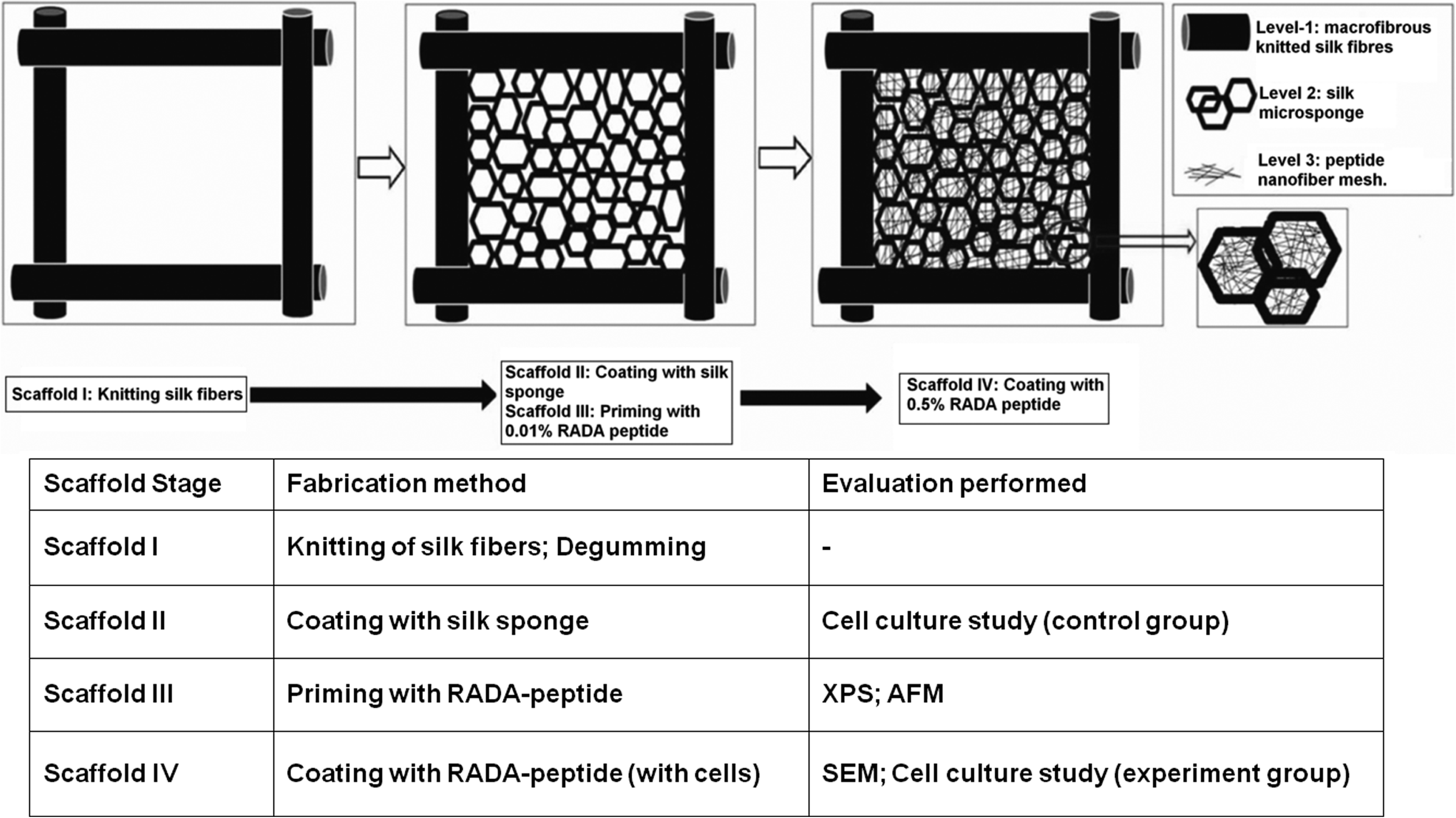
Schematic outlining the fabrication process of the hybrid silk/RADA-based fibrous scaffold with a triple hierarchical structure. XPS, X-ray photoelectron spectroscopy; AFM, atomic force microscopy; SEM, scanning electron microscope.
Scaffold fabrication and characterization
The knitted silk scaffolds were fabricated from B. mori silk using previously described methods. 19 Briefly, knitted scaffolds of 50 mm×50 mm dimension were fabricated using raw silk fibers on a knitting machine (Silver-reed SK270; Suzhou). Scaffold I was prepared by degumming the knitted scaffolds in a 0.1% (w/v) Na2CO3 and 0.1% (w/v) sodium dodecyl sulfate solution at 95°C–100°C for more than an hour to remove the sericin. 13 Scaffold II was created by reinforcing Scaffold I with a silk sponge to fill the pores of the knitted structure.4,11,19 This was performed by immersing Scaffold I in a 2% (w/v) silk solution, freezing at −20°C for 12 h, and freeze drying for 36 h. The freeze-dried scaffolds were treated with a 90/10 (v/v) methanol/water solution for 10 min, air dried for 24 h, and then cut into 10 mm×10 mm pieces for subsequent use.
Scaffold II was primed with RADA peptide using carbodiimide chemical crosslinking. Briefly, Scaffold II pieces were soaked in isotonic phosphate-buffered saline (PBS) for 30–45 min to hydrate the surface and to induce the surface rearrangement to bring forth hydrophilic functional groups. Carboxyl groups from aspartic and glutamic acids were activated by a reaction with 1-ethyl-3(dimethylaminopropyl)carbodiimide hydrochloride (EDC)/N-hydroxysuccinimide (NHS) solution (0.5 mg/mL of EDC with 0.7 mg/mL NHS in PBS buffer) for 15 min at room temperature. After treatment with EDC/NHS, the scaffolds were rinsed with distilled water and immersed in 0.01% (w/v) RADA peptide (Ac-RADARADARADARADA-CONH2; Sigma) solution for 2 h at room temperature. After rinsing in distilled water for 5 min, the RADA-primed scaffolds (Scaffold III) were freeze dried overnight and characterized by X-ray photoelectron spectroscopy (Kratos Analytical XPS; AXIS HSI) and multimodal atomic force microscopy (AFM, Nanoscope IV controller; Veeco Instrument) for morphological and elemental composition analysis of the scaffold surface.
Scaffold III pieces were soaked in 60 μL of 0.5% (w/v) RADA peptide solution for 10 min, gently transferred into fresh PBS, and rinsed with distilled water after 30 min to fabricate Scaffold IV. The surface morphology of Scaffold IV was evaluated by scanning electron microscopy (SEM; JEOL JSM-5600LV and JSM-6701), after formaldehyde fixation, graded dehydration in ethanol, critical point drying, and sputter coating with gold.
Cell culture and seeding
Bone marrow-derived mesenchymal stromal cells (BMSCs) were obtained from New Zealand White rabbits using a previously described protocol approved by the NUS Institutional Animal Care and Use Committee, the National University of Singapore. Scaffolds of 10 mm×10 mm (for biological assessment) and 20 mm×30 mm (for mechanical testing) dimensions, sterilized by exposure to formaldehyde gas, were seeded with P2 BMSCs at a density of 5×10⁵ cells/cm2 of scaffold area, and cultured in the wells of non-treated multi-well culture plates (NUNC). BMSCs were suspended in 0.5% (w/v) RADA peptide in 295 mM sucrose solution and transferred onto the scaffolds using the gravitational seeding technique. 19 After 10 min, the cell-seeded scaffolds were transferred into fresh PBS for 2 h to allow the self-assembly of the peptides. As a control, Scaffold II (hybrid scaffolds without RADA peptide) was seeded with the same number of cells. The cells were allowed to attach on the scaffold for 2 h, after which the cell-seeded scaffolds were cultured for 3 weeks in high glucose Dulbecco's Modified Eagle Medium (DMEM) (DMEM-HG) with 10% fetal bovine serum (FBS) and antibiotics, with the culture medium being changed twice a week.
Cell proliferation and metabolism
Cell proliferation was assessed by quantifying DNA content using PicoGreen assay (Quant-iT; Invitrogen) after 7, 14, and 21 days of culture (n=3, at each time point). 6 Briefly, the samples were washed with PBS, freeze dried, treated with lysis buffer, and homogenized. The supernatant obtained after centrifuging the homogenate was mixed with PicoGreen dye, and fluorescence intensity was measured at 520 nm wavelength using a fluorescence microplate reader after an excitation at 485 nm.
Cell metabolism was measured after 7, 14, and 21 days of culture using the AlamarBlue metabolic assay (BioSource International). Briefly, BMSCs-seeded scaffolds (n=4/group) were incubated in DMEM-HG supplemented with 5% FBS and 10% (v/v) AlamarBlue dye for 3 h. The absorbance of the culture media at 570/600 nm was measured in triplicate using a 96-well plate microplate reader. Using a culture medium supplemented with 10% AlamarBlue dye as a reagent blank, the percentage of AlamarBlue reduction, which is proportional to the cell viability in the sample, was calculated according to the formula provided by the vendor.
Morphological characterization of cell-seeded scaffolds
To visualize the presence and distribution of live cells, cell-seeded scaffolds after 10 and 20 days of culture were stained with fluorescein diacetate (FDA; Molecular Probes, Invitrogen Corporation). The scaffolds were incubated in 6 μg/mL FDA/PBS solution at room temperature for 3 min, rinsed twice with fresh PBS, and the green-stained live cells were visualized immediately using an inverted fluorescence microscope (IX71 Inverted Research Microscope; Olympus). Cellular morphology in the cell-seeded scaffolds was also characterized using SEM. The samples were harvested after 10 and 20 days of culture, formaldehyde fixed, freeze dried for 24 h, sputter coated with gold, and observed by SEM. The histology of the cell-seeded scaffolds after 21 days of culture was also studied by hematoxylin/eosin (H/E) staining and light microscopy.
Total RNA extraction, cDNA synthesis, and real-time reverse transcriptase-mediated polymerase chain reaction analysis
The gene expression for three ligament-specific ECM proteins (type I and III collagens, tenascin-C) was analyzed to evaluate the fibroblastic differentiation of the seeded BMSCs. 20 After 10 and 20 days of culture, total RNA was extracted from the cell-seeded scaffolds (n=3/group) using an RNeasy Mini Kit (Qiagen). Quantitative/reverse transcriptase-mediated PCR (Q-RT-PCR) was performed using SYBR-Green chemistry for type I and III collagens, and tenascin-C, with glyceraldehyde 3-phosphate dehydrogenase (GAPDH) as the housekeeping gene. The primer sequences (Table 1) were obtained from published literature.6,20,21 cDNA synthesis and PCR expansion (using iScript and iQ SYBR Green Supermix; Bio-Rad Laboratories) were performed in an iCycler iQ detection system (Bio-Rad Laboratories). The amplification was performed in triplicate, data were analyzed for relative expression using the ΔΔCt method, and the results were normalized to the gene expression levels in Scaffold II on day 10.
GAPDH, glyceraldehyde 3-phosphate dehydrogenase.
Total collagen and soluble GAGs assays
On the 7th, 14th, and 21st day of culture, the total soluble collagen synthesized and secreted into the culture medium was determined by the SirCol Assay (Biocolor) using previously described methods. 5 The culture medium was replaced by fresh DMEM with 5% FBS, 2 days before the day of the assay, to ensure that only freshly synthesized soluble collagen was assayed. The absorbance of PicroSirius red-stained collagen was read at 540 nm (TECAN Microplate Reader, Magellan Instrument; Control and Data Analysis Software) to obtain the concentration of collagen in the medium, which was then multiplied with the total volume of the medium collected from the respective scaffolds (n=4) to provide an estimate of the total amount of collagen secreted per scaffold over 2 days. DMEM-HG with 5% FBS was used as a “reagent blank” during the assay.
The total insoluble collagen deposited on the scaffolds was tested at the end of the study duration (day 21). Scaffolds (n=3/group) were rinsed in PBS and digested in 500 μL of 0.25 mg/mL pepsin solution with 0.5% Triton ×100 and 0.25 M HCl at 37°C for 2 h. The digested solutions were neutralized by NaOH, and the collagen levels were determined by SirCol Assay as just mentioned.
The total soluble sulfated GAGs synthesized and secreted into the culture medium were determined by the Blyscan Assay (Biocolor) following the assay manufacturer's protocol. The culture medium was harvested on days 7, 14, and 21. The absorbance measured at 656 nm, with 550 nm as the reference wavelength, was matched with a standard calibration curve to determine the GAG content in the media (n=5).
Biomechanical testing
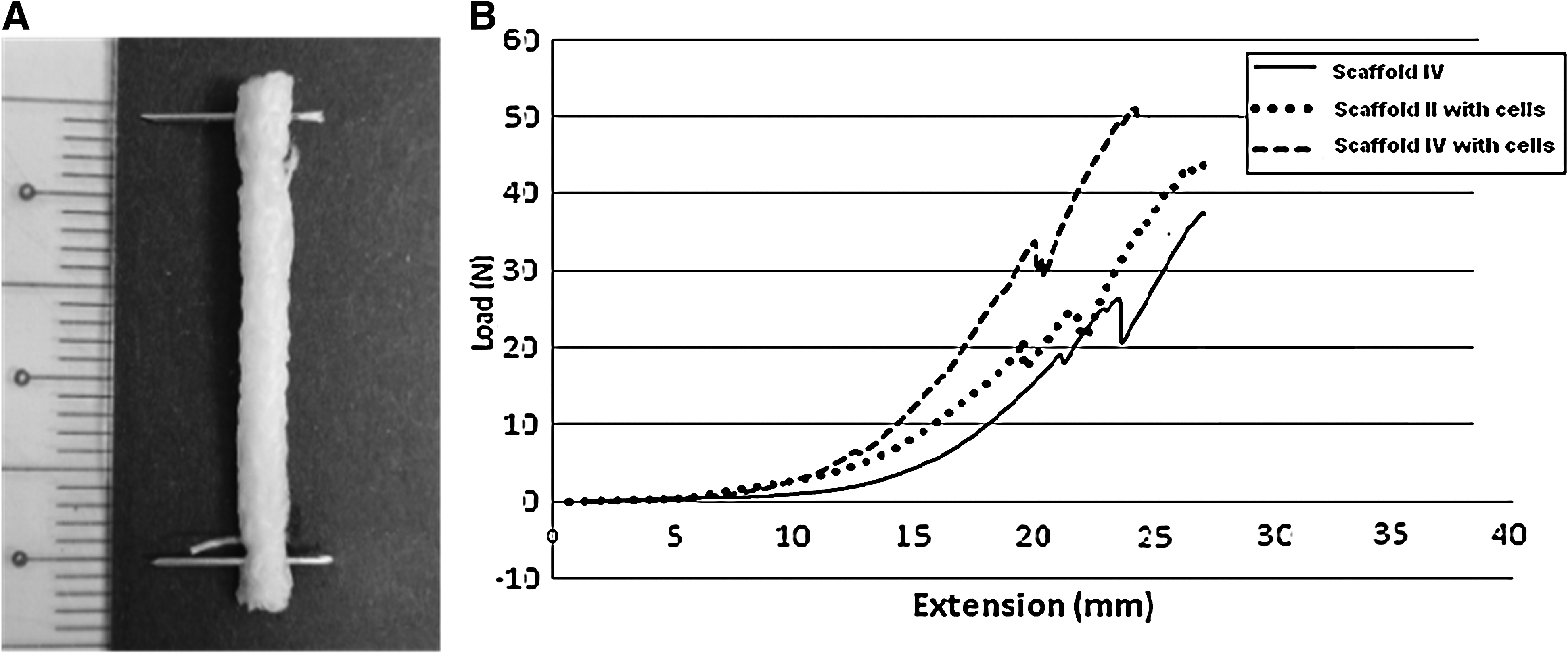
Biomechanical testing was performed on the cell-seeded scaffolds (n=4/group) after 21 days of culture, using a universal testing machine (Instron 3345 Tester; Instron). The unseeded Scaffold IV that had been similarly incubated in culture media for 3 weeks was also tested as an additional control. The scaffolds (30 mm×20 mm dimension) were rolled up along their long axis to obtain cylindrical specimens (average test length of 25 mm and diameter of 4 mm; Fig. 9) and stretched to failure, without any pretension or preconditioning, at a crosshead speed of 10 mm/min, while keeping them moist by spraying 1× PBS. The failure load and linear stiffness were determined from the load-displacement curves, and the scaffold's biomechanical properties were compared against those of mature ligament tissue (rabbit ACL; positive control). 12
Mechanical testing of
Statistical analysis
All data were expressed as means±standard deviation. Multiple comparisons were performed using one-way analysis of variance and posthoc Tukey tests; pairwise comparisons (gene expression data) were performed using two-tailed Student t-tests (SPSS 13.0 software package). p<0.05 was accepted as statistically significant.
Results
Scaffold fabrication and characterization
SEM imaging of Scaffold II showed a web-like microporous silk sponge, possessing interconnected pores of 50–200 μm diameter, coated on the knitted silk structure (Fig. 2A). XPS analysis showed a change in the elemental composition on the silk scaffold surface after priming with the RADA peptide (Table 2). The elemental composition (in atomic conc.%) of carbon decreased from 62.2% to 60.4%, nitrogen increased from 16.6% to 17.9%, and oxygen increased from 21.3% to 21.7%, after RADA priming. AFM microscopy indicated a change in the surface roughness of the scaffolds (Fig. 2B). While Scaffold I had a mean surface roughness (Ra) of 3.8±0.9 nm, after RADA coating, Scaffold III had a significantly higher mean roughness of 4.8±0.76 nm (p<0.05) (Fig. 2B). SEM imaging of Scaffold IV showed that the scaffold was coated with a nanofibrous mesh of the RADA peptide, with structural connections between peptide nanofibers and the silk sponge/fibres (Fig. 2C). The morphology of Scaffold IV, thus, comprised of three levels of structures: macrofibrous knitted silk fibers, a microporous sponge, and a nanofibrous peptide mesh (the diameter of the nanofibers was about 10–20 nm).

Cell proliferation and metabolism
PicoGreen assays showed that cell numbers increased significantly (3.5±0.24 μg/scaffold, 3.9±0.87 μg/scaffold, and 5.8±0.27 μg/scaffold on day 7, 14, and 21) in Scaffold IV during 3 weeks of culture; the increase was not significant in the control Scaffold II (2.8±0.51 μg/scaffold, 3.5±0.96 μg/scaffold, and 4.7±0.52 μg/scaffold at day 7, 14, and 21). At all time points, cell numbers were significantly higher in Scaffold IV than in Scaffold II (Fig. 3A).
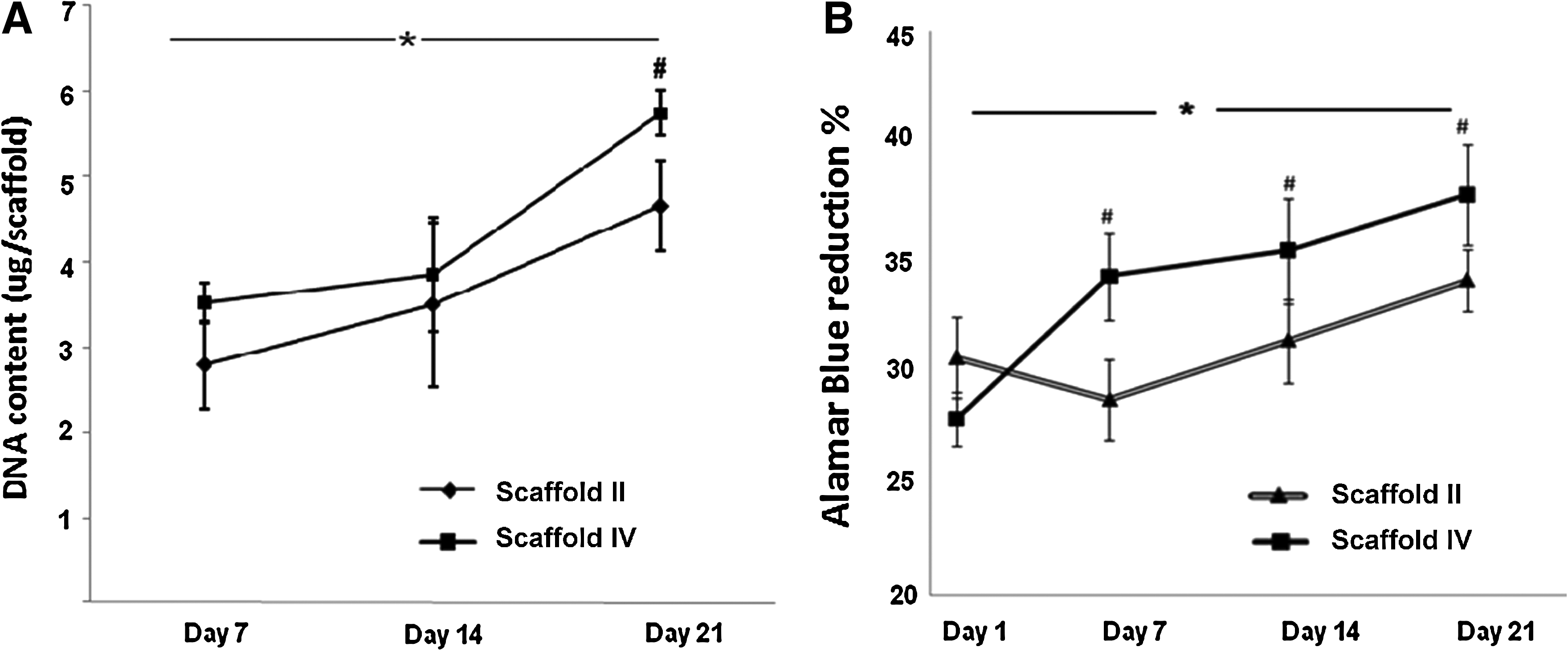
AlamarBlue assays showed that cell metabolism/viability increased significantly in both scaffold groups during 3 weeks of culture. (Posthoc tests show that the increases were significant between each time point in Scaffold IV, whereas in Scaffold II, the increases were significant only between days 1 and 7 and day 21.) While there was no difference between the two groups on day 1, at all later time points, cell metabolism/viability was significantly higher in Scaffold IV than in Scaffold II (paired t-test, p<0.05) (Fig. 3B).
Morphological characterization of cell-seeded scaffolds
Fluorescence microscopy after FDA staining showed higher cell density and more abundant cell proliferation in Scaffold IV compared with Scaffold II on days 10 and 20 (Fig. 4). SEM imaging corroborated the fluorescence microscopy evidence, showing better growth and proliferation in Scaffold IV compared with Scaffold II (Fig. 5). BMSCs proliferated to form sheet-like structures by day 10, and the sheets were confluent, covering most of the Scaffold IV surface by day 20 (Fig. 5B).

Fluorescence photomicrographs of fluorescein diacetate-stained BMSCs showing better cell proliferation in Scaffold IV
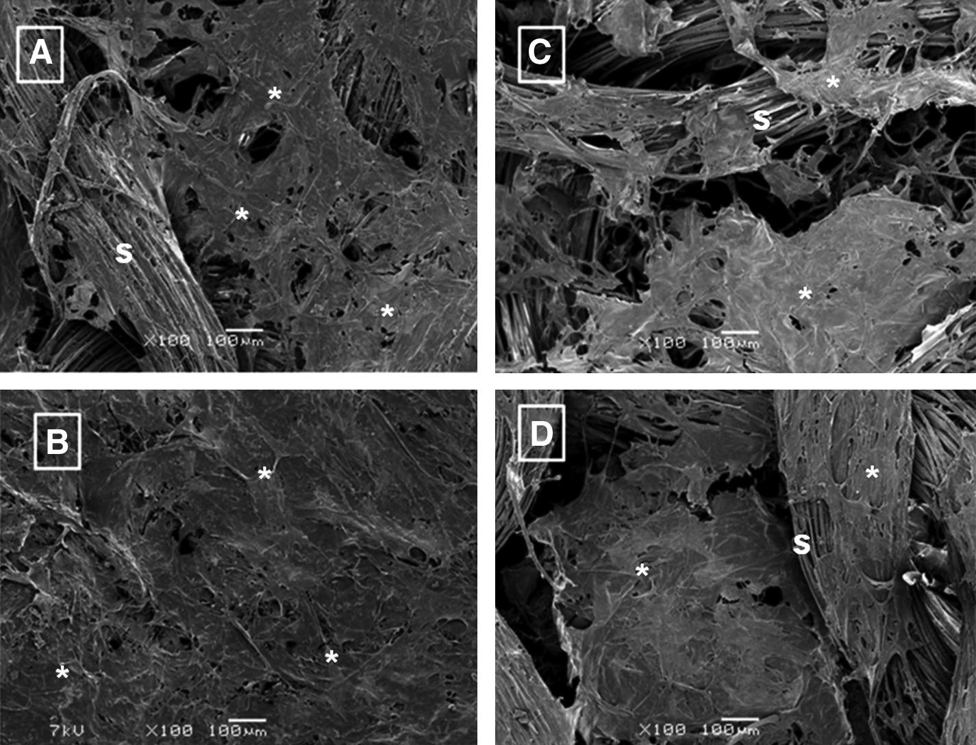
SEM photomicrographs showing better BMSC adherence and proliferation in Scaffold IV
H/E staining and histology revealed that BMSCs had a similar fibroblastic morphology in both the scaffolds. The cells attained a denser distribution, comprising thicker cell-sheet-like structures in Scaffold IV compared with Scaffold II, after 21 days of culture (Fig. 6).
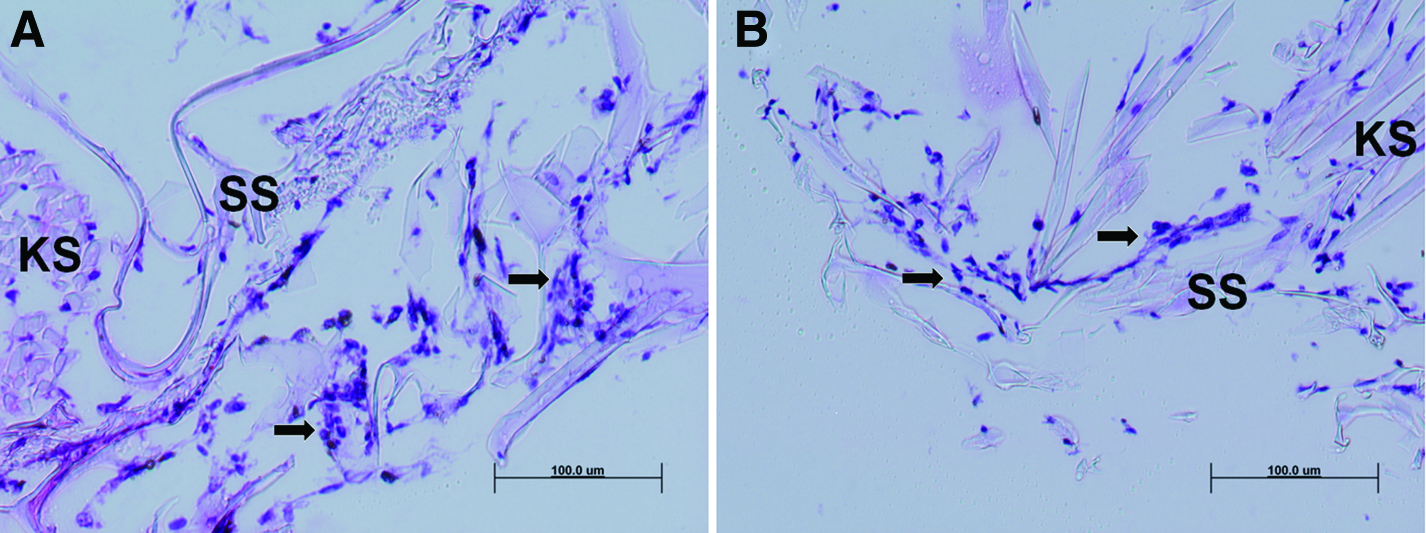
Histology after hematoxylin/eosin staining showing a higher BMSC density (indicated by arrows) in Scaffold IV
Real-time RT-PCR analysis of ligament-related genes
Real-time RT-PCR showed that the gene expression of type I collagen, type III collagen, and tenascin-C increased significantly between days 10 and 20 of culture. While collagen I expression was significantly higher in Scaffold II compared with Scaffold IV on day 10, the expression increased to similar levels in both groups by day 20. There were no significant differences in collagen III gene expression between the two groups at both the time points. Tenascin-C gene expression was significantly higher in Scaffold IV compared with Scaffold II at both the time points (Fig. 7).
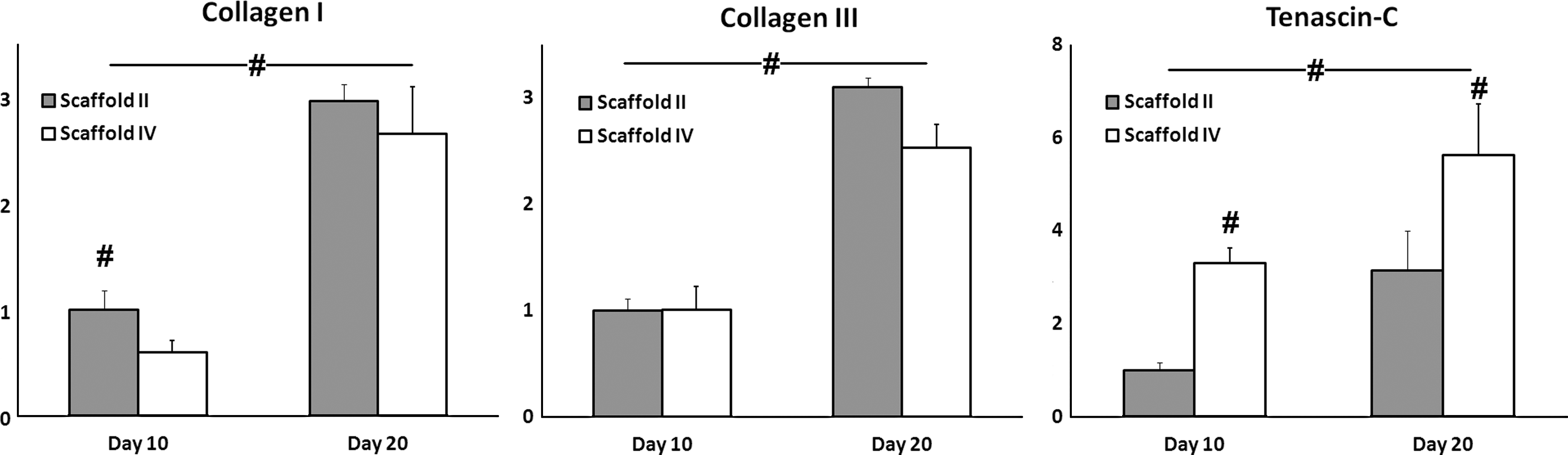
Normalized expression levels of ligament-related ECM genes (type I collagen, type III collagen, and tenascin-C) in BMSC-seeded Scaffolds II and IV after 10 and 20 days of culture (#p<0.05). Expression levels of the three genes increased significantly between days 10 and 20 of culture, with similar expression levels of type I and type III collagen in the two scaffold groups on day 20, while tenascin-C expression was significantly higher in Scaffold IV compared with Scaffold II at both the time points.
Total collagen and soluble GAG assays
Total soluble collagen produced by both groups of BMSC-seeded scaffolds increased significantly during 3 weeks of culture (p<0.05, Fig. 8A). (The increases were significant between day 7–14 and day 7–21 in Scaffold II, and between day 7 and 21 in Scaffold IV.) At all time points, BMSC-seeded Scaffold IV produced significantly more collagen (7.1-, 2.2-, and 2.2-fold higher at day 7, 14, and 21, respectively) than BMSC-seeded Scaffold II controls. After 21 days of culture, the total insoluble collagen deposited in Scaffold IV was also significantly higher than that in Scaffold II (p<0.05, Fig. 8B).

The total soluble GAG produced by both groups of BMSC-seeded scaffolds also increased significantly during 3 weeks of culture (p<0.05, Fig. 8C). (The increases were significant between day 7–21 in Scaffold II, and between day 7–14 and day 7–21 in Scaffold IV.) BMSC-seeded Scaffold IV produced more GAG (3.7, 1.8, and 1.6-fold higher at day 7, 14, and 21, respectively) than BMSC-seeded Scaffold II controls, the difference being significant on days 7 and 21.
Biomechanical properties
After 21 days of culture, BMSC-seeded Scaffold IV had a maximum tensile load of 37.9±9.2N, which was marginally higher than the maximum tensile load of BMSC-seeded Scaffold II (35.5±2.5N) and unseeded Scaffold IV (31.3±4.2N). The differences between the three groups were not statistically significant. There were no significant differences in the toe stiffness (0.2–0.3 N/cm), linear stiffness (4.3–4.9 N/cm), and elongation at maximum load (17.3–20.6 mm) between the different scaffold groups.
Discussion
The choice of appropriate biomaterials and scaffold design is crucial for success in tissue-engineering strategies. Scaffolds for engineering load-bearing tissues such as bone, cartilage, tendon, or ligament should not only be adequately strong, but also be biocompatible to permit seeded cells to lay down new tissue. Ligaments derive their characteristic mechanical properties from the triple hierarchical structure of type collagen I fibrils in their ECM. 22 This motivated us to create a scaffold that possessed a hierarchical structure, paralleling the collagen hierarchy in natural ECM. The different structural components in this scaffold system achieve distinct functions: The microfibrous knitted silk structure likely provides mechanical properties that are necessary for ligament tissue regeneration, the microporous silk sponge covers the pores of the knitted scaffold and increases the scaffold surface area, and the self-assembling peptide nanofibers coat the silk surface to further increase the surface area and to provide a biocompatible and biomimetic nanofibrillar substrate for the seeded cells.
Silk, due to its excellent mechanical properties, biocompatibility and slow degradation profile, is a promising candidate as a scaffolding biomaterial for engineering musculoskeletal tissues.3,8,9,18,23,24 Surface modification and coating strategies have been applied to tailor silk scaffolds that influence cellular response and enhance the formation of specific tissue types. Silk scaffolds have been coated with natural ECM proteins such as collagen and GAGs to create a biomimetic interface that improves cell adhesion, proliferation, and new tissue formation. 25 Earlier studies from our group have shown that silk sponge-coated knitted silk scaffolds (Scaffold II in this study) promote BMSCs differentiation into ligament lineage.4,11 However, the microstructural morphology of Scaffold II did not completely mimic the natural ECM microenvironment; to be truly biomimetic, a scaffold's fibers should be nanoscalar (similar to collagen fibrils) and significantly smaller than the resident cells. 14 The natural ECM-like nanostructure of the nanofiber scaffolds derived from self-assembling peptides such as RADA16 has been shown to improve cell attachment and proliferation and has encouraged the use of these scaffolds in engineering diverse tissues ranging from cartilage and bone to liver and nerve.26,27
As hypothesized, the RADA-coated hybrid silk scaffold developed in this study combined the advantages of the silk scaffold and peptide nanofibers and further enhanced BMSC attachment, metabolism, proliferation, and differentiation toward ligament lineage. Furthermore, the noncytotoxic environment of self-assembling peptides allowed the use of the peptide solution as a vehicle for cell delivery. Unlike gravitational cell seeding, where seeded cells can be lost from the scaffold surface, cells delivered in peptide solution likely remain in situ, as RADA peptide nanofibers are stable for 2–4 weeks.26,28 The large surface area presented by their nanofibrous structure and Arginine-Glycine-Aspartic-like cell-adhesion motifs in RADA peptides promote cell attachment to the scaffold14,15; L-amino acids released during peptide degradation can also potentially promote cell proliferation (reference). The RADA-coated hybrid silk scaffold promoted the fibroblastic differentiation of BMSCs, as indicated by significant increases in the expression levels of type I and III collagen (predominant collagen types in healing tendon)29,30 and tenascin-C (an important tendon matrix regulatory protein and an early marker of embryonic tendon/ligament formation).4,31,32 This repertoire of increased cell proliferation, ECM production, metabolism, and fibroblastic differentiation indicates the potential of the BMSC-seeded scaffold for ligament regeneration. An earlier work from our group has shown that the control scaffold (Scaffold II) used in this study could provide the necessary mechanical support for ligament regeneration both in vitro and in vivo.11,12 The maximum tensile load of the BMSC-seeded Scaffold IV was 21% higher than unseeded Scaffold IV and 7% higher than BMSC-seeded Scaffold II. These differences were, however, not statistically significant, likely due to the small sample size tested (n=4) or the relatively small contribution of the newly laid matrix during short-term culture to the mechanical properties of the silk scaffold. The biomechanical properties of all scaffold groups were also significantly lower than those of native rabbit ACL (maximum tensile load: 131.8±17.6N; linear stiffness: 47.1±14.8 N/cm; elongation at maximum load: 3±1 mm). 12 Scaffold strength and stiffness can potentially be achieved through scaling up of the scaffold dimensions, optimization of scaffold geometry, and choice of appropriate biomaterials. 13
There were several limitations in the study with scope for future work. First, the short duration (3 weeks) of the study was likely not long enough for the generation and maturation of new ligament tissue. Ligament tissue formation was demonstrated only through cell morphology, proliferation, gene expression, and ECM protein assays showing the fibroblastic differentiation of BMSCs. A longer study would potentially permit the formation of more mature ligaments with histological and improved biomechanical properties. Second, while we could create a scaffold with a triple hierarchical structure, it lacked the concentric and parallel organization observed in native ligament ECM, with the silk sponge component being essentially random in structure. Aligned scaffolds can direct cell growth and differentiation and help in ligament regeneration7,33–38; the freeze-drying technique can potentially be controlled to produce aligned sponge scaffolds. 39 Last, the scaffold developed in this study provided only structural cues to the seeded cells. Biochemical cues such as growth factors (basic fibroblast growth factor, transforming growth factor, and epidermal growth factor) have been incorporated into scaffolds to enhance the fibroblastic differentiation to enhance the fibroblastic differentiation of BMSCs and improve ligament regeneration.6,19,40 RADA-peptide-based scaffolds can be loaded with appropriate growth factors, 41 and/or functionalized to direct stem cell differentiation and enhance ligament regeneration. 16
Conclusions
A novel scaffold possessing a triple structural hierarchy comprising macrofibrous knitted silk fibres, a silk microsponge, and a peptide nanofiber mesh was developed by coating self-assembled RADA peptide nanofibers on a silk-based microfiber-reinforced-sponge scaffold. Compared with the control (microfiber-reinforced-sponge scaffold), BMSC-seeded RADA-coated scaffolds showed enhanced cell proliferation and metabolism during 3 weeks of culture, with cells attaining a fibroblastic morphology. The gene expression levels of key fibroblastic ECM proteins (type I collagen, type III collagen, and tenascin-C) increased significantly over the culture period, with tenascin-C expression being significantly higher in the RADA-coated scaffolds. BMSC-seeded RADA-coated scaffolds also produced increasing amounts (amount produced being higher than in control scaffolds) of total collagen and GAGs during 3 weeks of culture, and possessed 7% higher maximum tensile load compared with BMSC-seeded control scaffolds. Since self-assembled nanofibers can potentially be tailored to deliver biochemical cues that direct the fibroblastic differentiation of seeded stem cells, the RADA-coated hybrid silk scaffold system could be further developed for use in ligament tissue-engineering applications.
Footnotes
Acknowledgment
The study was funded by a research grant from the National Medical Research Council (NMRC), Singapore.
Disclosure Statement
No competing financial interests exist.