
Abstract
Inspired from biological systems, small synthetic organic molecules expressing the hydrogen bonding arrays of the DNA bases guanine and cytosine were prepared, and their self-assembly into rosette nanotubes (RNTs) was investigated. Due to their unique biological, physicochemical, and mechanical properties, RNTs could serve as the next generation of injectable orthopedic materials. In this study, a self-assembling module (termed twin base linkers or TBL) was synthesized, and the corresponding RNTs were used as bioactive components in composites of poly (2-hydroxyethyl methacrylate) (pHEMA) and hydroxyapatite (HA) nanoparticles (termed TBL/HA/pHEMA). The properties of these composites were characterized for solidification time, surface morphology, mechanical properties, and cytocompatibility. The experimental conditions were optimized to achieve solidification within 2–40 min, offering a range of properties for orthopedic applications. Composites with 20 wt% HA nanoparticles had a compressive strength (37.1 MPa) and an ultimate tensile stress (14.7 MPa) similar to that of a natural vertebral disc (5–30 MPa). Specifically, the TBL (0.01 mg/mL)/HA(20 wt%)/pHEMA composites improved long-term functions of osteoblasts (or bone-forming cells) in terms of collagen synthesis, alkaline phosphatase activity, and calcium deposition. Moreover, this composite inhibited fibroblast adhesion, thus decreasing the potential for undesirable fibrous tissue formation. In summary, this in vitro study provided evidence that TBL/HA/pHEMA composites are promising injectable orthopedic implant materials that warrant further mechanistic and in vivo studies.
Introduction
Despite intensive research in orthopedic tissue engineering, injectable materials that can adequately provide physiological and mechanical properties similar to natural bone are scarce.1–3 Previous studies have shown that to bond an implant with juxtaposed bone tissue, it is necessary to first form an apatite layer on an implant surface.9,10 Therefore, many types of bioactive ceramics, including hydroxyapatite (HA) and glass–ceramic A-W (an MgO-CaO-SiO2 glass matrix containing crystalline apatite and wollastonite), have been added to injectables to improve cytocompatibility and mechanical properties. While improving bone growth, adding HA alone to current injectables did not yield the desired effects. As a result, new bioactive injectable materials are clearly needed.
Supramolecular materials provide an opportunity to create and tailor the properties of such novel injectable materials.11–14 The rosette nanotubes (RNTs)15–24 are distinct for their helical nanotubular supramolecular architecture, and their multifunctionality, which enables biomimetic functions and versatile cargo-carrying capabilities.25,26 These features, along with their low toxicity,27–31 and the ability to synthesize nanotubes of any desired dimensions and function,15,32–38 have led to their use in various tissue-engineering applications.39–48 Maintained by electrostatic, hydrophobic, and stacking interactions, the dimensions of RNTs can be tailored by modifying their building blocks or by functionalizing their surface.15–24,32–38 For example, in a previous study, one type of RNT (Fig. 1) enhanced initial protein adsorption (vitronectin and fibronectin) and subsequent cell (osteoblasts, chondrocytes, and endothelial cells) functions. 39 Moreover, hydrophobic drugs (tamoxifen 25 and dexamethasone 26 ) were loaded into another type of RNT, and released over an extended period of time. Especially for orthopedic implant applications, RNTs featuring a lysine amino acid on their surface enhanced osteoblast adhesion significantly (>37% on titanium at a coating concentration as low as 0.001 mg/mL). 48 Moreover, once functionalized with RGD, the RNTs showed dramatically enhanced cytocompatibility properties for bone growth. 43 To further explore the use of RNTs in orthopedics, in this study, TBL/HA/poly (2-hydroxyethyl methacrylate) (pHEMA) composites were optimized in terms of solidification time, surface characteristics, mechanical properties, and cytocompatibility properties. This study indicated that TBL/HA/pHEMA composites are promising injectable materials for orthopedic applications, since they thermally and rapidly solidify into materials with compressive and tensile properties similar to those of natural bone while promoting osteoblast functions. These properties are expected to accelerate the healing process.

Schematic illustration of the stepwise self-assembly process of the TBL molecule
Materials and Methods
Synthesis of HA nanoparticles and TBL
A wet chemistry method was used to synthesize HA nanoparticles, according to Equation 1, which involved stirring (NH4)2HPO4 (Sigma-Aldrich) and Ca(NO3)2 (Sigma-Aldrich) in an NH4OH (Fisher Scientific) solution (pH>10).
For a narrow size distribution, the reaction was carried out in a flask submerged in an ice–water bath. First, an (NH4)2HPO4 solution (0.6 M, 30 mL) was added to dH2O (300 mL) whose pH was preadjusted to 10 using NH4OH. A solution of Ca(NO3)2 (30 mL, 0.6 M) was then added dropwise at 3 mL/min, and the resulting mixture was stirred slowly for 10 min. The precipitate was recovered by centrifugation (5000 rpm for 5 min), washed with dH2O (3×400 mL), and recovered each time by centrifugation. The calcium phosphate precipitates obtained were then treated hydrothermally in a Teflon liner (Parr Instrument) at 200°C for 20 h. After the hydrothermal step, the precipitates were washed with dH2O once and placed in an oven at 80°C overnight.
The TBL-building block was synthesized according to our previously reported synthetic strategy.20,39 Then, the twin base linker (TBL)-building block was dissolved in dH2O to achieve a 4 mg/mL stock solution. Solutions were sterilized by filtration through a 0.22-μm syringe filter.
Preparation of TBL/HA/pHEMA composites
A mixture of 2-hydroxyethyl methacrylate (HEMA) monomer (5 mL; Polysciences), deionized water, and HA nanoparticles was sonicated (VWR, power 5) for 20 min to give 0, 2, 10, and 20 wt% HA/pHEMA solutions, followed by the addition of TBL (0.01 mg/mL) and the initiator 2,2′-azobisisobutyronitrile (AIBN, 3 mg/mL; Sigma-Aldrich). Finally, the composites were heated in an oven at 60°C until the samples solidified completely. After polymerization, the TBL/HA/pHEMA composites were sterilized by soaking in 70% ethanol for 20 min and were exposed to ultraviolet light overnight before cell experiments.
Solidification
The solidification times of the TBL/HA/pHEMA composites (98 wt% HEMA, 2 wt% HA, 3 mg/mL AIBN, and 0.01 mg/mL TBL) were studied using four different heating methods. Samples of this final solution (1 mL each) in 1.5-mL centrifuge tubes were placed in (1) an oven (VWR) at 60°C, (2) a water bath at 60°C, (3) a water bath at 60°C with sonication (VWR, power level 5), and (4) a microwave (Emerson, 700W, power level 10). Solutions were checked every 1–2 min. When there was no liquid that could be drawn into a 1000-μL pipette, the time was recorded as the solidification point for the specific heating method.
TBL/HA/pHEMA composite characterization
TBL/HA/pHEMA samples [100% HEMA, 3 mg/mL AIBN, 0.01 mg/mL TBL, and HA (2%, 10%, and 20%)] were solidified by heating in an oven at 60°C for 2 h. The sample surfaces were first coated with a layer of gold–palladium using a PS-2 coating unit (International Scientific Instruments) under argon for 2 min. Then, the surface of the composites was characterized by a scanning electron microscope (LEO 1530-VP) at a 5-kV accelerating voltage.
Mechanical testing
The compressive properties of the TBL/HA/pHEMA composites were tested following the ASTM standard D695. TBL/HA/pHEMA composites were prepared following the above methods, and then poured into the appropriate compression mold to polymerize (60°C, 2 h). An Instron 5882 mechanical testing system was used to fabricate the compressive curve of cylindrical samples (12.7 mm in diameter and 25.4 mm in height) at a crosshead speed of 1.3 mm/min under dry conditions.
The tensile properties of the composites were determined following the ASTM standard D638. For this, the TBL/HA/pHEMA composite solution was placed into dog-bone shaped molds (3.18 in width for a narrow section, 4 mm in thickness, and 7.62 mm in gage length) and then into an oven (60°C, 2 h). The Instron 5882 mechanical testing system was used to determine the tensile properties of samples at a speed of 5 mm/min under dry conditions.
Osteoblast adhesion and proliferation studies
For cell studies, the TBL/HA/pHEMA samples were prepared in a mold with a 12-mm diameter and 2-mm thickness by heating in an oven at 60°C for 2 h. Osteoblast density and fibroblast density on composites were studied separately. Human fetal osteoblasts (ATCC and CRL-11372) at passage numbers 8–11 were cultured in Dulbecco's Modified Eagle's (DMEM; GIBCO)/Ham F-12 media supplemented with 10% fetal bovine serum (FBS; Hyclone) and 1% penicillin/streptomycin (P/S; Hyclone). Rat skin fibroblasts (FR, ATCC, and CRL-1213) were cultured in the Eagle's Minimum Essential Medium (EMEM; ATCC, 30–2003) supplemented with 10% FBS and 1% P/S under standard cell culture conditions for 4 h. To compare the results of the cell density with each type, the cell-seeding number was kept the same (3500 cells/cm2). All subsequent steps were identical for both cell lines. The composites were rinsed using PBS to remove nonadherent cells, and the remaining cells were fixed using 10% normal buffered formaldehyde (Fisher Scientific) for 10 min and 0.1% Triton X-100 (Sigma-Aldrich) for 5 min. Osteoblasts and fibroblasts were then stained with rhodamine-phalloidin (staining F-actin; Molecular Probes) to examine cell spreading and were further stained with DAPI (Invitrogen). The fluorescent cells were acquired with a 20× objective lens of a Zeiss Axiovert 200 M fluorescence microscope. The number of cells in five random fields per sample was counted using ImageJ. Each experiment was repeated three times and in triplicate. The cell adhesion density was indicated as the average number of cells/cm2 on each sample surface.
For osteoblast proliferation studies, similar to the above procedures, osteoblasts were seeded on composites at 1500 cells/cm2 and were cultured in DMEM/F-12 Ham media supplemented with 10% FBS and 1% P/S for 1, 3, and 5 days. After the prescribed time periods, adherent cells were fixed, stained, and counted under a fluorescence microscope as described above. All cellular experiments were run in triplicate and repeated at least three times for each substrate.
Total protein synthesis
Total protein content in the cell lysates was measured using a commercial BCA™ Protein Assay Reagent Kit (Pierce Biotechnology) and following the manufacturer's instructions. Aliquots from the supernatants of the protein-containing cell lysates (150 μL) were mixed with the reagent solutions and incubated at 37°C for 2 h. Optical absorbance was measured at 562 nm on a spectrophotometer (SpectraMax 340PC; Molecular Devices), and the protein concentrations were determined using a standard curve of known concentrations of albumin versus absorbance run in parallel with the experimental samples.
Alkaline phosphatase activity
The alkaline phosphatase activity in the cell lysates prepared above was evaluated by a commercially available Alkaline/Acid Phosphatase Assay kit (Upstate). Aliquots from the supernatants of cell lysates (20 μL) were mixed with NiCl2 (5 μL), bovine serum albumin (5 μL), phosphopeptide stock solution (5 μL) and pNPP ser/thr assay buffer (45 μL) in one well of a 96-well microplate. Then, aliquots (25 μL) of above mixtures (in triplicate) were transferred into a new plate and were incubated for 15 min at 37°C. A Malachite Green solution (100 μL) was added to detect alkaline phosphatase activity. The optical absorbance at 650 nm of the samples was measured on a spectrophotometer (SpectraMax 340PC; Molecular Devices) and analyzed with SoftMax Pro 5 software. Alkaline phosphatase synthesized by osteoblasts cultured on the substrates of interest was calculated according to a standard curve of known concentrations of inorganic phosphate versus optical absorbance run in parallel with the experimental samples. The final alkaline phosphatase activity was normalized to the substrate area and expressed as pmoles of converted pNPP/min×cm2.
Calcium deposition
Calcium deposition, as one of the most important indicators of osteoblast differentiation, was measured using a calcium reagent kit (Pointe Scientific). Briefly, after osteoblasts were lyzed and removed, the extracellular matrix on all substrates was immersed in an HCl solution (0.6 N) at 37°C for 24 h. After the prescribed time period, the amount of dissolved calcium present in the acidic supernatant was measured by reacting with o-cresolphthalein complexone to form a purple color solution. The optical absorbance of these samples was measured at 570 nm on a spectrophotometer (SpectraMax 340PC; Molecular Devices). Total calcium deposition was calculated from standard curves of known calcium concentrations versus absorbance run in parallel with the experimental samples. Calcium deposition values were normalized by the substrate area and expressed as mg/cm2.
Statistical analysis
Numerical data were analyzed with the Student's t-test to make pairwise comparisons. Statistical significance was considered at p<0.05.
Results and Discussion
Composite solidification studies
Three heating/mixing methods were tested in this study: water bath sonication, convection oven, and microwave oven. The water bath sonication method provides faster heat transfer relative to a convection oven. 49 However, water baths require specific molds for sample preparation. The sonication method promotes a more uniform and rapid mixing of the composite compared to the oven and the water bath methods. Moreover, sonication accelerates the polymerization, increases mass transfer, and produces more homogeneous molecular weight distributions, which may affect the resulting polymer structure and mechanical properties. 49 For radical polymerization, the microwave method shortens the reaction time by several orders of magnitude. 50 According to Wiesbrock and coworkers, 50 microwave-assisted polymerization, oven, or water-bath methods have similar temperature-conversion profiles, polymer molecular weights, and product distribution. The microwave method, however, is not suitable when considering sample preparation for mechanical studies, since the latter requires specific equipment and molds. 51
Table 1 gives the solidification time for the various methods tested here. Using water bath sonication or microwave heating, TBL/HA/pHEMA solidification times decreased significantly (<2 min) compared with those achieved using a conventional water bath (25–30 min) or oven (>40 min). Furthermore, the TBL/HA/pHEMA solidification process depended on the AIBN initiator concentration and the water ratio used in the composites. TBL/pHEMA composites with 20% or 30% water content were injectable after the solidification process. Figure 2 shows scanning electron microscopy images of the various composite surfaces, which demonstrate that the HA nanoparticles were embedded partially inside or on the pHEMA surface. The white structures correspond to HA nanoparticles and the grey areas to pHEMA. The difference in brightness is caused by the extent of electron scattering resulting from a difference in the electron density between HA and pHEMA. As the wt% of HA increased, the nanoparticle distribution on the polymer surface increased and with it the surface roughness increased (Fig. 2).52–56 Earlier reports have shown that HA particles incorporated in PMMA bone cements increased bioactivity and enhanced the bonding between bone and implants.5,53,56 Furthermore, composite surface nanoroughness achieved using nano-HA has also been correlated with enhanced protein adsorption and subsequently promoted osteoblast functions, which, in turn, could lead to enhanced osteointegration.42,53–57

Scanning electron microscopy images of TBL/HA/pHEMA composites.
Mechanical properties
Because of the larger volume required for these studies (3.2 mL/sample for compression studies and 3 mL/sample for tensile studies) relative to the solidification studies (1 mL/sample), the time required to achieve solidification was greater. For orthopedic applications, an optimal injectable material should have mechanical strength similar to the bone it will replace after solidification. Especially, for injection into vertebral discs, the mechanical properties (e.g., compressive strength) of pure pHEMA are inadequate. 57 Previous work has shown that the incorporation of inorganic materials (e.g., HA nanoparticles) into pHEMA improves mechanical properties.4,5,55 Figure 3 shows the compressive strength of the composites prepared in this study increased with increasing HA wt% (Fig. 4), from 9.2 to 37.1 MPa. The ultimate tensile strength, on the other hand, decreased with increasing HA wt%, from 36.7 to 14.7 MPa. The compressive strength of the 20 wt% HA TBL/HA/pHEMA composite was 37.1 MPa, which is similar to that of the vertebral disc (5–30 MPa). 56 The ultimate tensile strength of the TBL/pHEMA composites was 35.7 MPa, and the TBL/HA (2 wt%)/pHEMA composites reached 21.1 MPa, which was lower than the 20% water content (26.0 MPa) or 30% water content TBL/HA/pHEMA samples (27.3 MPa). The tensile strength of the TBL/HA(20 wt%)/pHEMA composite was 14.7 MPa, which is also similar to that of cervical vertebrea (ca. 10–15 MPa). 55 Therefore, mechanically speaking, the 20% HA- and TBL-incorporated pHEMA composites would be the best for load-bearing orthopedic applications, particularly in the spine.

Compressive strength of the TBL/HA/pHEMA composites. The samples were water-free and contained TBL (0.01 mg/mL), HEMA monomer solution, and varying amounts of HA (as indicated in the histogram). TBL/HA/pHEMA solutions were poured into the cylindrical mold (12.7 mm in diameter and 25.4 mm in height) to polymerize while heating at 60°C in an oven for 2 h. The Instron 5882 mechanical testing system was used to test the compressive strength at a crosshead speed of 1.3 mm/min under dry conditions. Data=Mean±SEM; n=3. *p<0.05 compared to 2 wt% HA+TBL pHEMA composites; ‡p<0.05 compared to 10 wt% HA+TBL pHEMA composites; #p<0.05 compared to 20 wt% HA+TBL pHEMA composites.
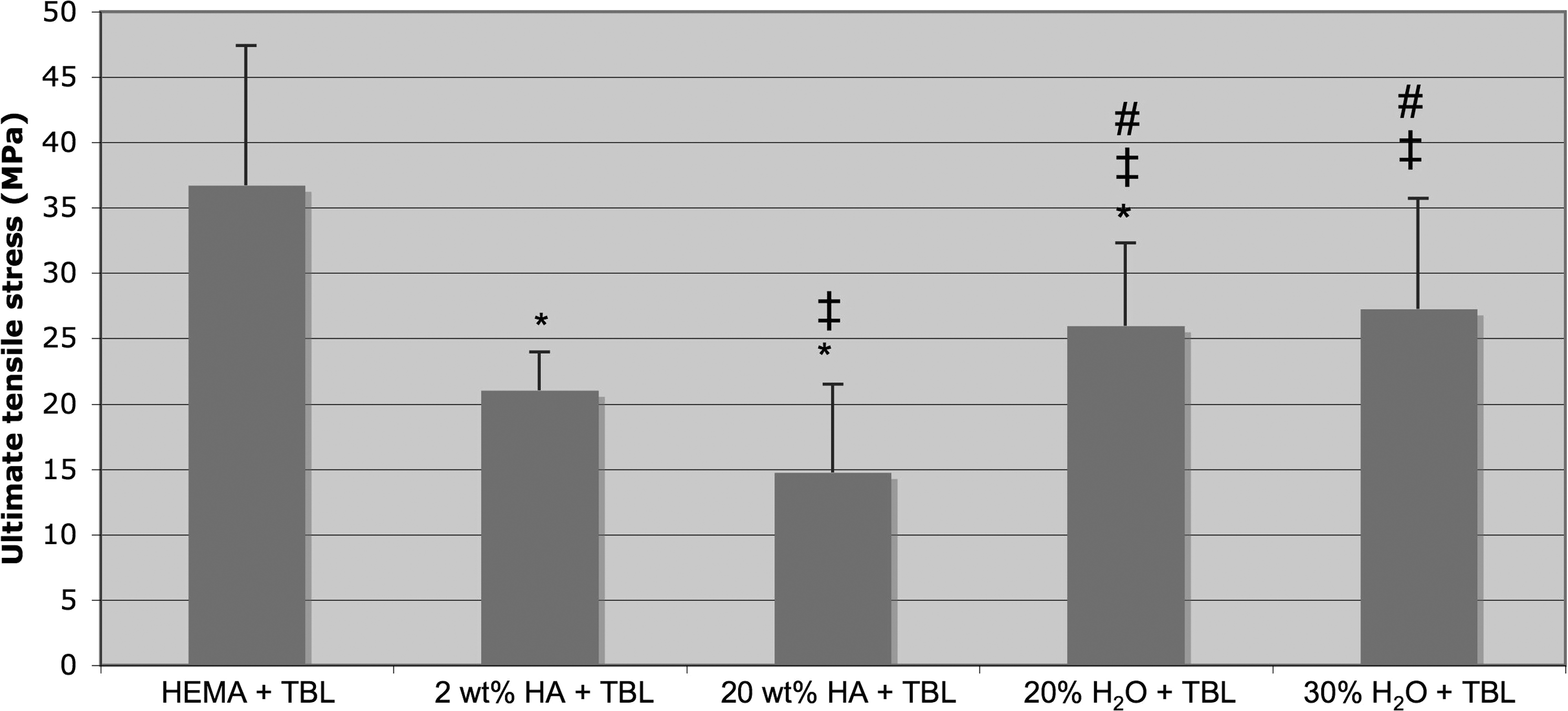
Ultimate tensile strength of the TBL/HA/pHEMA composites. All composites contain TBL (0.01 mg/mL) and the indicated HA or H2O content. HEMA+TBL, 2 wt% HA+TBL, and 20 wt% HA+TBL composites contained no water. The TBL/HA/pHEMA composite solution was placed into dog-bone shaped molds and then into an oven at 60°C for 2 h. The Instron 5882 mechanical testing system was used to determine the tensile properties of samples at a speed of 5 mm/min under dry conditions. Data=Mean±SEM; n=3. *p<0.05 compared to HEMA+TBL pHEMA composites; ‡p<0.05 compared to 2 wt% HA+TBL pHEMA composites; #p<0.05 compared to 20 wt% HA+TBL pHEMA composites.
Cytocompatibility properties
Most importantly, all of the composites containing RNTs (0.01 mg/mL of TBL) enhanced osteoblast adhesion compared to composites without TBL. In particular, the TBL/HA(20 wt%)/pHEMA composites had the highest osteoblast adhesion density, which was 43% and 13.4% greater than pure pHEMA controls and the TBL/HA(2 wt%)/pHEMA composites, respectively (Figs. 5 and 6). On the other hand, the TBL/HA(20 wt%)/pHEMA composites had the lowest fibroblast adhesion density compared to the other samples. Fibroblasts are responsible for the formation of fibrous tissue and compete with bone-forming osteoblasts for space at the interface between the implant device and the injectable material, thus potentially causing insufficient bonding with juxtaposed bone. These results are promising, especially considering that no growth factors were used in this study.
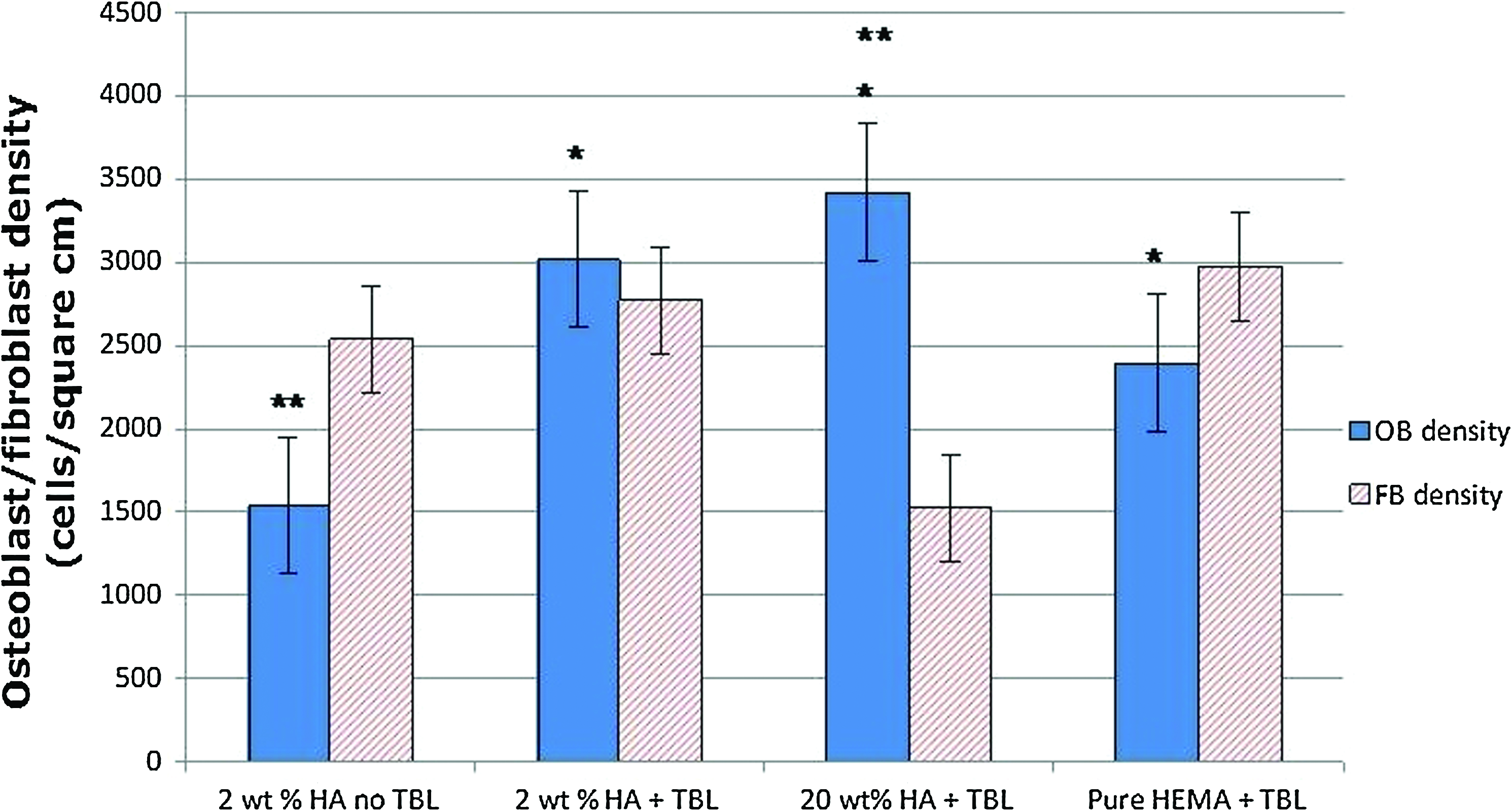
Osteoblast (OB) and fibroblast (FB) cell adhesion on pHEMA composites containing 2 wt% HA, 2 wt% HA and TBL, 20 wt% HA and TBL, or TBL for 4 h. All the composites were water-free and contained TBL (0.01 mg/mL) and HA as indicated in the histogram. Values are mean±SEM; n=3. *p<0.05 compared to 2 wt% HA no TBL for respective cells; **p<0.05 compared to fibroblasts on the respective sample. Color images available online at www.liebertonline.com/tea

Fluorescence microscope images of osteoblast morphologies on pHEMA containing
Here, we have demonstrated that RNTs incorporated into HA/pHEMA composites had higher osteoblast densities than HA/pHEMA composites (Fig. 7). The results also suggested that the RNTs contributed more to osteoblast proliferation than adding 2 wt% HA to the composites, although both enhanced the cytocompatibility properties of the composites compared to pure pHEMA. Therefore, the combination of RNTs and HA as additives to injectable pHEMA composites is a promising strategy for improving both mechanical and cytocompatibility properties.

Osteoblast cell density on pHEMA composites containing 2 wt% HA, 2 wt% HA and TBL, 20 wt% HA and TBL, and TBL after 1-, 3-, and 5-day culturing. All the composites were water-free and contained TBL (0.01 mg/mL) and HA as indicated in the histogram. Values are mean±SEM; n=3. *p<0.05 compared to 2 wt% HA at respective time periods; **p<0.05 compared to the previous time point for respective sample. Color images available online at www.liebertonline.com/tea
In addition, collagen synthesis by osteoblasts was the highest (0.02, 0.10, and 0.15 μg/cm2) on TBL/HA(20 wt%)/pHEMA composites compared to all other substrates investigated after 7, 14, and 21 days (Fig. 8). HA (2 wt%)/pHEMA composites exhibited greater collagen synthesis compared to pure TBL/pHEMA substrates after 14 and 21 days. Similarly, TBL/HA (20 wt%)/pHEMA composites promoted greater alkaline phosphatase activity by osteoblasts relative to HA(2 wt%)/pHEMA, TBL/HA(2 wt%)/pHEMA, and TBL/pHEMA composites (Fig. 9). Osteoblasts showed lower alkaline phosphatase activity on TBL/HA(2 wt%)/pHEMA composites compared to TBL/pHEMA composites.
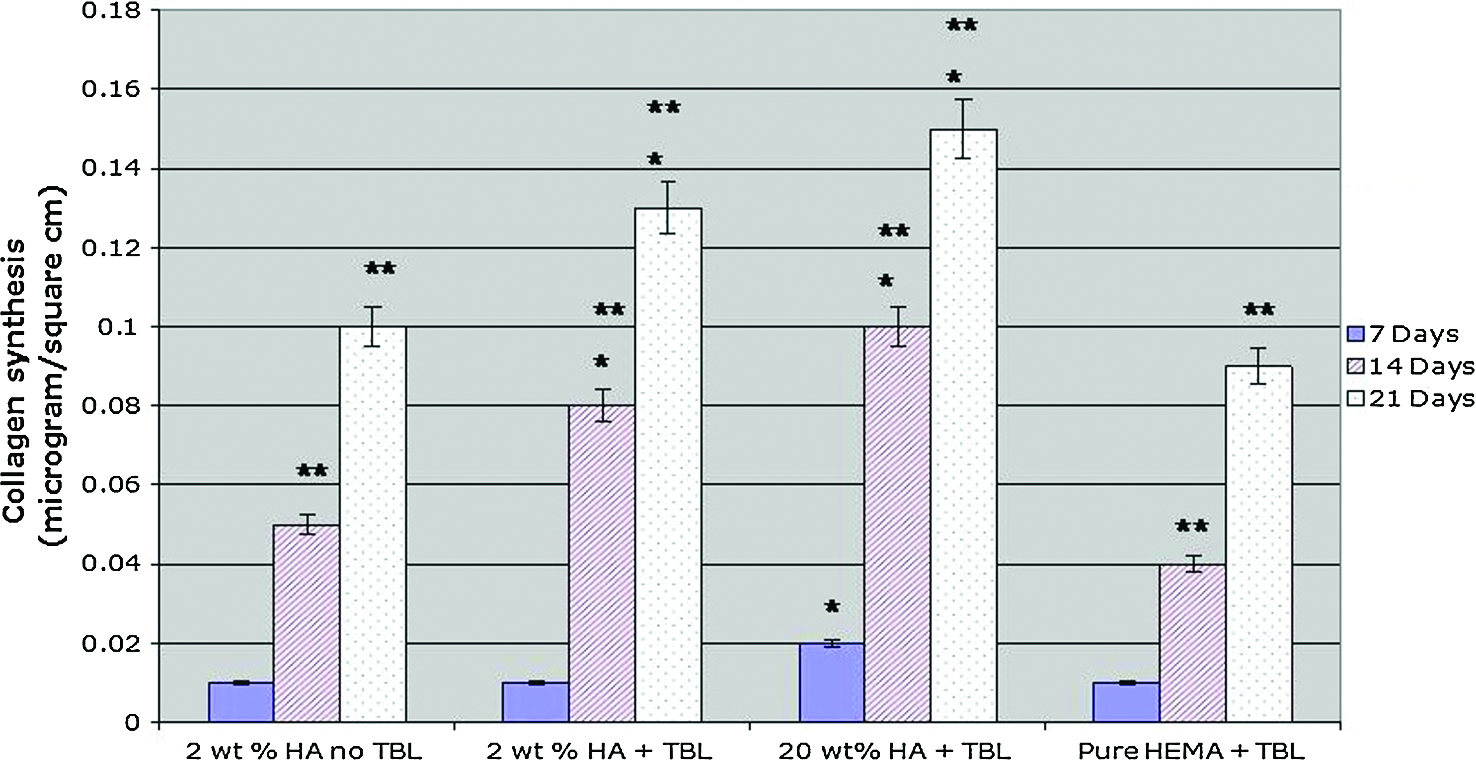
Production of collagen by osteoblasts after culturing for 7, 14, and 21 days. All the composites were water-free and contained TBL (0.01 mg/mL) and HA as indicated in the histogram. Values are mean±SEM; n=3. *p<0.05 compared to 2 wt% HA for respective time periods; **p<0.05 compared to the previous time point for respective sample. Color images available online at www.liebertonline.com/tea
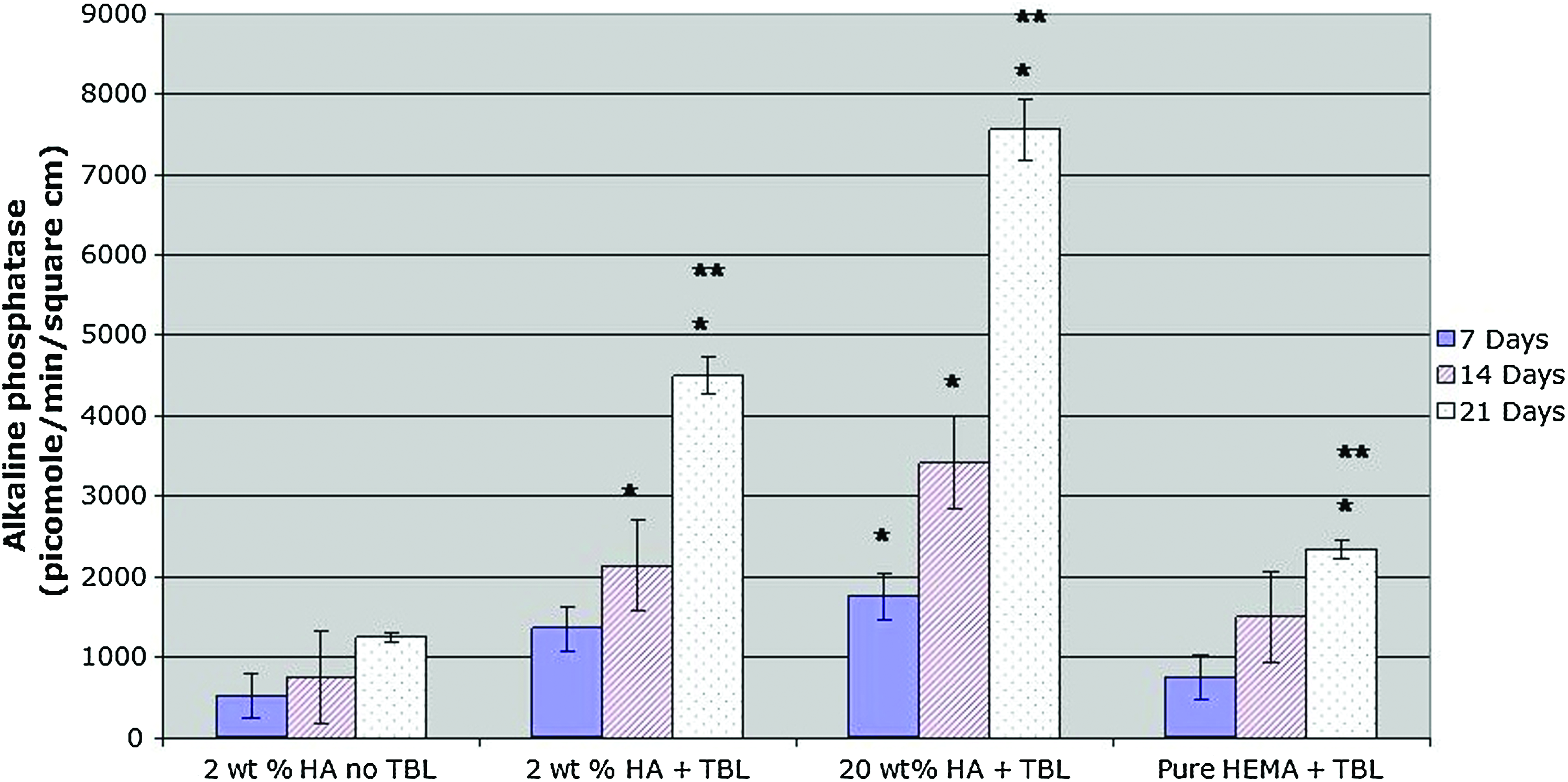
Alkaline phosphatase activity of osteoblasts after culturing for 7, 14, and 21 days. All composites were water-free and contained TBL (0.01 mg/mL) and HA as indicated in the histogram. Values are mean±SEM; n=3. *p<0.05 compared to 2 wt% HA for respective time periods; **p<0.05 compared to previous time points for respective sample. Color images available online at www.liebertonline.com/tea
Lastly, calcium deposition by osteoblasts was greater (4.2 μg) on TBL/HA(20 wt%)/pHEMA composites after 21 days of culture compared to all other composites tested (Fig. 10). Therefore, the TBL(0.01 mg/mL)/HA(20 wt%)/pHEMA composites solidified by heating in an oven at 60°C increased osteoblast adhesion, proliferation, and osteoblast long-term functions (i.e., collagen synthesis, alkaline phosphatase synthesis, and calcium deposition), and as a result, they may serve as a bioactive injectable orthopedic implant material.
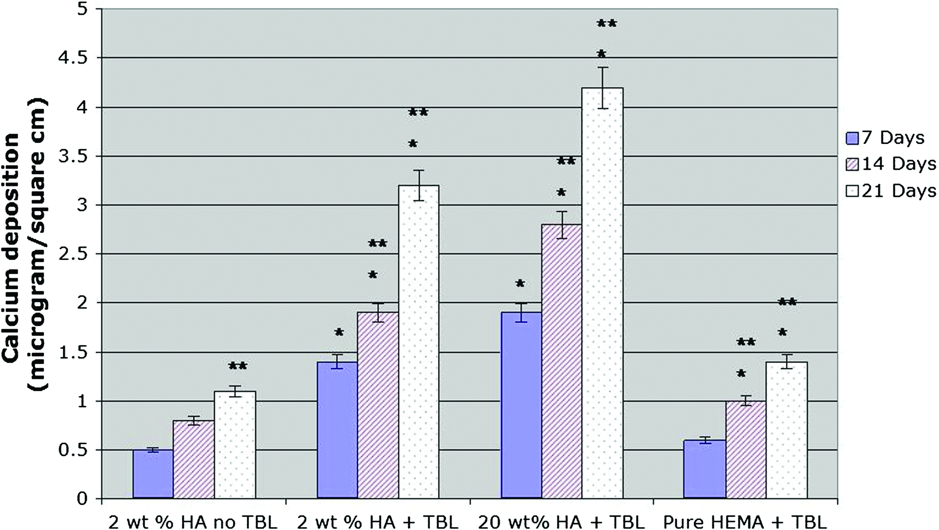
Calcium deposition by osteoblasts after culturing for 7, 14, and 21 days. All composites were water-free and contained TBL (0.01 mg/mL) and HA as indicated in the histogram. Values are mean±SEM; n=3. *p<0.05 compared to 2 wt% HA for respective time period; **p<0.05 compared to previous time points for respective sample. Color images available online at www.liebertonline.com/tea
Conclusions
Composites of RNTs assembled from TBL, HA, and pHEMA were prepared, and their solidification, mechanical, and cytocompatibility properties were investigated. The compressive strength of the composites increased with HA nanoparticle content (from 9.2 to 37.1 MPa) and decreased with water content (from 9.2 to 3.0 MPa). The addition of HA nanoparticles to TBL/pHEMA composites achieved mechanical strength (up to 37.1 MPa for compressive strength and 35.7 MPa for ultimate tensile strength) similar to vertebral discs. RNTs assembled from TBL and HA nanoparticles effectively increased osteoblast activity while decreasing fibroblast adhesion. Importantly, osteoblast adhesion improved on composites of RNTs, HA (20 wt%), and pHEMA compared to formulations with lower HA content or no RNTs. Moreover, HA(20 wt%)/TBL/pHEMA composites stimulated osteoblasts to (1) synthesize more collagen, (2) increase alkaline phosphatase activity, and (3) induce more calcium deposition. Based on these results, the composite solidified by heating at 60°C and made from HA (20 wt%), RNTs assembled from TBL (0.01 mg/mL), and pHEMA with no water incorporation was the optimal formulation here and, thus, warrants further studies as an injectable orthopedic implant material.
Footnotes
Acknowledgments
The authors acknowledge Audax Medical, Inc., for financial assistance, and Canada's Natural Science and Engineering Research Council, Canada's National Research Council, and the University of Alberta.
Disclosure Statement
No competing financial interests exist.