
Abstract
Engineering functional cartilaginous grafts using stem cells isolated from osteoarthritic human tissue is of fundamental importance if autologous tissue engineering strategies are to be used in the treatment of diseased articular cartilage. It has previously been demonstrated that human infrapatellar fat pad (IFP)–derived stem cells undergo chondrogenesis in pellet culture; however, the ability of such cells to generate functional cartilaginous grafts has not been adequately addressed. The objective of this study was to explore how environmental conditions regulate the functional development of cartilaginous constructs engineered using diseased human IFP–derived stem cells (FPSCs). FPSCs were observed to display a diminished chondrogenic potential upon encapsulation in a three-dimensional hydrogel compared with pellet culture, synthesizing significantly lower levels of glycosaminoglycan and collagen on a per cell basis. To engineer more functional cartilaginous grafts, we next explored whether additional biochemical and biophysical stimulations would enhance chondrogenesis within the hydrogels. Serum stimulation was observed to partially recover the diminished chondrogenic potential within hydrogel culture. Over 42 days, stem cells that had first been expanded in a low-oxygen environment proliferated extensively on the outer surface of the hydrogel in response to serum stimulation, assembling a dense type II collagen–positive cartilaginous tissue resembling that formed in pellet culture. The application of hydrostatic pressure did not further enhance extracellular matrix synthesis within the hydrogels, but did appear to alter the spatial accumulation of extracellular matrix leading to the formation of a more compact tissue with superior mechanically functionality. Further work is required in order to recapitulate the environmental conditions present during pellet culture within scaffolds or hydrogels in order to engineer more functional cartilaginous grafts using human osteoarthritic FPSCs.
Introduction
MSCs are beginning to be used clinically in the treatment of focal cartilage defects.43,44 While the use of MSCs has been shown to improve repair compared with untreated controls, critically normal hyaline cartilage does not regenerate, rather a hyaline-like or fibro-cartilagenous tissue forms.45–47 Chondrogenic predifferentiation of MSCs within collagen gels, leading to improved functionality of the engineered graft prior to implantation, has been shown to improve the outcome in long-term sheep studies, 48 resulting in superior repair compared with undifferentiated MSCs or chondrocytes. 49 While promising, the quality of repair observed in these animal model studies using predifferentiated MSCs is still variable,48,49 suggesting that engineering a more functional graft may be required prior to implantation. Further, it remains to be elucidated whether such predifferentiated cartilaginous grafts can be engineered using stem cells isolated from diseased human donors. If autologous MSCs are to be ultimately used for regeneration of damaged or diseased articular cartilage in osteoarthritic joints, then it will be necessary to first identify the optimal culture conditions that lead to the development of functional cartilaginous tissues using such cell sources.
Human infrapatellar fat pad (IFP) contains multipotent stem cells (FPSCs) that can be easily harvested arthroscopically, 23 and that possess at least comparable chondrogenic capacity to chondrocytes isolated from adult articular cartilage. 24 We have previously demonstrated that functional cartilage tissue can be engineered using immature porcine FPSCs32,33,35; however, it remains unclear whether such grafts can be engineered using stem cells isolated from human osteoarthritic IFP tissue. The IFP of patients with OA has been shown to have an inflammatory phenotype, 50 which may impact the ability of stem cells isolated from this tissue to generate a functional cartilaginous tissue. The objective of this study was to first compare chondrogenesis of FPSCs in pellet culture to that in agarose hydrogels that are commonly used to engineer functional cartilaginous grafts. The influence of the oxygen tension during monolayer expansion of FPSCs on the subsequent properties of these cartilaginous grafts was assessed. We then explored how biochemical (serum) and biophysical (HP) cues would influence the functional development of cartilage tissues engineered using human FPSCs.
Materials and Methods
Cell isolation and expansion
Ethical approval for the study was obtained from the institutional review board of the Mater Misericordiae University Hospital with IFP tissue being obtained from five patients with knee OA at joint arthroplasty (1 male and 4 females, 50–79 years old). Fat pad was maintained in sterile phosphate-buffered saline (PBS) and transferred immediately to the Trinity Centre for Bioengineering for further processing. Fibrous tissue was carefully removed from the fat pad. Remaining tissue was weighed, washed thoroughly in PBS, and diced followed by incubation under constant rotation at 37°C with high-glucose Dulbecco's modified Eagle's medium (hgDMEM; GlutaMAX™) (GIBCO, Biosciences) containing 1% penicillin (100 U/mL)–streptomycin (100 μg/mL) and collagenase type II (4 mL solution/g tissue, 750 U/mL; Worthington Biochemical, LanganBach Services) for 4 h. Cells were filtered through serial cell sieves (Falcon, Sarstedt) with pore sizes from 100 μm, 70 μm, to 40 μm. The isolated FPSCs were seeded at a density of 5000 cells/cm2 and cultured in expansion medium [hgDMEM GlutaMax supplemented with 10% v/v fetal bovine serum (FBS) and penicillin (100 U/mL)–streptomycin (100 μg/mL) (all from Gibco, Biosciences)]. The expansion medium was also supplemented with 5 ng/mL fibroblast growth factor-2 (FGF-2) (Prospect-Tany TechnoGene Ltd.) that has previously been shown to enhance proliferation and chondrogenesis of porcine FPSCs. 51 Cells were expanded to passage two (P2) and the oxygen level during cell expansion was maintained at either 20% or 5% O2. All cultures in this study were kept at 37°C. For the HP study (described below in 'HP Loading’ section), a superlot of FPSCs pooled from three different donors was used. For studies other than the HP Loading study, FPSCs isolated from individual donors were used, with replicate studies undertaken using FPSCs isolated from separate donors undertaken as described in the Results section.
Colony-forming unit–fibroblast assay
For the colony-forming unit–fibroblast (CFU-F) assay, freshly isolated cells were plated in 58 cm2 petri dishes at a density of 135 cells/cm2 and maintained with or without FGF-2 (5 ng/mL) at either 20% O2 or 5% O2. Triplicate dishes were plated for all conditions. After 12–14 days, cells were fixed with 2% paraformaldehyde (PFA), stained with 1% crystal violet (Sigma-Aldrich), and colony numbers (>50 cells) were counted. Total colony number and colony diameter were determined using ImageJ software (Rasband, W.S., Image J, U.S. National Institutes of Health, Bethesda, MD). For the purposes of comparison between groups, colony size was defined as the average diameter of the 10 largest colonies formed.
Chondrogenesis in pellet culture
Pellets were formed from culture-expanded cells by centrifuging 250,000 cells (P2) in 1.5 mL conical microtubes at 650 g for 5 min. The pellets were maintained in chondrogenic medium (CM): hgDMEM GlutaMax supplemented with penicillin (100 U/mL)–streptomycin (100 μg/mL) (Invitrogen), 100 μg/mL sodium pyruvate, 40 μg/mL L-proline, 50 μg/mL L-ascorbic acid-2-phosphate, 4.7 μg/mL linoleic acid, 1.5 mg/mL bovine serum albumin, 1× insulin-transferrin-selenium, and 100 nM dexamethasone (all from Sigma-Aldrich). The CM was additionally supplemented with 10 ng/mL recombinant human transforming growth factor-beta3 (TGF-β3; Prospect-Tany TechnoGene Ltd.) or 10 ng/mL TGF-β3+10% FBS (Lot no. 10270; Gibco). Each pellet was cultured in 1 mL of medium and the medium was changed twice a week. The oxygen level for pellet culture was maintained at either 20% O2 or 5% O2. All pellets were cultured for 3 weeks with pellets assessed at days 0 and 21.
Chondrogenesis in agarose hydrogels
FPSCs (P2) were suspended in 2% agarose hydrogels (type VII; Sigma-Aldrich). All hydrogels were seeded at a density of 10×106 cells/mL, except for the agarose gels subjected to HP (both free swelling [FS] controls and loaded constructs), which were seeded at a higher density of 20×106 cells/mL in an attempt to improve the overall functionality of the construct. The agarose cell suspension was cast in a stainless steel mould and cored using a biopsy punch to produce construct cylinders (Ø5mm×1.5 mm). Constructs were maintained in CM at 5% O2 for a period of 42 days with medium exchanges performed twice weekly. Gel samples were assessed at days 0, 21, and 42.
HP loading
Gel constructs were subjected to HP at a magnitude of 10 MPa and a frequency of 1 Hz for 2 h/day and 5 days/week for the final 4 weeks of the 6-week differentiation period in a custom HP bioreactor. 42 Six cell-seeded gels were transferred to sterilized, heat-sealed bags filled with 15 mL of medium for the 2-h loading period. The bags for HP loading were placed into a water-filled pressure vessel while FS control groups (also contained in heat-sealed bags) were placed in an open water bath, both maintained at 37°C. After 2 h, all constructs were removed from heat-sealed bags and cultured in tissue culture polysterene plate at 37°C and 5% O2.
Mechanical analysis
Constructs were mechanically assessed using a protocol described previously. 52 Briefly, constructs from each group were tested in unconfined compression between impermeable platens using standard Zwick testing machine with a 5 N load cell (Zwick Z005; Roell). A ramp and hold cycle with a ramp displacement of 1 μm/s until 10% strain was applied and maintained until equilibrium was reached (30 min). At this point, dynamic tests were performed with a cyclical strain amplitude of 1% at 1 Hz. Dynamic moduli were calculated as the ratio of the determined stress amplitude to the applied strain amplitude.
Biochemical analysis
Pellet and gel samples were digested in papain (125 μg/mL) in 0.1 M sodium acetate, 5 mM cysteine HCl, and 0.05 M EDTA (pH 6.0) (all from Sigma-Aldrich) at 60°C under constant rotation for 18 h. Total pellet DNA content was measured using a Quant-iT™ PicoGreen® dsDNA kit (Molecular Probes, Biosciences) with a lambda DNA standard and the DNA content of gels was quantified using the Hoechst Bisbenzimide 33258 dye assay (Sigma-Aldrich). Proteoglycan content was estimated by quantifying the amount of sulfated glycosaminoglycan (sGAG) in constructs using the dimethylmethylene blue dye-binding assay (Blyscan, Biocolor Ltd.), with a chondroitin sulfate standard. Total collagen content was determined by measuring the hydroxyproline content. Samples were hydrolyzed at 110°C for 18 h in concentrated HCL (38%) and assayed using a chloramine-T assay 53 with a hydroxyproline-to-collagen ratio of 1:7.69. 54
Histology and immunohistochemistry
Pellets and gels were fixed in 4% PFA, embedded in paraffin, and sectioned (5 μm). Sections were stained with 1% alcian blue 8GX (Sigma-Aldrich) in 0.1 M HCl for sGAG, and picrosirius red for collagen. The deposition of collagen types I and II was identified through immunohistochemistry.42,55 Briefly, sections were quenched of peroxidase activity for 20 min (PBS was used to rinse sections between steps) and treated with 0.25 U/mL chondroitinase ABC (Sigma) in a humidified environment at 37°C for 1 h to enhance permeability of the extracellular matrix by removal of chondroitin sulfate. After incubation with 10% goat serum to block nonspecific sites, the primary antibody of mouse monoclonal anti-collagen type I diluted 1:400 or mouse monoclonal anti-collagen type II diluted 1:100 (Abcam) was applied for 1 h at room temperature. Then, the secondary antibody (anti-mouse IgG biotin conjugate; Sigma-Aldrich) was added for another hour. Color was developed using the Vectastain ABC reagent (Vectastain ABC kit; Vector Laboratories) for 45 min and exposure to peroxidase DAB substrate kit (Vector laboratories) for 5 min. Slides were dehydrated through ethanol and xylene and mounted with Vectamount medium (Vector Laboratories). Human ligament and cartilage were included as controls for collagen type I and collagen type II, respectively.
Statistics
Numerical and graphical results are presented as mean±standard deviation (3–4 samples). Statistics were performed using R (The R Foundation for Statistical Computing, Vienna, Austria). Groups were analyzed for significant differences using a linear model for analysis of variance with multiple factors and interactions between these factors were also examined. Tukey's HSD test for multiple comparisons was used as post-tests. Significance was accepted at a level of p<0.05.
Results
The chondrogenic potential of FPSCs is diminished in hydrogel culture in comparison to pellet culture
As we have reported previously, 56 expansion of FPSCs in the presence of FGF-2 significantly reduced the CFU-F colony number and increased the colony diameter (Supplementary Fig. S1 A, B; Supplementary Data are available online at www.liebertonline.com/tea). Further, the colony diameter in FGF-2-expanded stem cells was significantly larger when maintained at 5% O2 compared with 20% O2. Following expansion to passage 2, stem cells expanded in the presence of FGF-2 were differentiated at either 5% O2 or 20% O2 using a pellet culture model. Stem cells expanded at 5% O2 formed larger pellets (Fig. 1A) and were more proliferative during chondrogenesis, as evident by a higher DNA content at day 21 (Fig. 1B), with total sGAG and collagen accumulation greatest for cells expanded and differentiated at 5% O2 (Fig. 1C). When normalized to DNA content the levels of sGAG and collagen within the pellets were comparable for stem cells expanded at either 5% O2 or 20% O2, but were always greatest for differentiation at 5% O2 (Fig. 1D). These general results were observed for stem cells isolated from other donors, with the only exception being that for some donors differentiation at 5% O2 was found to suppress total collagen production (see Supplementary Fig. S2).

The effect of oxygen tension during expansion and differentiation on chondrogenesis of infrapatellar fat pad–derived stem cells (FPSCs) in pellet culture for 21 days. Pellet morphology
In an attempt to engineer a cartilaginous graft, FPSCs were next seeded into agarose hydrogels and the levels of matrix accumulation (on a per cell basis) after 21 days in culture at 5% O2 were compared with that in pellets. Both sGAG and collagen syntheses were lower in hydrogel culture in comparison to pellet culture, with noticeably weak staining for both alcian blue and type II collagen, despite the attainment of a spherical cellular morphology within the hydrogel system (Fig. 2). This was observed for stem cells expanded at either 5% O2 or 20% O2. Further, the greater proliferative potential of stem cells expanded at 5% O2 that was observed in pellet culture was diminished in hydrogel culture.
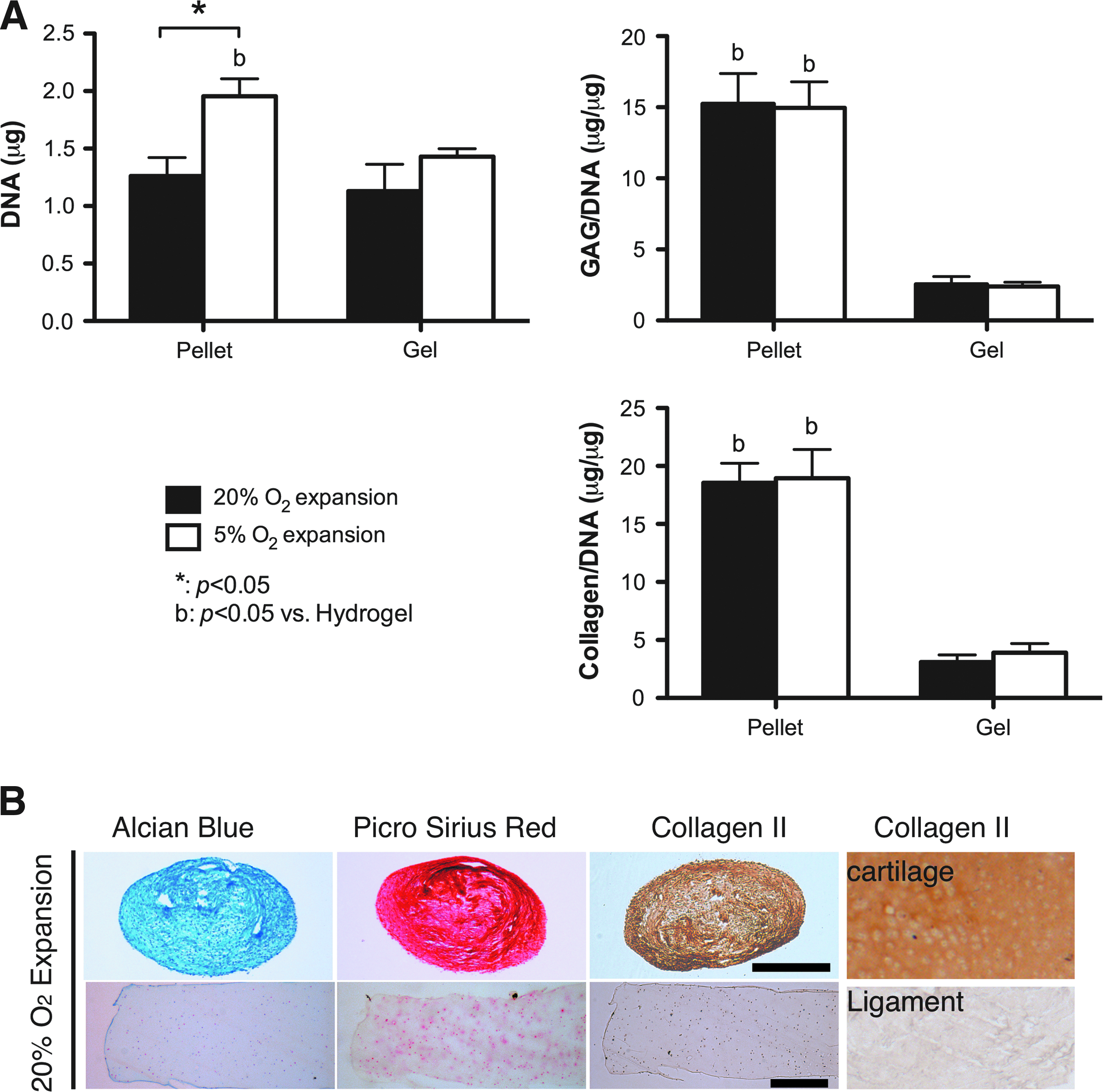
A comparison of the chondrogenic potential of FPSCs maintained in pellets or in agarose hydrogels (10 million cells/mL) for 21 days.
Serum stimulation promotes cell proliferation in both pellets and hydrogels and enhances matrix accumulation within hydrogels
In an attempt to provide a further stimulus to FPSCs embedded in agarose hydrogels to synthesize cartilage-specific extracellular matrix, constructs were additionally stimulated with 10% serum. In pellet culture this led to greater cell proliferation but no increase in matrix synthesis (Fig. 3A). In contrast, the presence of serum significantly enhanced sGAG synthesis within cell-seeded hydrogels, although synthesis levels were still lower than that observed in pellets. Collagen synthesis within hydrogels, when normalized to DNA content, was not affected by serum supplementation. Again, the oxygen levels during expansion did not appear to affect levels of matrix accumulation within cell-seeded hydrogels over 21 days of in vitro culture.
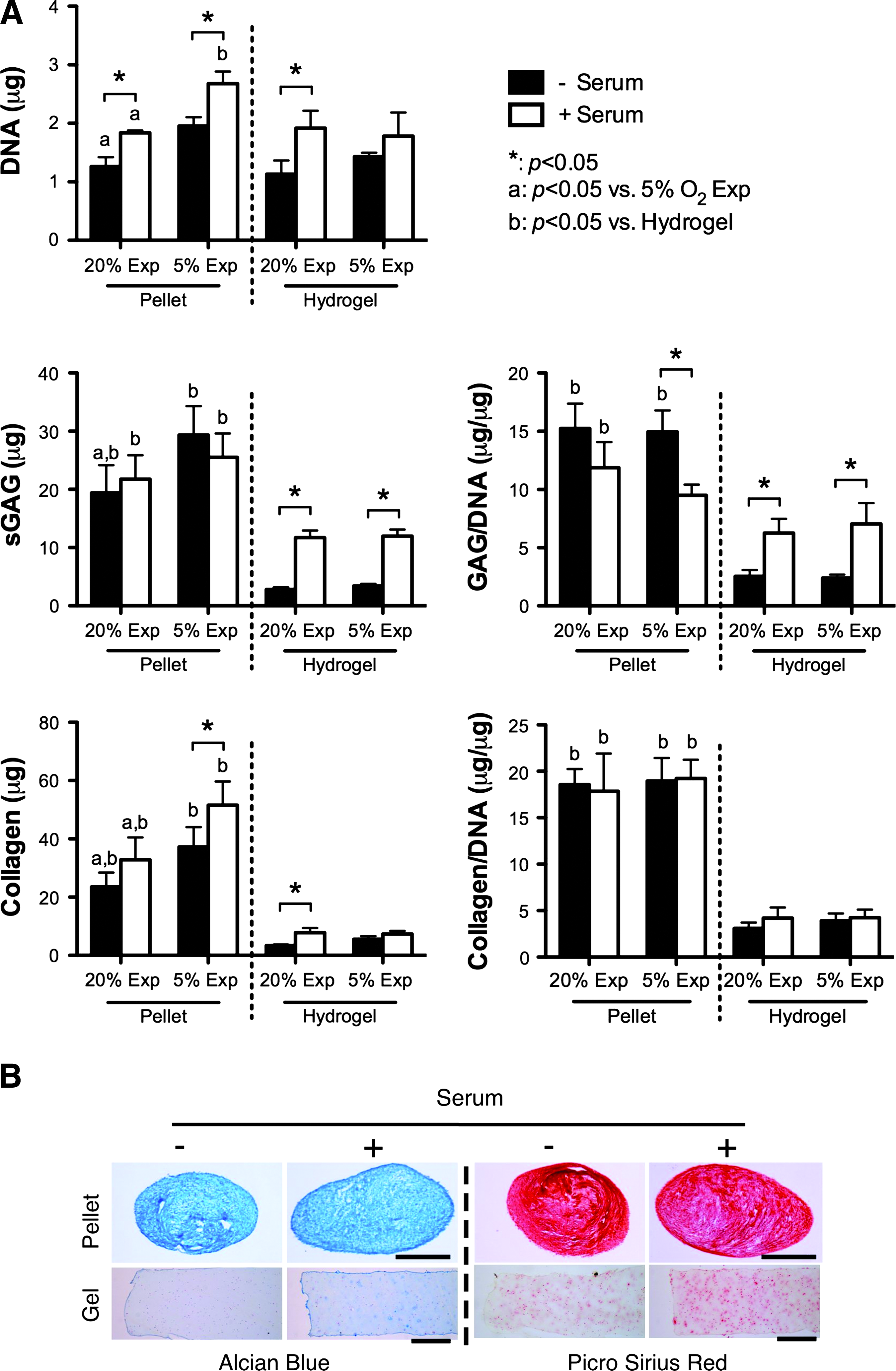
Effect of serum on FPSCs maintained in pellets or in agarose hydrogels (10 million cells/mL) for 21 days.
Oxygen conditions during the expansion of FPSCs influences the long-term development of cartilaginous grafts
We next explored the development of cartilaginous grafts engineered using FPSCs embedded in agarose hydrogels over a longer period of 42 days of in vitro culture. Unlike the findings for constructs cultured for 21 days, the DNA content within hydrogels dramatically increased for stem cells that were first expanded at 5% O2 and differentiated in the presence of serum (Fig. 4A), mirroring the findings first observed in pellet culture (Fig. 3). Expansion at 5% O2 also led to noticeably higher levels of total GAG and collagen accumulation within constructs supplemented with serum. Histological analysis of these constructs revealed that these higher levels of extracellular matrix accumulation were not due to dramatically greater synthesis within the hydrogels, but rather due to the formation of a dense layer of highly cellular cartilaginous tissue that formed on the surface of these constructs (Fig. 4B). This self-assembled tissue that formed on top of the hydrogels stained positively for type II collagen and weakly for type I collagen (Fig. 4C).

Effect of serum on the FPSCs maintained in agarose hydrogels (10 million cells/mL) for 42 days.
The application of HP enhances the mechanical functionality of cartilaginous grafts by generating a more compact tissue
In an attempt to further enhance chondrogenesis, cell-seeded hydrogels were subjected to 10 MPa of cyclic HP (1 Hz for 2 h/day, 5 days/week). HP did not influence total DNA, sGAG, or collagen content within the hydrogels, although it did appear to lead to a more homogeneous matrix distribution in gel constructs, with staining within the hydrogel appearing slightly more intense in constructs subjected to HP (Fig. 5B). Significant cartilaginous tissue outgrowth was observed on the surface of the hydrogels, particularly those additionally supplemented with serum. In the absence of serum, HP resulted in constructs of smaller height (Fig. 5C). When mechanically evaluated, these constructs were stiffer than controls maintained in FS conditions (Fig. 5D).
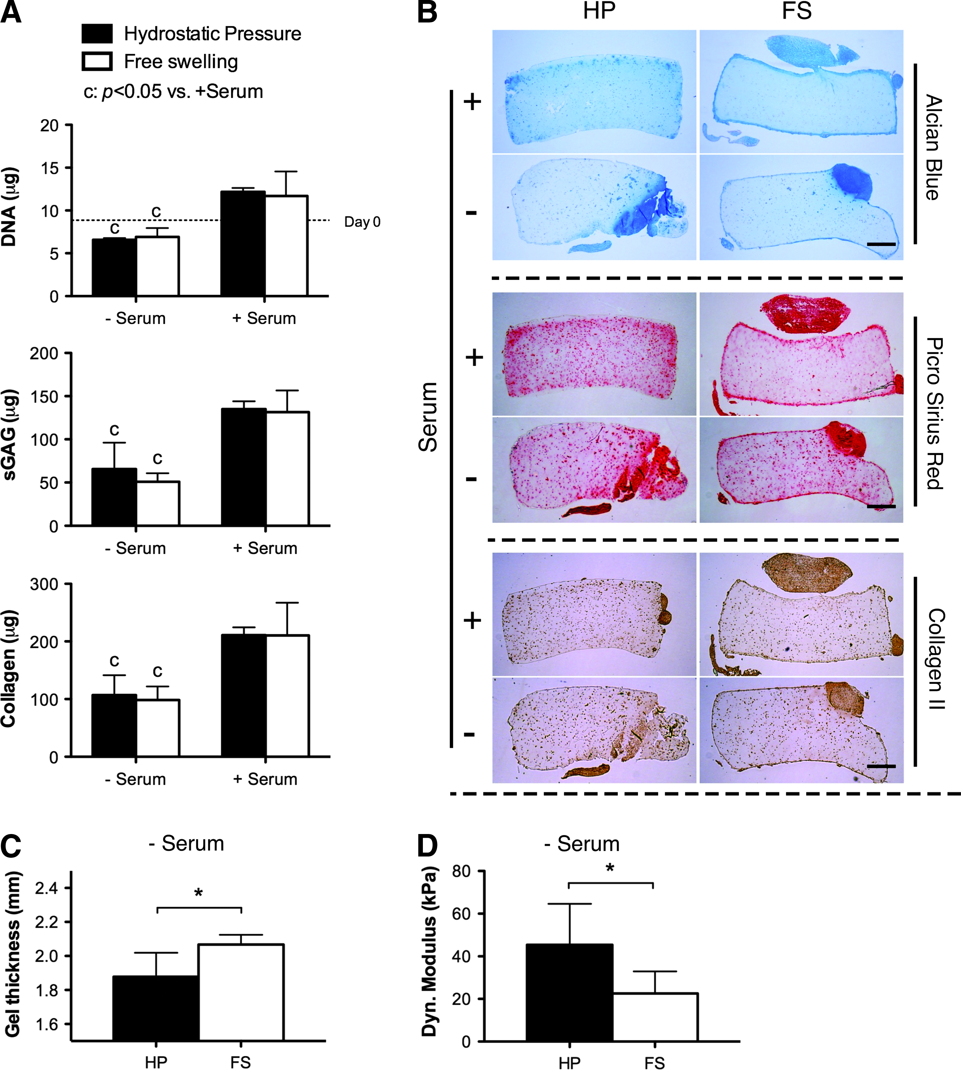
Effect of hydrostatic pressure and serum on chondrogenesis of FPSCs maintained in agarose hydrogels (20 million cells/mL) for 6 weeks. Hydrostatic pressure loading was applied for the final 4 weeks of culture.
Discussion
Chondrogenic priming of MSCs to generate a more functional cartilage-like graft prior to implantation has been shown to improve the quality of repair in animal model studies of cartilage regeneration.48,49 Translating such therapies to treat damaged and diseased articular cartilage in man will most likely necessitate the development of tissue engineering strategies to generate functional cartilaginous grafts using autologous stem cells. The objective of this study was to explore how environmental factors during both the expansion and differentiation of diseased human FPSCs would influence their chondrogenic capacity following encapsulation into three-dimensional hydrogels. Despite previous findings that agarose hydrogels support robust chondrogenesis of stem cells isolated from skeletally immature animals,32,33,35 we found that the chondrogenesis of diseased human FPSCs was diminished in these hydrogels compared with pellet culture. Stimulation of the cell-seeded hydrogels with potent biochemical (serum) or biophysical (HP) stimuli did lead to improvements in cartilaginous matrix synthesis and construct functionality, respectively, although the inherent chondrogenic capacity observed in the pellet environment was never recapitulated.
When expanded in the presence of FGF-2, FPSCs maintained at 5% O2 formed larger colonies, suggesting a greater proliferative potential at this lower oxygen tension. Enhanced proliferation has previously been reported for MSCs maintained at a low oxygen tension.57–60 This proliferative potential during monolayer expansion was maintained during chondrogenic differentiation, both in pellet culture, but also in hydrogel culture once FPSCs were stimulated with serum. FPSCs did not appear to proliferate within the hydrogel, but rather on the construct surface. Cells cannot directly adhere to agarose, which coupled with the fact that the FPSCs are completely encapsulated within the hydrogel leads to limited proliferation. However, FPSCs can potentially migrate out onto the hydrogel surface, adhere to newly synthesized extracellular matrix, and then proliferate as they are no longer encapsulated in agarose, a process that appears to be accelerated in the presence of serum. Similar cellular outgrowth and capsule formation has been observed in chondrocyte-seeded hydrogels supplemented with serum.61,62 Interestingly, previous studies have reported that these layers are rich in type I collagen and therefore termed fibrous capsules, although the layers formed by the FPSCs stain positively for type II collagen, suggesting that specific experimental factors, such as the presence of TGF-β3 in the medium or maintenance at a low oxygen tension during differentiation, promoted a more cartilaginous phenotype.
Encapsulation of FPSCs in a three-dimensional hydrogel led to a diminished chondrogenic potential compared with pellet culture. It is well established that agarose can help support a chondrogenic phenotype for culture-expanded chondrocytes, 7 and has been widely used as a hydrogel for cartilage tissue engineering using chondrocytes,63–65 bone-marrow-derived MSCs,66,67 adipose-derived stem cells, 31 and FPSCs,33,35,55,68 although many of these studies used cells isolated from young animals. A key difference between the high-density pellet culture system 11 and hydrogel encapsulation in agarose is the difference in cell–cell and cell–matrix interactions. Cellular condensation and expression of proteins associated with cell–cell interactions, such as N-cadherin, are known to be important at the onset of chondrogenesis.69,70 The pellet culture system also supports paracrine signaling important for chondrogenesis. The absence of ligands within the agarose hydrogel to facilitate integrin-mediated binding of FPSCs to their local substrate may also play a role in diminishing their chondrogenic potential. Alternative approaches to engineering functional cartilaginous grafts that recapitulate aspects of the pellet culture system, such as the self-assembly process, 71 may provide promising approaches when using diseased human FPSCs.
Serum has previously been shown to influence the growth and development of engineered cartilaginous constructs.61,72–76 Supplementing the CM with serum was observed to enhance cartilaginous extracellular matrix accumulation in FPSC-seeded hydrogels, although synthesis levels on a per cell basis were still lower than that in pellet culture. The abundance of growth factors present in serum, such as TGF-β superfamily, insulin-like growth factor-1, and platelet-derived growth factor, may be playing a role in this enhanced biosynthetic activity. Serum also contains adhesive glycoproteins, such as fibronectin,77,78 which have numerous biological activities, including mediating cell–cell adhesion and enhancing cell–substrate anchoring and spreading. 79 Further, by facilitating the proliferation of FPSCs on the surface of the hydrogels, serum may also be indirectly enhancing chondrogenesis by allowing greater cell–cell interactions in this region of the engineered tissue, thereby mimicking aspects of the pellet culture system known to support more robust chondrogenensis of diseased human FPSCs.
The application of HP did not enhance overall levels of matrix accumulation within FPSC-seeded constructs. This is in contrast to the results of previous studies demonstrating that cyclic HP can enhance chondrogenesis of bone marrow42,80–85 and adipose-derived 86 stem cells. These discrepancies could potentially be explained by differences in phenotype between stem cells isolated from different tissues, or alternatively due to differences in cell–cell and cell–scaffold interactions between different studies that may play a central role in mechano-transduction of biophysical signals such as HP. The cell-seeding density can also impact the response to biophysical cues,87,88 and may potentially explain discrepancies in reported responses to HP. It should be noted, however, that while the application of HP did not increase overall levels of matrix accumulation, it did appear to influence the spatial distribution of matrix within the construct. HP has previously been shown to result in the formation of more compact pellets generated using bone-marrow-derived MSCs, 84 and influence the spatial accumulation of matrix in MSC-seeded hydrogels. 42 Histological analysis suggests that the application of HP preferentially supports the accumulation of matrix components within the hydrogel as opposed to on the construct surface. In hydrogels not additionally stimulated with serum, the application of HP was also observed to suppress outgrowth and/or swelling of the construct, as evident by a smaller construct thickness. This may explain why these constructs were stiffer than their FS counterparts, as compaction of an engineered cartilaginous tissue can lead to improvements in biomechanical functionality. 89 Therefore the application of cues, such as HP, may play a key role in mechanically priming cartilaginous constructs engineered using FPSCs prior to implantation. Further optimization of these environmental conditions is required as the mechanical properties of these engineered grafts are still two orders of magnitude lower than normal articular cartilage.
In conclusion, we have demonstrated that encapsulation of diseased human FPSCs into agarose hydrogels in an attempt to engineer a functional cartilaginous graft can suppress their inherent chondrogenic potential. While this can be partially recovered through the application of biochemical or biophysical stimuli, their chondrogenic capacity is still diminished, and the resulting mechanical properties of the engineered grafts are still noticeably lower than native articular cartilage. Further work is required in order to recapitulate the environmental conditions present during pellet culture within hydrogels or scaffolds in order to engineer fully functional cartilaginous grafts using stem cells derived from human osteoarthritic IFP tissue.
Footnotes
Acknowledgments
Funding for this study was provided by Irish Research Council for Science, Engineering & Technology under enterprise partner scheme with Sports Surgery Clinic Dublin (IRCSET-SSC-2010-01), a European Research Council Starter Grant (StemRepair–Project number: 258463), and a President of Ireland Young Researcher Award (08/Y15B1336). The authors would like to thank Andrew Steward for his technical support with HP loading.
Disclosure Statement
The authors have nothing to disclose.
References
Supplementary Material
Please find the following supplemental material available below.
For Open Access articles published under a Creative Commons License, all supplemental material carries the same license as the article it is associated with.
For non-Open Access articles published, all supplemental material carries a non-exclusive license, and permission requests for re-use of supplemental material or any part of supplemental material shall be sent directly to the copyright owner as specified in the copyright notice associated with the article.