
Abstract
We investigated the therapeutic effects of a craniosynostosis-associated molecule, NEL-like molecule-1 (NELL1; NEL [a protein strongly expressed in neural tissue encoding the epidermal growth factor-like domain]), on osteolysis induced by polyethylene (PE)-particle debris. We used a murine calvarial osteolysis model with in vivo adenovirus (Ad)-mediated gene transfer. In total, 76 female Balb/c mice were randomly assigned to four groups for treatment 1 day postoperation: SHAM (injected with 0.1 mL saline without implantation of particles); PE control (injected with 0.1 mL saline after implantation of particles); PE+(Ad-GFP-NELL1) (injected with 0.1 mL Ad-GFP-NELL1 in saline after implantation of particles); and PE+(Ad-GFP) group (injected with 0.1 mL Ad-GFP in saline after implantation of particles). Green fluorescent protein (GFP) and NELL1 delivery in vivo after the injection were validated by optical imaging at 10 day postop, and then, all mice were sacrificed for analysis by three-dimensional (3D) microcomputed tomography (micro-CT), real-time polymerase chain reaction (PCR), histology, and biomechanical testing. Exogenous NELL1 and GFP were expressed in the osteolysis area for at least 9 days after the Ad-GFP-NELL1 injection. Serial 3D micro-CT images and testing of bone volume, bone mineral density, trabecular thickness, bone surface density, and connectivity density revealed that the new bone promoted with the Ad-GFP-NELL1 injection could almost compensate the PE-induced osteolysis and regenerate significantly better than with the Ad-GFP treatment. The expression of osteopontin (OPN) was significantly higher with Ad-GFP-NELL1 transduction among all the samples. Real-time PCR examination confirmed the augmented expression of OPN, Runx-2, and receptor activator of nuclear factor-kappa B ligand (RANKL). The elastic modulus was significantly greater with Ad-GFP-NELL1 than with the PE and/or Ad-GFP group (p<0.01). We found no transgene-associated toxic effects. Ad-GFP-NELL1 gene transfer effectively reversed the calvarial osteolysis and could be considered a new treatment for osteolysis through promoting bone regeneration.
Introduction
NEL-like molecule-1 (NELL1; NEL [a protein strongly expressed in neural tissue encoding the epidermal growth factor-like domain]) is a novel growth factor that is believed to specifically target cells committed to the osteochondral lineage. 6 Compared with bone morphogenetic protein (BMPs), NELL1 may be more specific to bone cells.7,8 In vivo studies of grafting NELL1 protein-coated PLGA scaffolds into rat calvarial defects revealed the osteogenic potential of NELL1 to induce bone regeneration equivalent to BMP-2, 9 and a later study illustrated that an injection of adenovirus (Ad)-NELL1+Ad-BMP-2 stimulated a synergistic increase in bone formation compared with Ad-BMP-2 alone in a nude mouse model. 10 In rat and sheep fusion models, NELL1 could significantly improve the spinal fusion rate.11,12 In our previous research, NELL1 could promote high-quality bone regeneration in a rat femoral distraction osteogenesis model. 6 To date, no evidence exists of whether NELL1 is effective in treating particle-induced osteolysis.
Due to the characteristics of NELL1, we hypothesized that it may be used as a potential therapeutic agent that treats debris-associated osteolysis. We used a calvarial osteolysis model that tests whether the NELL1 protein delivered by an Ad direct injection could promote new bone formation that heals the osteolytic lesion rather than simply suppresses further bone loss and its possible underlying mechanisms.
Materials and Methods
Generation of Ad harboring human NELL1 cDNA with green fluorescent protein tag
From pcDNA3.1-NELL1 (a gift of Dr. Kang Ting, University of California, Los Angeles), we made a recombinant Ad vector carrying NELL1 and Green fluorescent protein (GFP) (Ad-GFP-NELL1) as was done in our previous study. 6
Particles
Commercial PE particles13,14 (Ceridust 3615, 6.5–8.5 μm, mean diameter 7 μm) were processed under sterile conditions, washed for 24 h twice in 75% ethanol at room temperature, and dried in a vacuum desiccator. The particles were subdivided for separate use, sterilized by Co60, and stored at 4°C. The absence of endotoxin was confirmed by a Limulus assay (Limulus Amebocyte Lysate QCL-1000; Cambrex).
Animals
All experiments were performed in accordance with the General Hospital of Chinese People's Liberation Army Animal Care and Use Committee guidelines. Female Balb/c mice (6–8 week old) were purchased from the Experimental Animal Center of the Military Medical Science Institute. The animals were housed in a light- and temperature-controlled environment and given food and water ad libitum.
Surgical technique and calvarial osteolysis protocol
In total, 76 Balb/c mice were anesthetized with an intraperitoneal injection of 10% chloral hydrate (3.0 mL/kg; Drug Manufacturing Room of CPLA) after 1 week acclimation. Then, the hair was removed by cream, and a 1×1 cm area of calvarial bone was exposed by making a midline sagittal incision over the calvaria. Ten milligrams of PE particles were spread over the area, leaving the periosteum intact. The incision was then closed with the help of a simple 6-0 silk suture.15,16 One day after the surgery, the animals were randomly divided into four groups for treatment (n=19 in each group): SHAM control; PE control; PE+(Ad-GFP-NELL1); and PE+(Ad-GFP). The SHAM-operated animals were injected with 0.1 mL saline without the implantation of particles and designated as SHAM controls, while the PE controls were mice injected with 0.1 mL saline after the implantation of particles. Saline or gene delivery done by Ad in 1×10⁹ plaque-forming units diluted in 0.1 mL saline was directly injected subcutaneously into the calvaria. All solutions were injected at 1 day postoperatively.
Optical imaging
Five mice randomly chosen from each group were anesthetized with an intraperitoneal injection of 10% chloral hydrate at 10 days after surgery to monitor in vivo transfection with an adenoviral injection. Before imaging began, the heads of the mice were shaded by black paper to diminish the autofluorescence, except for the glabrous area shaved by cream. From each specimen, we obtained a fluorescent image (measurement in photons, related to 560 nm; exposure time: 1 s; BERTHOLD NC100). Then, the mice were sacrificed, and specimens were fixed in 4% paraformaldehyde in phosphate-buffered saline (PBS) for the next histological evaluation.
Microcomputed tomography evaluation
The remaining mice were sacrificed 10 days after the surgery, by over-anesthetizing with 10% chloral hydrate, and the calvaria were fixed in 4% paraformaldehyde for 48 h. Then, each calvarium underwent scanning at 27 μm resolution, 80 kV, and 450 μA for 88 min by Micro-CT (System: RS-9; GE Healthcare). Serial axial images were used to generate three-dimensional (3D) reconstructions, and calibration was performed with a phantom, according to the manufacturer's instructions.
The center of the standardized region of interest (ROI) was defined as the crossing point of each mouse's anterior fontanel at the surface layer (Y). A 5×1×4 mm (X, Y, Z) region was used for calculating the bone parameters (Fig. 3A), including bone volume (BV), bone mineral density (BMD), trabecular thickness (Tb.Th.3D), bone surface density (BS/BV),6,17 and connectivity density.18–20 A fixed, global threshold of 1200HA/m3 was used to distinguish mineralized from unmineralized and poorly mineralized tissue.
Histological evaluation
After microcomputed tomography (micro-CT) scanning, the specimens were decalcified in 10% w/v ethylenediamine tetraacetic acid (EDTA) that was changed twice a week for 2 weeks at 37°C. The demineralized skulls underwent paraffin embedding and serial sectioning at 5-μm intervals before being rinsed and placed in ice-cold PBS. Routine hematoxylin and eosin (HE) and Toluidine blue staining were performed for a histological analysis.
Immunohistochemistry evaluation
For osteopontin (OPN) analysis, sections were first incubated with primary antibody (Rabbit monoclonal; Abcom) at a 1/100 dilution recommended by the manufacturer in a humidified chamber at 4°C overnight, and then, with 50 μL MaxVision™ reagents as a secondary antibody (Goat Anti-Rabbit IgG; Maxim Ltd.) for 15 min. The sections were counterstained with Mayer's hematoxylin for the identification of nuclei and mounted with Permount after DAB development. Photomicrographs were taken with a BX51 Olympus microscope and Picture Frame software. OPN productions were further quantified with imagepro-plus software.
Real-time polymerase chain reaction
Total RNA from the skulls (n=5 in each group) was extracted using Trizol (Invitrogen), according to the manufacturer's instructions, and cDNA was synthesized by the use of a ReverTra Ace qPCR RT Kit (Toyobo). Polymerase chain reaction (PCR) involved the sequences for NELL1 (forward, 5′-CACAGCTGCACCTGCAAACCG-3′, reverse, 5′-GCGGGAATGGTTGTGGCACTCAA-3′), OPN (forward, 5′-CTCGATGTCATCCCTGTTGC-3′, reverse, 5′-TGCCCTTTCCGTTGTTGTC-3′), receptor activator of nuclear factor-kappa B ligand (RANKL; forward, 5′-CACCATCAGCTGAAGATAGT-3′, reverse, 5′-CCAAGATCTCTAACATGACG-3′), and Runx-2 (forward, 5′-GTTCAACGATCTGAGATTTGTG-3′, reverse, 5′-GGGAGGATTTGTGAAGACTG-3′) with the ABI Prism 7500 sequence detector (PE-Applied Biosystems). With the Ct value of GAPDH (forward, 5′-TCACCACCATGGAGAAGGC-3′, reverse, 5′-GCTAAGCAGTTGGTGGTGCA-3′) as an internal control, the comparative quantification of gene expressions of the different groups of samples was calculated according to the formula provided in the manufacturer's manual. The liver and lung expressions of NELL1 detection were also detected.
Biomechanical testing
For the indentation test, skulls (n=5, each) were stored at −20°C in PBS with protease inhibitor until the day of testing. The skulls were fixed on a model made of dental base acrylic resin powder, whose stiffness is much higher than calvaria, to avoid side disfiguration and slipping during the test. The specimens were compressed at the cross point of the anterior fontanel at a speed of 0.015 mm/s to a displacement of 0.25 mm. The load and displacement were recorded during the procedure. The elastic modulus was calculated as the slope of the linear portion of the stress–strain curve between 40% and 90% of the deformation. Young's modulus was calculated from the following equation:
where s is the slope of the load-displacement curve in its linear region, v is Poisson's ratio (assumed to be 0.2 here), and d is 1 mm, the diameter of the indenter. 21
Statistical analysis
Statistical analysis among the groups was performed by the one-way ANOVA, using the SPSS software package. The level of significance was set at p<0.05.
Results
Operative outcome
All mice tolerated the surgical procedure well, except two mice in the PE+Ad-GFP group that died because of the anesthesia. All incisions healed, with no obvious inflammation complications. There were no detectable adverse effects and tumors from the administration of adenoviral vehicles.
Identification of in vivo transduction with Ad-GFP-NELL1 and Ad-GFP
At 9 days after the injection, optical imaging was performed to prove the successful adenoviral transduction. The superimposed charge-coupled device clearly revealed GFP expression in the Ad-GFP-NELL1 and Ad-GFP mouse heads, with a high intensity of light emission (Fig. 1A). The mean grays of Ad-GFP-NELL1 mice were defined as onefold, with the data for SHAM, PE, and Ad-GFP groups being 0.60±0.02, 0.64±0.02, and 0.95±0.11, respectively (p<0.05, Fig. 1B). The figures indicated sustained NELL1 and GFP protein expression for at least 9 days.
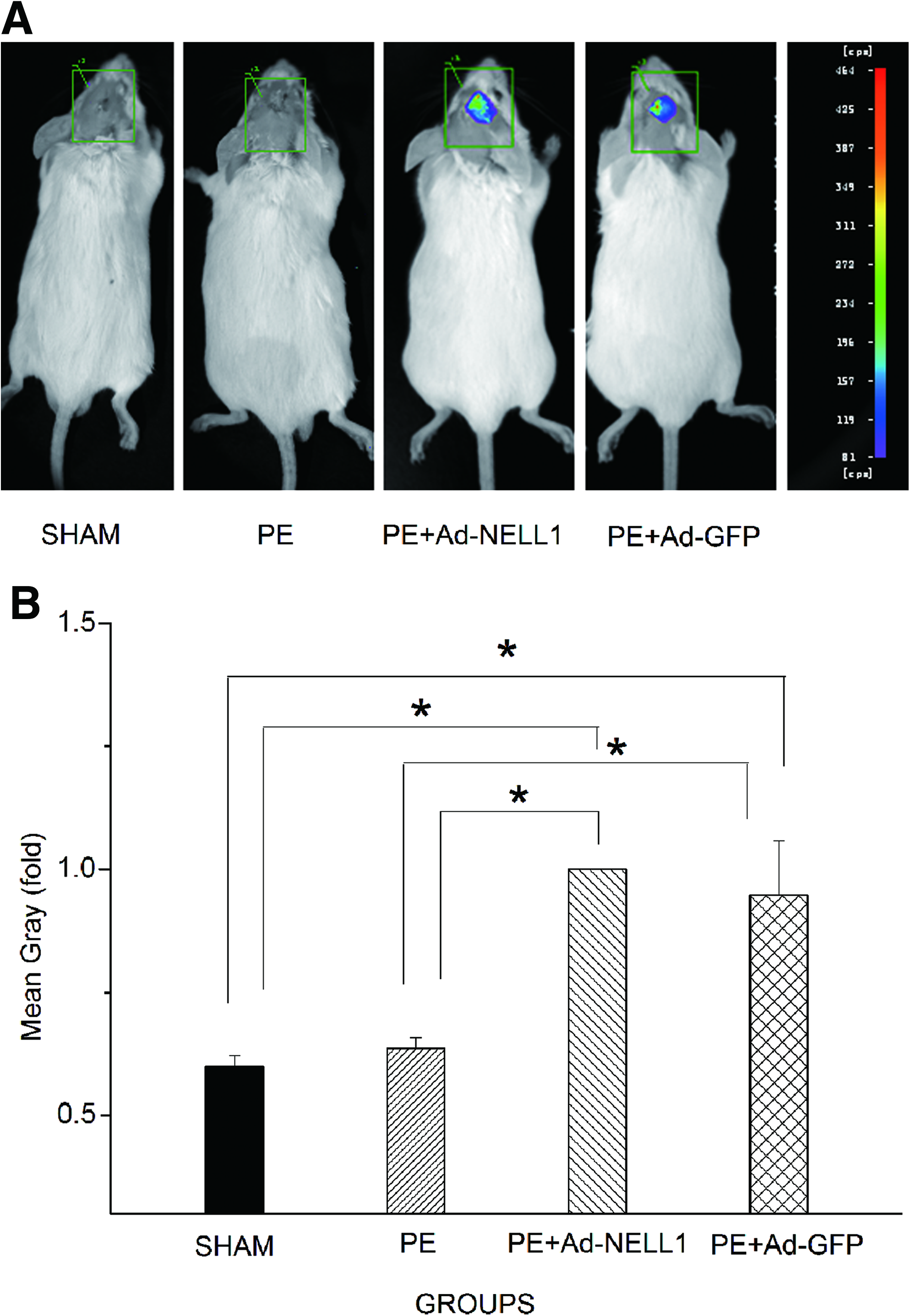
Effective adenoviral NELL1 gene delivery to calvarial lesion.
Micro-CT evaluation
Three-dimensional micro-CT images of mice skulls were reconstructed showing the successful induction of the osteolysis of murine calvaria by Ceridust 3615 PE particles in the current model (Fig. 2A, B). In addition, the calvarial bones with a continuous and smooth surface were revealed in the PE-implanted samples with Ad-GFP-NELL1 treatment compared with those with Ad-GFP treatment (Fig. 2C, D) or PE controls (Fig. 2B).
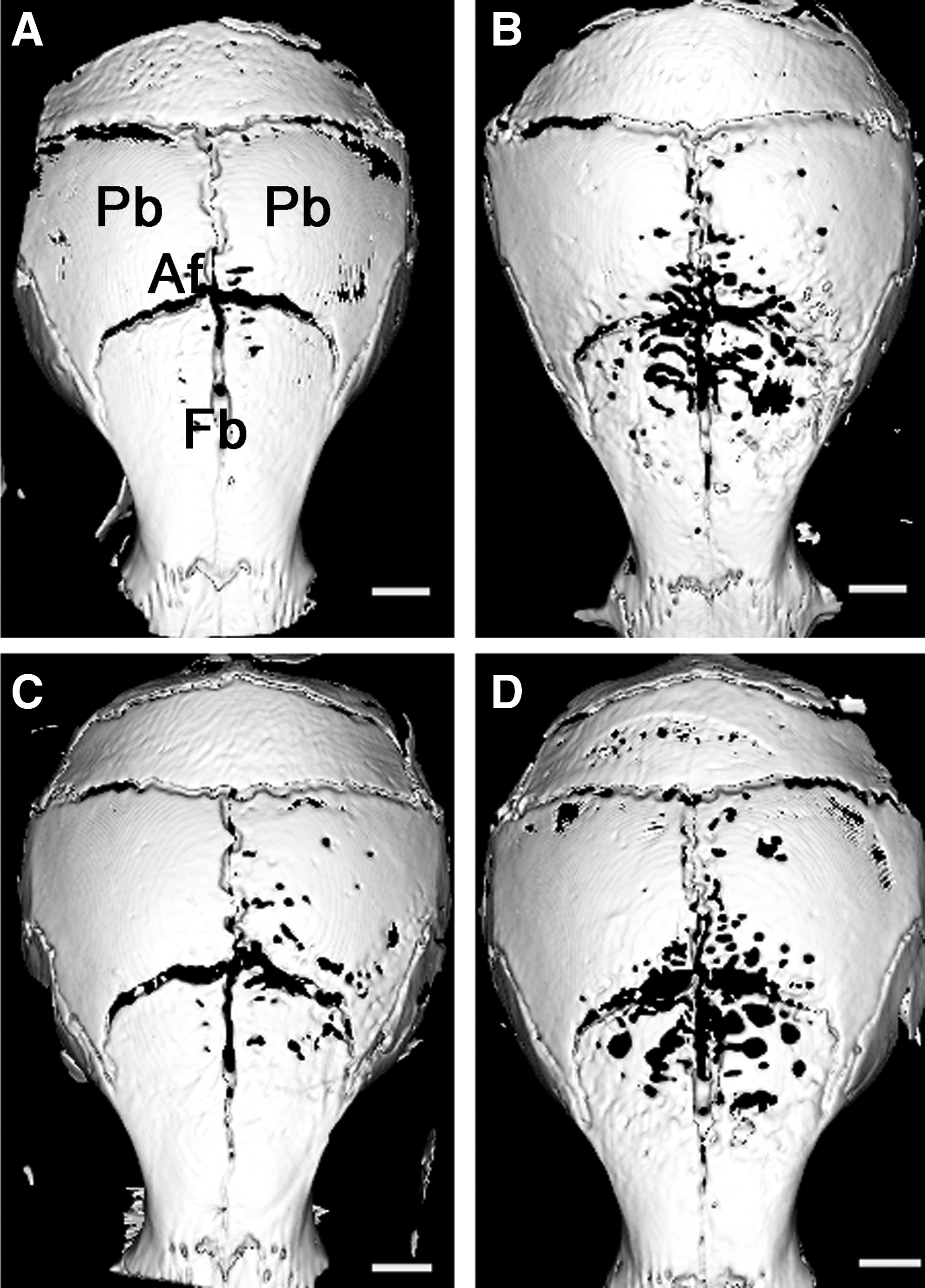
Three-dimensional micro-CT reconstruction images for skulls from four different treatment groups.
Based on an equivalent ROI (Fig. 3A), BV significantly increased with PE+Ad-GFP-NELL1 than with PE+Ad-GFP and PE treatment (p<0.01, Fig. 3B), with no differences between the PE+Ad-GFP and SHAM groups (p>0.05). Similarly, the BMD for the PE+Ad-GFP-NELL1 group, close to the SHAM group, was much higher than that for the PE+Ad-GFP and PE control groups (p<0.05, Fig. 3C). The homoplastic tendency emerged in the analysis of Tb.Th.3D (Fig. 3D), an important trabecular bone parameter. BS/BV, as a two-dimensional (2D) parameter, could indicate osteolysis more representatively. All values were lower for SHAM and PE+Ad-GFP-NELL1 groups than for PE+Ad-GFP and PE control groups (p<0.01, Fig. 3E), so NELL1 increased the BV and density, specifically indicating its capacity of compensation for the osteolysis. Data for another 2D parameter, connectivity density, further confirmed these results (Fig. 3F).
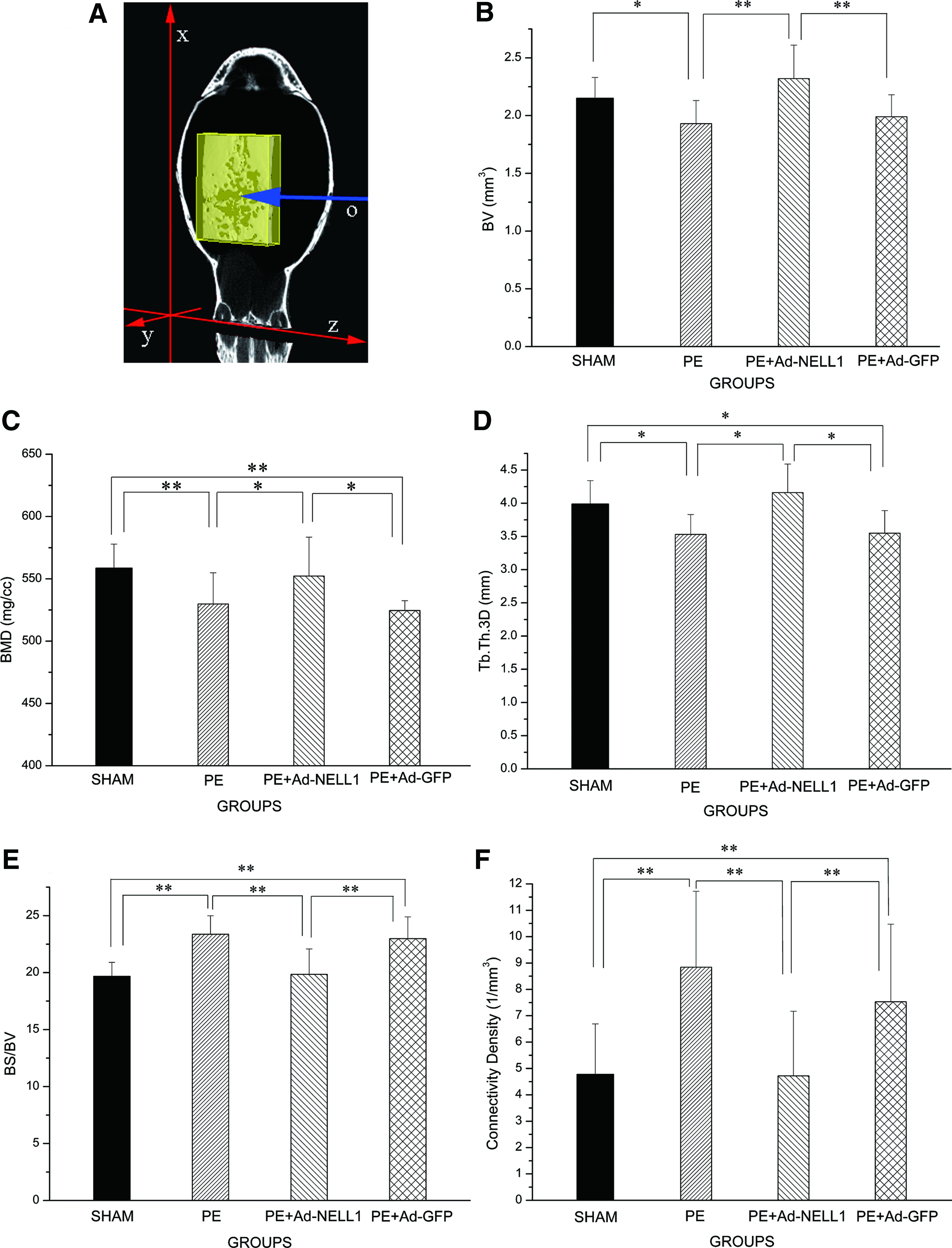
The quantitative analysis of micro-CT in the standardized ROI.
Histology
A histological evaluation of the murine calvaria model of wear debris-induced osteolysis on HE sections clearly indicated that PE treatment resulted in abundant bone resorption as compared with SHAM treatment. This effect was reduced with PE+Ad-GFP-NELL1 (Fig. 4). Despite the inflammation, the new bone with NELL1 well compensated for the bone loss observed in the Toluidine blue staining. Combined with the micro-CT analysis data, it indicated that the new bone mass was much more in the PE+Ad-GFP-NELL1 group than in the PE+Ad-GFP and PE control groups.
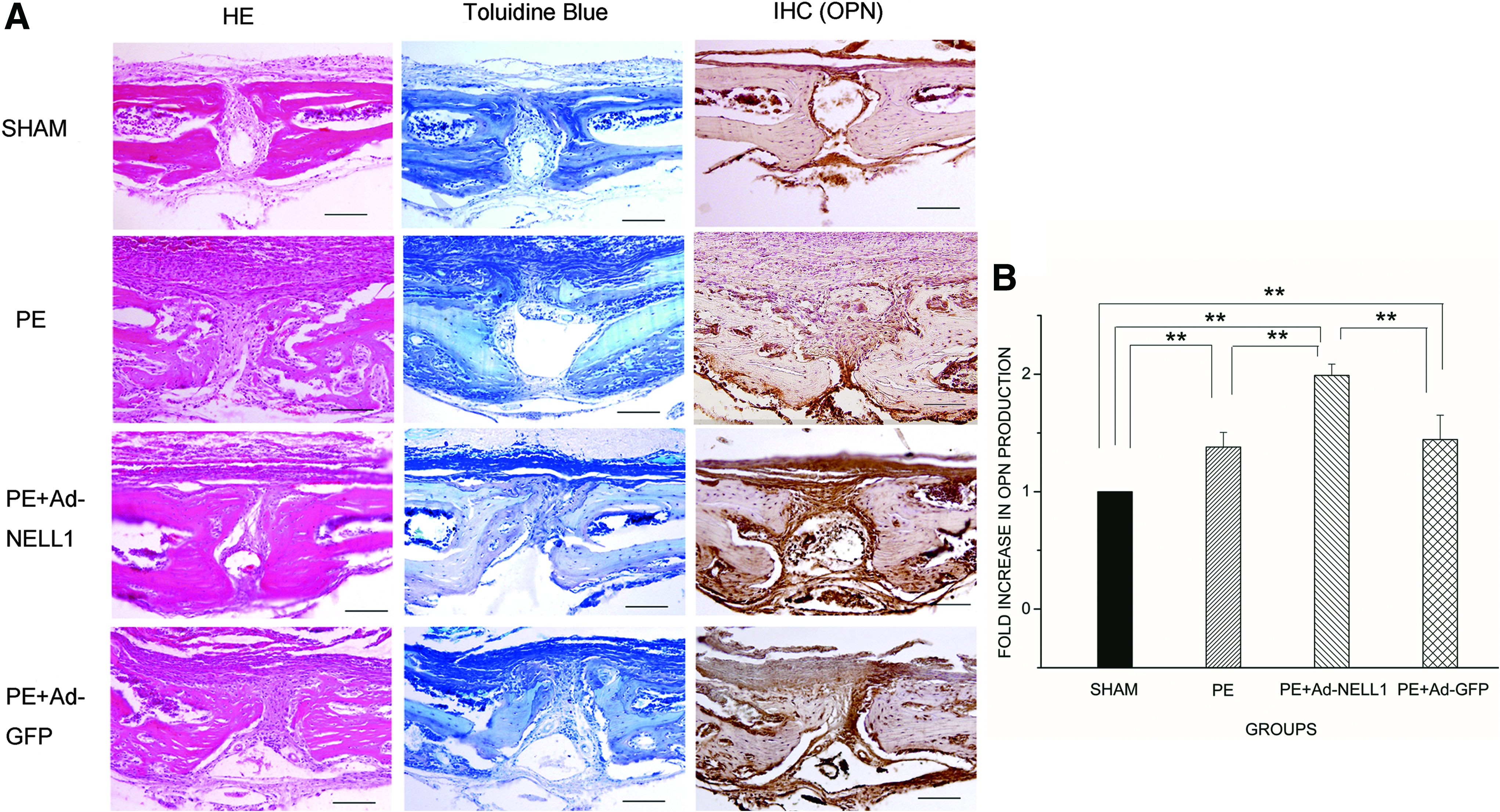
Histological and immunohistochemical evaluations of calvarial tissue in the ROI for micro-CT analysis.
Immunohistochemistry of OPN, a bone-relevant marker gene, was performed to expose the mechanisms of NELL1 compensating the osteolysis. Positive staining was detected on the surface of new bone in all bone regeneration areas (Fig. 4A). Consistent with the previous results, 6 there was a significantly higher OPN level exhibited in osteoblasts and periosteum with PE+Ad-GFP-NELL1 samples than with other treatments (Fig. 4B).
Real-time PCR
Neither was NELL1 mRNA detected in SHAM, PE, and PE+Ad-GFP mice, nor was NELL1 detected in NELL1 mouse livers or lungs (Fig. 5A). This result indicates that the high transduction efficiency of Ad-GFP-NELL1 has been achieved in our model, and there is no systemic dissemination of adenoviral NELL1 through a regional injection. The levels of the osteogenesis genes, OPN and Runx-2, were higher with Ad-GFP-NELL1, compared with that of RANKL, an essential mediator required to promote osteoclastogenesis (Fig. 5B).
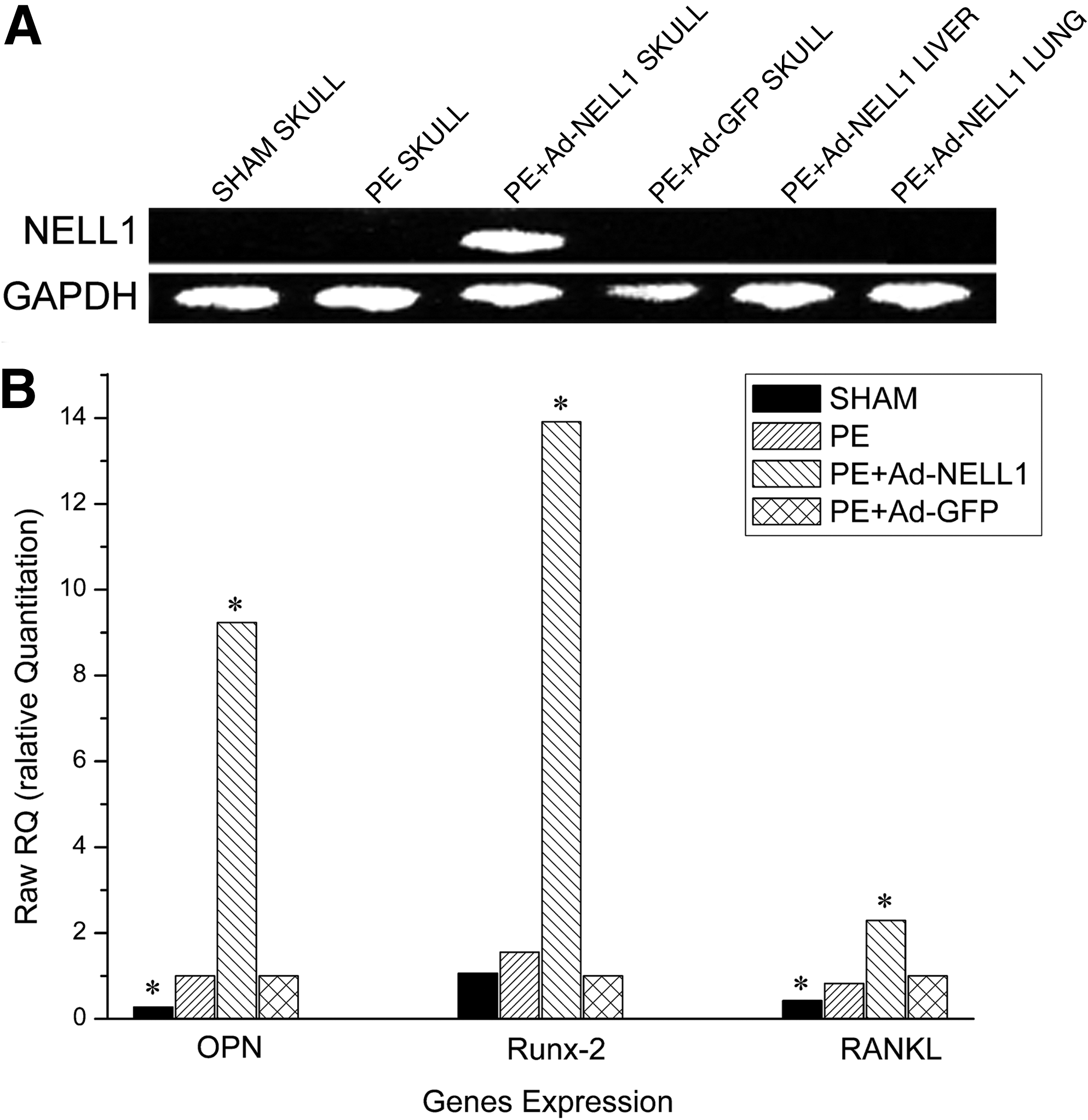
Gene expression of NELL1, osteoblastic, and osteoclastic molecules. Panel
Biomechanical properties
In indentation analysis, both the load and displacement of bone were almost linearly changed (data not shown). The slope of the curve (40% and 90%), stiffness, was calculated as 4.94±0.75, 0.63±0.20, 2.61±0.13, and 0.85±0.10 N/mm for SHAM, PE, PE+Ad-GFP-NELL1, and PE+Ad-GFP groups, respectively. The Young's modulus values were 4.74±0.72, 0.60±0.20, 2.50±0.12, and 0.82±0.09 MPa, accordingly. The elastic modulus was significantly higher for SHAM than for the other three groups, while that for the NELL1 group was much higher than that for the PE and PE+Ad-GFP groups (p<0.01, Fig. 6).
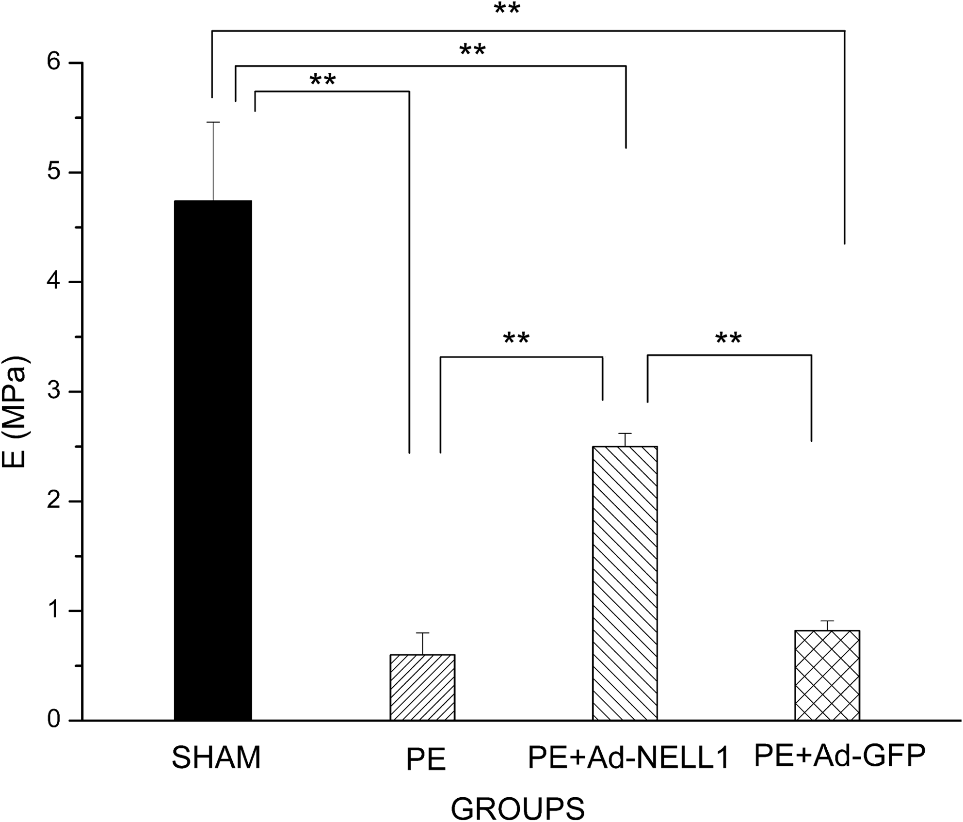
The quantified analysis of four groups. Young's modulus in the skulls' anterior fontanel areas (n=5, per group; **p<0.01).
Discussion
This study was designed to determine the therapeutic effects of NELL1 gene transfer in treating PE particle-associated periprosthetic bone osteolysis, using a well-established murine calvarial model. We found that the PE particles stimulation significantly increased the bone loss, but marked bone regeneration occurred with Ad-GFP-NELL1 transduction. Moreover, the biomechanical properties of the NELL1-treated mouse skulls were significantly improved. Thus, NELL1 is a potential therapeutic or preventive agent that treats osteolysis by regenerating bone.
Although the mechanisms are complicated, the positive correlations between the particles and osteolysis have been proved. Studies have suggested that wear debris may promote the aseptic inflammation at first, and then recruit inflammatory cells; the subsequently secreted factors such as interleukin (IL)-6 and tumor necrosis factor-α may cause bone resorption, which leads to prosthesis failure.22,23 Although Campbell et al. have proved that the clinical retrieved size of a particle is the submicron, which may be ideal for animal models, 24 Green et al. evaluated the in vitro response of murine peritoneal macrophages to PE particles of definitive size distributions at varying volume doses, and demonstrated that the most biologically active PE particles are in the phagocytosable size range 0.3–10 μm. 13 The volume of applied particles is also a critical factor in addition to the particle size for the induction of different cellular responses. For instance, the expression level of VEGF, but not IL-8, in the cells exposed to 0.5 mg/mL Ceridust 3615 was equal to that in cells exposed to 0.5 mg/mL retrieved PE particles. 25 The tissue reaction to PE particles revealed the formation of fibrous granulomatous tissues centered around the sagittal suture area, which was accompanied by massive bone resorption and the formation of osteoclasts bordering the cortex. 14 Attenuation of the debris-associated inflammation and reversion of implants surrounding bone resorption could be considered a therapeutic strategy. However, once the bone has been lost, the method is limited. In the current study, we investigated whether regenerated bone mass could neutralize the bone resorption, and form a new balance to decrease, or prevent, the osteolysis.
NELL1, as a novel osteoinductive factor, has been identified as being capable of promoting osteochondrogenic cell differentiation and mineralization in vitro and bone regeneration in multiple animal models.6,9,10,12 The absence of the protein's application in the osteolysis field resulted in this study. Micro-CT is a useful and promising method that precisely evaluates the amount of osteolysis induced by particle loading. Using an equivalent ROI, we found that the NELL1 enhanced bone formation, increased BV, and also repaired the structures of the bone resorption area. The decreased BS/BV indicated that the new bone mass was fixed and orderly organized rather than irregularly disorganized, which is consistent with other results. 17 Connectivity density further validated the results. In the osteolysis models of this experiment, we found that major bone resorption occurred in the frontal and anterior part of parietal bones where our ROI was defined. The threshold chosen by the 3D view could effectively differentiate the four treatment groups.
On histology, OPN was higher with the Ad-GFP-NELL1 transduction. Furthermore, the mRNA expression of OPN was detected concordantly by real-time PCR. Runx-2 was upregulated with NELL1, which disagreed with a previous study. 26 The possible explanation is that there may be a potential pathway between NELL1 and Runx-2 through osteoclasts, in which NELL1 elevates Runx-2. RANKL, an essential mediator that is secreted by osteoblasts through osteoblast and osteoclast interaction, 27 binds to its physiologic signaling receptor, RANK, on the membranes of macrophages and osteoclast precursors, subsequently providing the signals required for their survival, maturation, and activation. 28 The data shown in Figure 5 suggest that NELL1 may play an important role between osteoblasts and osteoclasts in the RANKL-RANK pathway, as a novel mechanism in the osteogenesis. The definite effect on osteoclasts needs confirmation in future studies. Despite remarkable results in structure and molecular analysis, the biomechanical properties of NELL1 specimens could not match those of the SHAM ones. We hypothesized that if the therapeutic time were prolonged, then the differences would disappear. Similar results were found in a rat femoral distraction osteogenesis model. 6 However, in the calvarial osteolysis model, the investigation was limited because of the autotherapy in 20 days. Therefore, another long-time osteolysis model in rat tibia has been ongoing in our lab.
In the well-established calvarial osteolysis model, Ad-GFP-NELL1 is effective for treating wear debris-associated osteolysis by promoting bone regeneration. It could be a new avenue for osteolysis research and application. Further studies are needed to verify the therapeutic effect in a long-term osteolysis model, which is closer to the clinical process. Besides, a better understanding of the molecular mechanisms of the therapeutic influences and of the safety concerns are needed, for a more effective method for using NELL1 in clinical stages.
Footnotes
Acknowledgment
This work was supported by the National Natural Science Foundation of China (No. 30872621, No. 30872633, and No.30930092) and the Special Project of the “Twelfth Five-year Plan” for Medical Science Development of PLA (BWS11J025).
Disclosure Statement
K.T. and X.Z. are cofounders of Bone Biologics, Inc., which licensed the patent of Nell1 in bone regeneration filed from UCLA.