
Abstract
Cells in culture deposit a complex extracellular matrix that remains intact following decellularization and possesses the capacity to modulate cell phenotype. The direct application of such decellularized matrices (DMs) to 3D substrates is problematic, as transport issues influence the homogeneous deposition, decellularization, and modification of DM surface coatings. In an attempt to address this shortcoming, we hypothesized that DMs deposited by human mesenchymal stem cells (MSCs) could be transferred to the surface of polymeric scaffolds while maintaining their capacity to direct cell fate. The ability of the transferred DM (tDM)-coated scaffolds to enhance the osteogenic differentiation of undifferentiated and osteogenically induced MSCs under osteogenic conditions in vitro was confirmed. tDM-coated scaffolds increased MSC expression of osteogenic marker genes (BGLAP, IBSP) and intracellular alkaline phosphatase production. In addition, undifferentiated MSCs deposited significantly more calcium when seeded onto tDM-coated scaffolds compared with control scaffolds. MSC-seeded tDM-coated scaffolds subcutaneously implanted in nude rats displayed significantly higher blood vessel density after 2 weeks compared with cells on uncoated scaffolds, but we did not observe significant differences in mineral deposition after 8 weeks. These data demonstrate that DM-coatings produced in 2D culture can be successfully transferred to 3D substrates and retain their capacity to modulate cell phenotype.
Introduction
The cell-instructive cues presented by the extracellular matrix (ECM) offer an alternative and potentially more translational approach toward accelerating osteogenic differentiation of MSCs. Proteins and proteoglycans present in native bone ECM play a vital role in modulating the proliferation, migration, and differentiation of bone forming cells.11,12 These same proteins and peptide motifs have also demonstrated the capacity to directly modulate MSC phenotype when coated onto synthetic cell culture substrates in both two- and three-dimensions.13–15
Decellularized matrix (DM) coatings represent a novel approach to confer bioactivity to synthetic substrates through the addition of a complex, physiologically relevant ECM to biomaterial surfaces. DM coatings are produced by first culturing matrix-depositing cells upon a substrate, followed by the removal of those cells such that the underlying matrix remains intact. Subsequently, cultured cell populations are then able to directly interact with residual ECMs representative of different cell and tissue types. DM coatings have previously demonstrated the capacity to both accelerate the osteogenic differentiation of MSCs and to extend their multipotency to higher passages in culture.16–18
The successful application of DM coatings to 3D constructs currently requires customized perfusion flow bioreactors to minimize DM heterogeneity resulting from poor oxygen and nutrient diffusion to matrix-depositing cells.19,20 This technique may prove costly and time consuming in the eventual scale up of DM coatings to customized therapeutic implantable materials of differing geometries. In an attempt to address this issue, we recently developed a novel approach toward the preparation of DM-coated culture surfaces. 21 DMs deposited on standard tissue culture plastic (TCP) can be removed, solubilized, and transferred to alternate substrates, while retaining their cell-instructive properties.
We therefore hypothesized that DMs transferred from TCP (transferred DMs [tDMs]) would retain the capacity to modulate MSC osteogenic differentiation when used as a coating on 3D polymeric constructs. To test our hypothesis, we coated scaffolds formed of poly(lactide- co-glycolide; PLG), a common biodegradable polymer utilized in bone tissue engineering, with tDMs. We then analyzed the ability of tDM-coated scaffolds to accelerate the osteogenic differentiation of MSCs under osteogenic culture conditions in vitro through an analysis of multiple markers of differentiation. We also assessed the capacity of tDM-coated constructs to drive MSC differentiation in vivo when implanted in an ectopic site. As the induction of MSCs toward the osteogenic lineage in culture has been previously shown to boost their capacity to form bone in vivo, 22 we also analyzed whether osteogenic differentiation could be enhanced by seeding osteogenically induced MSCs on tDM-coated substrates.
Materials and Methods
Scaffold preparation
Macroporous scaffolds were fabricated using a gas foaming/particulate leaching method as described. 23 Briefly, microspheres composed of PLG (85:15 DLG 7E; Lakeshore Biomaterials) were prepared using a double-emulsion process. Lyophilized PLG microspheres were mixed with NaCl particles (250–425 μm in diameter) and compressed into solid disks (final dimensions: 8.5 mm diameter and 1.5 mm thickness) in a custom-made stainless steel die using a Carver press (Fred S. Carver) for 1 min. Compressed disks were then exposed to high pressure CO2 gas (5.5 MPa) for 16 h followed by rapid pressure release. NaCl particles were leached from scaffolds by submersion in distilled H2O for 24 h. Scaffolds were functionalized with 0.1 N NaOH for 1 min and placed in a sealed 50 mL Steriflip (Millipore) conical tube with 95% EtOH under gentle vacuum for 30 min for sterilization. Scaffolds were then rinsed twice under vacuum with sterile phosphate-buffered saline (PBS) and used immediately.
Cell culture
Human bone marrow-derived MSCs (Lonza) were expanded in minimum essential alpha medium (α-MEM w/L-glutamine, w/o ribo/deoxyribonucleosides; Cat. #12000-022; Invitrogen) supplemented with 10% fetal bovine serum (FBS; JR Scientific) and 1% penicillin and streptomycin (P/S; Mediatech). Cells were cultured under standard conditions and utilized at passages 4–6. Supplemented medium (SM) containing 50 μg/mL ascorbate-2-phosphate was utilized on all cells for one passage prior to experimental use to prime cells for enhanced matrix deposition. 24 Osteogenically induced cells were cultured in osteogenic medium (OM) further supplemented with 10 mM β-glycerophosphate and 10 nM dexamethasone for 5 days prior to use.
Preparation of DMs
DMs were prepared as previously described. 18 Briefly, MSCs were seeded on six-well plates at 50,000 cells/cm2 and cultured in SM (α-MEM, 10% FBS, 1% P/S, and 50 μg/mL A2P) for 15 days under standard culture conditions with media changes performed every 3 days. Wells were then rinsed with PBS and treated with 0.5% Triton X-100 (Sigma Aldrich) in 20 mM NH4OH in PBS for 5 min at 37°C. Following an additional PBS rinse, wells were treated with DNAse (Sigma; 150 units/mL PBS) for 1 h at 37°C and again rinsed in PBS. Plates were then allowed to dry in a sterile biosafety cabinet for up to 12 h and stored at room temperature in the dark for up to 1 month prior to use.
Transfer of DMs to 3D scaffolds
Homogenized DM solutions were prepared using methods similar to that previously described. 21 Six-well DM-coated plates were scraped in the presence of sterile 0.02 N acetic acid (50 μL per well), DM contents transferred to microcentrifuge tubes, and sonicated (Sonics & Materials Vibra-Cell VCX130PB) on ice with 2–3 s pulses 10–15 times to mechanically homogenize DM contents. Quantification of tDM protein solutions was performed with a BCA protein assay (Thermo Scientific; # 23227) per manufacturer's instructions. DM contents were diluted to a final concentration of 500 μg per mL in acetic acid prior to deposition on macroporous scaffolds.
Sterilized scaffolds were placed on sterile gauze to remove excess PBS and transferred to the surface of a Steritop (Millipore) vacuum filter attached to a 500 mL glass bottle. Fifty microliters of DM solution (acetic acid only for sham-coated scaffolds) was then added drop wise to the surface of each scaffold via micropipettor, followed by the application of a gentle vacuum to the bottom of the scaffolds to ensure absorption of the DM solution. When they appeared dry (∼15–30 min), scaffolds were inverted and an equal volume of DM solution was applied to their opposite side. Scaffolds were then allowed to dry overnight under vacuum in a sterile biosafety cabinet. Scaffolds were rinsed in cell culture media (2×30 min) prior to cell seeding.
Cell seeding
tDM and sham-coated scaffolds were seeded with either undifferentiated or osteogenically induced MSCs. MSCs (7.5×10⁵ cells per scaffold) were suspended in 35 μL of media and applied drop wise to the scaffold surface. Scaffolds were placed in standard cell culture incubators (37°C, 5% CO2) for 3 h to allow cell attachment. Scaffolds were then transferred to 12-well plates containing 2 mL of their respective media (SM for undifferentiated cells; OM for osteogenically induced cells) per well and placed on an XYZ shaker (Stovall) overnight. Scaffolds intended for implantation in vivo were utilized the following morning. For scaffolds used for in vitro analysis, media was exchanged for OM the following day and every 3 days thereafter.
Scaffold characterization
Quantification of tDM protein solutions applied to scaffold surfaces was carried out by BCA protein assay per manufacturer's instructions. MSC-seeded scaffolds cultured in vitro were collected at various time points and imaged for morphological analysis. Scaffold porosity was analyzed using Archimedes' principle as previously described. 23 Scaffolds were also analyzed by SEM to assess matrix coverage and scaffold surface morphology. Scaffolds were gold sputter coated and imaged at 20× and 2500× using a Hitachi S3500-N.
DNA quantification
Total DNA present in scaffolds was determined using the Quant-iT PicoGreen dsDNA kit (Invitrogen). Briefly, cell-seeded scaffolds were rinsed in PBS, minced, and placed in passive lysis buffer (Promega). Following a freeze thaw cycle, the lysate was sonicated briefly, separated from the scaffold material via centrifugation, and quantified in comparison to a known standard curve.
Osteogenic marker analysis
Polymerase chain reaction
Total RNA from scaffolds was collected following 1 or 2 weeks in culture. Scaffolds were rinsed 1× in sterile PBS, minced with a sterile scalpel, and placed in 350 μL of RLT buffer (Qiagen) supplemented with 10 μL/mL β-mercaptoethanol. Total RNA was purified using the RNeasy Mini kit (Qiagen) and 400 ng of total RNA reverse transcribed with the QuantiTect Reverse Transcription Kit (Qiagen). Quantitative polymerase chain reaction (qPCR) was performed using TaqMan1 Universal PCR Master Mix (Applied Biosystems) on a Mastercycler1 realplex2 (Eppendorf); primers and probes for BGLAP (Hs01587814_g1), IBSP (Hs00173720_m1), MRPL13 (Hs00204173_m1), RUNX2 (Hs00231692_m1), SP7 (Hs01866874_s1), and VEGFA (Hs00900055_m1) were purchased from Applied Biosystems. Amplification conditions were 50°C for 2 min and 95°C for 10 min, followed by 40 cycles at 95°C for 15 s and 60°C for 1 min. Quantitative PCR results were normalized to RPL13 transcript level to yield ΔCt values.
Alkaline phosphatase and calcium quantitation
Intracellular alkaline phosphatase (ALP) from MSCs seeded on tDM and sham-coated scaffolds was quantified using a PNPP colorimetric assay at 405 nm as described. 25 ALP activity was normalized to DNA content determined as described above. Total calcium present on MSC-seeded scaffolds was measured using an OCOP colorometric assay similar to that previously described. 25 Briefly, minced scaffolds were incubated in 0.9 N H2SO4 overnight to solubilize surface calcium deposits. Calcium concentration in solution was then quantified and compared to a known standard curve.
Ectopic model of bone formation
Treatment of experimental animals was in accordance with University of California, Davis Animal Care guidelines and all National Institutes of Health animal-handling procedures. Ten-week-old male nude rats (n=6 per group) were anesthetized and maintained using 2% isoflurane with O2 flow at 2 L/min delivered through a mask. Four pockets were created in the dorsum, and scaffolds were subcutaneously implanted. Scaffolds were collected following euthanization at 2 and 8 weeks. Scaffolds were fixed overnight in 10% formalin followed by storage in 70% ethanol at 4°C. Scaffolds were demineralized (8 week only) overnight prior to histological analysis with CalciClear (National Diagnostics), then bisected, paraffin embedded, and sectioned at 5 μm.
Vessel quantification
Vessel quantification was performed 2 weeks postimplantation using hematoxylin and eosin (H&E) stained scaffold cross-sections at 100× magnification similar to that previously described. 23 Vessels were enumerated from H&E-stained sections by counting circular structures with well-defined lumens containing erythrocytes. The presence of human cells was determined by immunohistochemistry using antibodies for human nuclear antigen (HNA, MAB1281, Millipore; 1:20) and a mouse specific HRP/DAB detection kit (Abcam; ab64259).
MicroCT analysis
Scaffolds removed from animals after 8 weeks were assessed for mineralization prior to decalcification for histology. Bone volume fraction (BVF) and bone mineral density (BMD) were determined using microcomputed tomography. Bone tissue in the reconstructed images was determined by thresholding (>85 mg HA/cm3) to partition mineralized tissue from fluid and soft tissues. BVF was determined by dividing the number of pixels representing bone tissue (bone volume) by the number of pixels in the cylindrical segment (total volume). The mean density of the bone material (BMD) was calculated as the average density (mg HA/cc) of the BVF.
Statistical analysis
Results are expressed as mean±standard error of the mean. Statistical analyses were performed by ANOVA followed by Student–Newman–Keuls post hoc tests assessing significance to probability values (p)<0.05.
Results
Characterization of tDM-coated scaffolds
Macroporous PLG scaffolds were coated with 52.5±4.8 μg of tDM protein as determined by BCA protein quantification. DM decellularization, and the retention of several key proteins and proteoglycans present in bone ECM, has previously been established.18,21 tDM-coating of PLG scaffolds was characterized by microscopic- and macroscopic analysis. Scanning electron microscopy at 20× magnification revealed a well-distributed coating of DM material over the pores on the scaffold surface (Fig. 1A) when compared with uncoated scaffolds (Fig. 1B). Upon examination at higher magnification (2500×), we detected distinct differences in surface morphology between tDM-coated and uncoated scaffolds at the cellular scale (10 μm). At this magnification, the surfaces of coated scaffolds exhibited a rough topography (Fig. 1C), while control scaffolds were smoother (Fig. 1D). Coomassie brilliant blue total protein staining of scaffolds demonstrated relatively uniform distribution of protein throughout the scaffold surface (Fig. 1E), while little to no apparent staining was seen on uncoated scaffolds (Fig. 1F). Although surface porosity appeared slightly reduced following tDM-coating, an analysis of bulk scaffold porosity between tDM-coated and uncoated scaffolds revealed no significant differences (data not shown).

Characterization of transferred decellularized matrix (tDM) scaffold coating. SEM images of tDM-coated scaffolds at 20×
tDM-coated (tDM) and uncoated (C) scaffolds were then seeded with either undifferentiated (−) or osteogenically induced (+) MSCs. Cell-seeded scaffolds were monitored for morphological differences at 1, 3, and 5 weeks. Scaffold curling, thought to occur as a result of cell-mediated mechanical forces applied to the scaffold surface, was evident at increasing magnitudes for tDM scaffolds seeded with both cell types (Fig. 2A–C), while noncoated scaffolds displayed little to no curling (Fig. 2D–F). Total DNA quantification performed on each cell/scaffold combination revealed significantly more cells on tDM-coated scaffolds compared with control scaffolds (Fig. 2G).
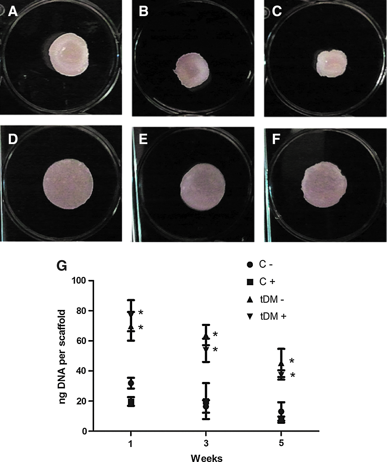
Analysis of the cellular deformation of tDM-coated scaffolds. Images of undifferentiated mesenchymal stem cell (MSC)-seeded tDM-coated and uncoated scaffolds at 1
In vitro osteogenic response to tDM-coated scaffolds
Undifferentiated and osteogenically induced MSCs were cultured on tDM and uncoated scaffolds and monitored for the expression of several different markers of osteogenic differentiation. qPCR analysis was utilized to assess MSC expression of osteocalcin (BGLAP) and bone sialoprotein (IBSP), two markers of mature osteoblast function. Osteocalcin expression was significantly enhanced in osteogenically induced MSCs cultured on tDM-coated scaffolds at 1 and 2 weeks (Fig. 3A). MSC bone sialoprotein expression was significantly enhanced by induction or the presence of a tDM-coating at 1 week, with only the tDM-coating significantly boosting expression after 2 weeks (Fig. 3B). The expression of RUNX2 and SP7 (osterix), two transcription factors related to osteogenic differentiation, was significantly lower in induced MSCs and those cultured on tDM-coated scaffolds after 2 weeks (Fig. 3C, D). Finally, MSC expression of vascular endothelial growth factor (VEGF) was assessed under each condition as a marker of the angiogenic potential of MSCs. While the combination of osteogenically induced MSCs and control scaffolds resulted in the lowest VEGF expression at 2 weeks, induced cells cultured on tDM-coated scaffolds displayed significantly greater VEGF expression (Fig. 3E).
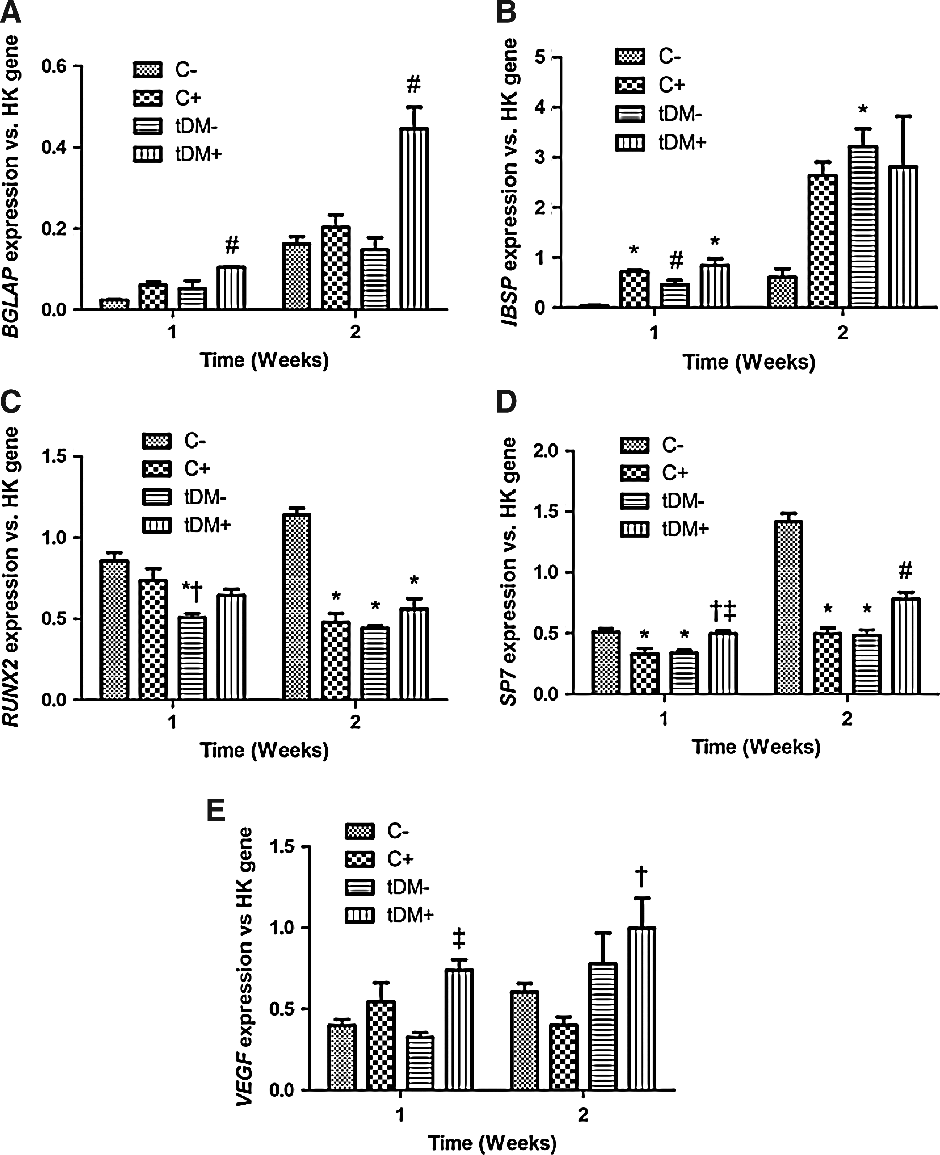
Quantitative polymerase chain reaction analysis of MSC gene expression. tDM-coated [tDM] and uncoated [C] scaffolds seeded with undifferentiated [−] or osteogenically induced MSCs [+] collected after 1 and 2 weeks. *p<0.05 versus C−; †p<0.05 versus C+; ‡p< 0.05 versus tDM−; #p<0.05 versus all other groups (n=3–6).
ALP activity within MSCs seeded on tDM- and uncoated scaffolds also displayed significant differences (Fig. 4). While the preconditioning of MSCs in OM for 5 days significantly upregulated ALP expression at the time of seeding, both MSC populations cultured on tDM-coated scaffolds had surpassed control-seeded cells in ALP expression by 1 week. MSCs also displayed significantly higher ALP expression when cultured on DM-coated scaffolds compared with control scaffolds at 3 and 5 weeks postseeding.
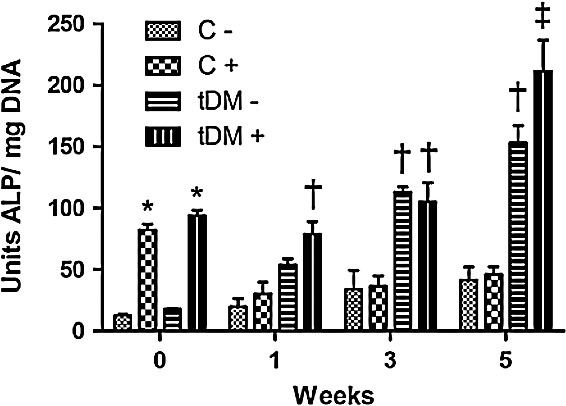
MSC intracellular alkaline phosphatase (ALP) quantification. tDM-coated [tDM] and uncoated [C] scaffolds seeded with undifferentiated [−] or osteogenically induced MSCs [+] collected at 0, 1, 3, and 5 weeks. *p<0.05 versus C- and tDM−; †p<0.05 versus C− and C+; ‡p<0.05 versus all other groups (n=4).
Total calcium deposition from undifferentiated and osteogenically induced MSCs cultured on tDM-coated and control scaffolds was quantified after 1, 3, and 5 weeks. Osteogenically induced MSCs cultured on DM-coated substrates deposited the most calcium over the 5-week culture period, significantly more than all other groups at both 3 and 5 weeks (Fig. 5). In addition, osteogenic induction of MSCs or the culture of MSCs on DM-coated scaffolds alone also resulted in significantly greater calcium deposition compared with undifferentiated MSCs on control scaffolds at week 5.

Quantification of total calcium deposition on MSC-seeded scaffolds. tDM-coated [tDM] and uncoated [C] scaffolds seeded with undifferentiated [−] or osteogenically induced MSCs [+] collected at 0, 1, 3, and 5 weeks. *p<0.05 versus C−; †p<0.05 versus all other groups (n=4).
In vivo osteogenic response to tDM-coated scaffolds
Undifferentiated and osteogenically induced MSCs seeded onto tDM-coated and control scaffolds were subcutaneously implanted in nude rats to assess the efficacy of the tDM-coating to modulate MSC activity in vivo. Histological sections taken from scaffolds retrieved at 2 weeks revealed significant differences in vessel formation (Fig. 6). Vessel density within the scaffold perimeter was significantly increased in scaffolds coated with tDM compared with uncoated scaffolds, and in scaffolds seeded with undifferentiated MSCs compared with osteogenically induced MSCs. MSC death or migration from the implant site appeared to have occurred by 2 weeks, as suggested by negative immunohistochemical staining for human nuclear antigen (data not shown).

Vessel quantification in implanted scaffolds. Control scaffolds seeded with undifferentiated
Excised scaffolds analyzed at 8 weeks postimplantation revealed no significant differences in scaffold mineralization. MicroCT scans of the scaffolds indicated low levels of mineral formation around the edges of the scaffolds, with a trend for increased mineralization on uncoated scaffolds (Fig. 7A, B) compared with tDM-coated scaffolds (Fig. 7C, D). Quantitative measurements of BVF also presented a trend for less mineralization in DM-coated scaffolds (Fig. 7E). A subsequent analysis of BMD for the mineral detected within each group indicated a trend for highest BMD in the undifferentiated MSC-seeded tDM-coated scaffolds; however, this result was not significant (p=0.086).

MicroCT analysis of scaffold mineralization. Image slices from uncoated scaffolds seeded with undifferentiated
Discussion
Current strategies to coat 3D substrates with cell-secreted matrices place matrix-depositing cells directly upon the target material surface.26,27 These cells may suffer from inadequate transport of oxygen and nutrients when not adjacent to the construct surface, potentially leading to the deposition of a heterogeneous ECM and even cell death prior to decellularization. While the use of bioreactors represents one approach to overcome this problem, this strategy requires customized, potentially costly equipment and relies on shearing forces, which themselves may alter matrix deposition and architecture. The goal of this study was to demonstrate that DM coatings engineered to modulate cell phenotype in 2D culture can be used to coat 3D implantable materials, thereby imparting them with cell-instructive properties.
We successfully transferred osteogenic MSC-deposited DM coatings from 2D tissue culture plates to 3D PLG scaffolds, and observed increased expression of several markers of MSC osteogenesis over a 5-week culture period. tDM-coated substrates enhanced MSC expression of osteogenic genes representative of mature osteoblast function (BGLAP and IBSP). While the expression of osteogenic transcription factors appeared to be reduced by surface interactions with tDM-coatings, this downregulation closely matched that which occurred in the control osteogenically induced cell group (C+), and therefore is likely indicative of temporal gene expression fluctuations observed in MSCs during osteogenic differentiation.25,28 In addition, ALP production, total calcium deposition, and the quantity of cells present within tDM-coated PLG scaffolds were significantly higher over the 5-week culture period. Increased cell numbers within tDM-coated scaffolds following seeding may result from increased scaffold surface area and/or the availability of matrix-related cell binding sites due to the presence of tDM. We previously demonstrated that MSCs differentially utilize distinct integrins when attaching to TCP compared with DM-coated TCP, and similar effects may be present in this 3D system. 21 Although total cell viability decreased over time for all groups, likely resulting from transport limitations associated with near static culture, the potential for tDM coatings to enhance cell survival merits further examination.
The increased expression of osteogenic markers from MSCs cultured on tDM-coated scaffolds demonstrates the utility of this technique in boosting stem cell differentiation in 3D culture. The combination of osteogenic signals from media supplements and ECM cues from tDM-coated substrates was more effective in driving MSC osteogenic differentiation than OM alone. While induction of MSCs in OM provided a slight boost in the expression of several osteogenic markers, this advantage was also further enhanced when combined with the presence of a tDM coating. Modulations in osteogenic gene expression, and enhanced ALP production from MSCs cultured on tDM-coated scaffolds, was indicative of robust differentiation at the cellular level. Further, total calcium deposition within scaffolds demonstrated the effectiveness of tDM coatings at improving the overall functional output of this bone forming construct.
While the pathway of DM efficacy in facilitating MSC differentiation is still poorly understood, there are likely several contributing factors. Cell-derived DMs mediate cell-substrate interactions upon coating of a material; therefore, physical interactions between cells and DMs likely play an important role in the activation of intracellular signaling pathways that determine cell fate. DM topography at the micro- and nanoscale may also contribute to the efficacy of the coating, as cell shape resulting from biomaterial surface properties can instruct cell phenotype. 29 The presence of DM coatings may partially mitigate the need for cells to remodel their local microenvironment following seeding, allowing them to react more quickly to their surrounding inductive cues. Residual growth factors present in DM coatings may also drive changes in cell phenotype. DM coatings also likely interact with serum factors present in the media or secreted from newly seeded cells and may boost the capacity of such factors to interact with the cell surface.
The tDM-coated scaffolds studied herein did not significantly enhance mineralization of MSC-seeded constructs in vivo. The choice of subcutaneous defect was made as a preliminary site of analysis due to its high vascularity in comparison to orthotopic defect sites. We also examined osteogenically induced cells in addition to the more commonly utilized undifferentiated MSCs, as they have previously demonstrated the capacity to form greater amounts of bone following implantation. 22 MSCs transplanted within tDM-coated scaffolds resulted in increased vascularity over the first 2 weeks of implantation, possibly due to increased MSC trophic factor release and/or more cells present within implanted constructs. This finding corresponds with our in vitro gene expression analysis, which indicated higher VEGF expression from MSCs cultured on tDM-coated scaffolds at 2 weeks. We also observed decreased vascularization when implanting osteogenically-induced MSCs, regardless of the substrate, potentially due to reductions in the secretion of proangiogenic factors by osteogenically differentiated MSCs. 30 Histological analysis of scaffold sections 2 weeks postimplantation revealed no discernable MSCs within the implant. These data suggest that cell death from hypoxia and/or cell migration away from the defect site are still serious limitations to consider when designing in vivo studies that measure DM efficacy in vivo. MicroCT data indicated a trend toward reduced mineralization of DM-coated scaffolds over 8 weeks. This distinct difference from our in vitro assays of scaffold mineralization may be due to the relative lack of endogenous osteogenic cues present in the subcutaneous environment. These data demonstrate the importance of considering the concentration of osteoinductive signals, whether substrate mediated or soluble cues, to drive osteogenesis. The incorporation of osteogenic cues such as growth factors or bioceramic materials may be useful in maximizing the osteogenic potential of tDM coatings in vivo. 27
The ability to engineer DM coatings on large 2D culture surfaces could potentially solve challenges related to cell survival and matrix homogeneity that occur when seeding matrix-depositing cells on 3D implantable constructs. Decellularization, modification, collection, and homogenization of DM materials carried out in a 2D environment would undoubtedly allow for the engineering of more precise and customizable DM solutions. These solution mixtures could be used to coat substrates of different sizes and shapes with concentration gradients, or even be mixed with other DMs to create ECM interfaces representative of physiologically adjacent tissues. 31 Possible disadvantages to this technique may include inherent differences in the DMs produced by cells cultured in 2D compared with those cultured in 3D. DMs secreted in 3D may possess structural or compositional characteristics that better allow them to manipulate cell behavior in the 3D environment. In addition, the method by which the cell-derived ECM is broken down prior to substrate coating will likely determine DM efficacy. 32 The conformation of proteins, proteoglycans, and ECM-bound growth factors that may be present within the DM must not be altered to the point of inactivity during transfer.
Conclusion
Biomaterial constructs coated with a cell-derived ECM can instruct cell phenotype. Current techniques used to apply such coatings are time consuming and expensive, as they require convective culture systems and direct contact between matrix-depositing cells and substrate surfaces. This study successfully demonstrates that a cell-derived ECM coating can be collected from a 2D culture substrate and transferred to a 3D implantable construct while retaining the capacity to instruct cell phenotype. This technique provides a new tool in advancing the ability of synthetic biomaterials to mimic the properties of native tissue.
Footnotes
Acknowledgments
The authors are grateful for financial support from The Hartwell Foundation (to J.K.L.) and the California Institute for Regenerative Medicine UC Davis Stem Cell Training Program (CIRM T1-00006 and CIRM TG2-01163).
Disclosure Statement
No competing financial interests exist.