
Abstract
The objective of this study was to design and prepare a new contrast agent of magnetic resonance (MR) imaging for the evaluation of therapeutic angiogenesis. Diethylenetriaminepentaacetic acid (DTPA) residue of a chelator was chemically introduced to dextran with a molecular weight of 74,000 (dextran-DTPA). Cyclic peptide containing an arginine-glycine-aspartic acid (RGD) sequence (cyclic RGD) with an inherent affinity for the αvβ3 integrin was then introduced to dextran-DTPA (Cyclic RGD-dextran-DTPA). Gd3+ was added to cyclic RGD-dextran-DTPA to prepare a dextran-based MR contrast agent (Cyclic RGD-dextran-DTPA-Gd). Cyclic RGD-dextran-DTPA-Gd had affinity for cells expressing the αvβ3 integrin and showed a higher longitudinal relaxivity compared with DTPA-Gd of an MR contrast agent clinically used. Right femoral, external iliac, and deep femoral and circumflex arteries and veins were surgically ligated to prepare a mouse model of hindlimb ischemia. A laser Doppler analysis and histological evaluation confirmed that hindlimb ischemia healed naturally and was accompanied by angiogenesis, while αvβ3 integrin was expressed in the ischemic-angiogenic region without any treatment. Mice at 7 days after vascular ligation were used as an angiogenesis model. When intravenously injected into mice with hindlimb ischemia, cyclic RGD-dextran-DTPA accumulated in the ischemic-angiogenic region and showed the MR ability to detect the ischemic-angiogenic region. It is concluded that cyclic RGD-dextran-DTPA-Gd is a promising material for evaluation of therapeutic angiogenesis.
Introduction
Molecular imaging is defined as the in vivo visualization of spatiotemporal distribution of molecular biological processes in the cell and tissue of interest.10,11 There is no doubt that the technology of molecular imaging plays an important role in the noninvasive evaluation of tissue regeneration. Molecular imaging includes the imaging probes and the corresponding imaging modality. So far, various types of imaging probe have been reported, such as Gd3+ and manganese ion Mn2+, 19 F, and iron oxide nanoparticles for magnetic resonance imaging (MRI),12,13 radioisotopes (99mTc, 111In, 123I, 18F, 64Cu, and 124I) or their derivatives for positron emission tomography (PET)14,15 and single photon emission computed tomography (SPECT),16–18 luciferase and β-galactosidase for bio-luminescence imaging,19,20 and quantum dots and near-infrared fluorescent (NIRF) dyes for fluorescence imaging.21,22 To maximize the efficacy of molecular imaging, DDS technologies contribute by increasing in the signal-to-noise ratio of the target tissue. It has been demonstrated that combination with nano-sized carriers, such as water-soluble polymers, polymer micelles, emulsions, and liposomes, enables imaging probes that simultaneously condense and are stably carried in the blood circulation, resulting in enhanced imaging efficacy.23,24 Furthermore, modification with targeting moietes such as antibody enables nano-sized carriers to be actively delivered to the tissue of interest.
Angiogenesis is defined as the formation of capillaries from existing vessels and is a fundamental process that is involved in various phenomena, including development, wound healing, tissue regeneration (physiological angiogenesis), and progression of chronic inflammation and tumor (pathological angiogenesis). 25 Angiogenesis occurs via several processes: (1) the degradation of extracellular matrix surrounding the existing vasculature; (2) the proliferation and migration of endothelial cells thereat as well as the attraction of blood-derived macrophages and circulating stem cells; and (3) the integration of endothelial cells, followed by tube formation.26,27 Many reports have been focused on the tumor angiogenesis imaging. 28 The hypoxia-inducible factor-1α,29,30 vascular endothelial growth factor (VEGF),31–33 matrix metalloproteinase,34,35 and αvβ3 integrin32,36,37 have been used as a molecule of interest. On the other hand, the angiogenic therapies against the ischemic disease have been actively developed and performed. 38 However, there are a few research reports39–42 regarding the molecular imaging for “therapeutic” angiogenesis. It is quite important to develop technologies and methodologies for accurately determining therapeutic effects.
In this study, an MRI contrast agent linked to dextran was prepared to evaluate the angiogenic process. A cyclic peptide containing an arginine-glycine-aspartic acid (RGD) sequence (cyclic RGD) with an inherent affinity for αvβ3 integrin activated during the proliferation and migration of endothelial cells43,44 and diethylenetriaminepentaacetic acid (DTPA) residues for chelation of Gd3+ were chemically introduced to dextran. The affinity of the dextran-based MRI agent for cells expressing the αvβ3 integrin receptor was investigated, while angiogenic imaging efficacy was evaluated in a mouse model of hindlimb ischemia.
Materials and Methods
Materials
Dextran with a weight-averaged molecular weight of 74,000, DTPA anhydride, and gadolinium chloride were purchased from Sigma Chemical Co., St. Louis, MO. A cyclic peptide with the sequence of ACRGDMFGCA (Cyclic RGD)45,46 was obtained from Activotec, Cambridge, UK. Other chemicals were obtained from Nacalai Tesque, Inc., Kyoto, Japan and used without further purification.
Preparation of contrast agents (Fig. 1)
First, DTPA anhydride (330 mg, 0.93 mmole) and 4-dimethylaminopyridine (16 mg) were added to 10 mL of dehydrated dimethyl sulfoxide containing 100 mg (1.9 mmole of hydroxyl [OH] groups) of dextran. The reaction solution was agitated at room temperature for 18 h to introduce DTPA residues to the OH of dextran, followed by dialysis against double-distilled water (DDW) for 2 days, and freeze drying to obtain DTPA-introduced dextran (dextran-DTPA). The amount of DTPA residues introduced was measured by conventional conductometric titration. Next, cyclic RGD (11 mg, 11 μmole of amino groups) was added to dextran-DTPA (50 mg, 210 μmole of carboxyl groups) in 2-morpholineoethanesulfate (MES)-buffered solution (0.1 M, pH 5.0), followed by adding coupling agents of 1-ethyl-3-(3-dimethylaminopropyl)-carbodiimide (EDC, 74 mg, 630 μmole) and N-hydroxy succinimide (NHS, 120 mg, 630 μmole). The reaction solution was agitated at room temperature for 3 h to introduce the amino groups of cyclic RGD to the carboxyl groups of dextran-DTPA, followed by dialysis against DDW for 2 days and freeze drying to obtain cyclic RGD-introduced dextran-DTPA (Cyclic RGD-dextran-DTPA). The amount of cyclic RGD introduced was measured by a trinitrobenzene sulfonic acid method. 47 Finally, 0.2 mL of gadolinium chloride solution (156 mg/mL, 84 μmole) was mixed with 1 mL of cyclic RGD-dextran-DTPA solution (5 mg/mL, 8.4 μmole) in 0.1 M MES-buffered solution (pH 6.0). The mixtures were agitated at room temperature for 3 h to chelate Gd3+ to the DTPA residues. The reaction solution was purified by a PD-10 column (GE healthcare UK Ltd., Buckinghamshire, UK) with DDW and freeze drying to obtain Gd3+-chelated cyclic RGD-dextran-DTPA (Cyclic RGD-dextran-DTPA-Gd). The amount of Gd3+ chelated was measured by an atomic absorption spectrophotometer (AA-6800, Shimadzu Corp., Kyoto, Japan). Gd3+-chelated dextran-DTPA (dextran-DTPA-Gd) was also prepared as a control.

Preparation procedure of cyclic RGD-dextran-DTPA-Gd. RGD, arginine-glycine-aspartic acid; DTPA-Gd, diethylenetriaminepentaacetic acid-gadolinium. EDC, 1-ethyl-3-(3-dimethylaminopropyl)-carbodiimide; NHS, N-hydroxy succinimide; MES, 2-morpho-lineoethanesulfate.
In vitro evaluation of contrast agents
5-([4,6-dichlorotriazin-2-yl]amino)fluorescein hydrochloride (4 mg/mL, 500 μL; Sigma Chemical, Co.) was added to cyclic RGD-dextran-DTPA (20 mg/mL, 500 μL). The solution pH was then adjusted to 10 by adding sodium hydroxide. 48 The reaction solution was agitated at room temperature for 2 h and purified on a PD-10 column with DDW, followed by freeze drying to obtain fluorescein-labeled cyclic RGD-dextran-DTPA. Fluorescein-labeled dextran-DTPA was also prepared as a control.
Human umbilical vein endothelial cells (HUVEC; American Type Culture Collection, Manassas, VA) expressing αvβ3 integrin were cultured at 37°C in a 95% air-5% carbon dioxide atmosphere in an endothelial cell basal medium (EBM®)-2 (Lanza, Walkersville, MD) supplemented with EGM®-2 SingleQuots® (Lanza) composed of human recombinant epidermal growth factor, heparin, hydrocortisone, fetal bovine serum (2 vol%), human recombinant basic fibroblast growth factor, ascorbic acid, human recombinant VEGF, recombinant insulin-like growth factor-1, gentamycin sulfate (50 mg/mL), and amphotericin-B (50 μg/mL). The medium was changed after 3 days of culture and every 3 days thereafter. When proliferated in a sub-confluence condition, the cells were used for the following experiments.
HUVEC were seeded at the density of 2.0×10⁵ cells/dish on 35 mm glass-bottom dishes (Matsunami Glass Ind., Ltd., Osaka, Japan) and cultured with 250 μg/mL of fluorescein-labeled cyclic RGD-dextran-DTPA or dextran-DTPA. After 12 h of incubation, cells were washed with cold phosphate-buffered saline and fixed with 4 wt% paraformaldehyde for 30 min. Cells nuclei were then stained with Hoechst 33342 (Molecular Probes, Inc., Eugene, OR). Imaging data for cells was collected on an Olympus Fluoview FV300 confocal laser scanning microscope (Olympus, Corp., Tokyo, Japan).
In vitro MRI acquisition of contrast agents was performed in a 7.0 T, 40 cm bore horizontal magnet (Kobelco and Jastec, Kobe, Japan) interfaced to a Bruker Avance console (Bruker Biospin, Ettlingen, Germany). A 72 mm diameter birdcage coil (transmission and reception; Bruker Biospin) was used for measurement of contrast agents. An aqueous solution of contrast agents with different Gd3+ ion concentrations (150 μL) was placed into a polymerization chain reaction (PCR) tube (200 μL) cluster plate (Simport Plastics Ltd., Beloeil, Canada). The PCR tube cluster plate was set in the center of the birdcage coil. Sample temperature was maintained at room temperature (∼19°C). The longitudinal relaxation time (T1)-weighted images were obtained using a conventional SE sequence with the following parameters: pulse repetition time (TR)=400 ms; echo time (TE)=9.57 ms; matrix size=256×256; field of view (FOV)=4.8×4.8 mm; slice thickness (ST)=2.0 mm; and number of acquisitions (NA)=1. The total acquisition time for three slices was 1.7 min. Two-dimensional multi-slice saturation-recovery MRI was carried out using a rapid acquisition with relaxation enhancement (RARE) sequence for T1 map calculation with the following parameters: TR=500, 750, 1000, 1500, 3000, and 5000 ms, TE=2.2 ms, matrix size=256×256, FOV=4.8×4.8 mm2, ST=2.0 mm, and NA=1. Total acquisition time for saturation-recovery MRI was 9.5 min. The longitudinal relaxivity (R1) was calculated by the following formula: R1=(1/T1− 1/T0)/C, where T0 is the longitudinal time of DDW, and C is the corresponding Gd3+ concentration.
Animal model
Hindlimb ischemia was created in 6-week-old male C57BL/6 mice (Japan SLC, Inc., Shizuoka, Japan) to prepare a model of natural angiogenesis from ischemia. After the mice had been anesthetized with an intraperitoneal injection of sodium pentobarbital (60 mg/kg), the right groin area was shaved and prepped with povidone-iodine. The entire right saphenous artery and vein and the right external iliac artery and vein along with the deep femoral and circumflex arteries and veins were ligated, cut, and excised to obtain a model of severe hindlimb ischemia. To confirm the model preparation and natural healing through the angiogenesis, hindlimb blood perfusion (n=4) was scanned by a laser Doppler perfusion image (LDPI) analyzer (Moor Instruments Ltd., Devon, UK) at 1 and 7 days after treatment. To eliminate the influence of the surgical procedure, the average blood perfusion in the bilateral feet was evaluated. To minimize influential variables including ambient light and temperature, perfusion was expressed as a percentage of the blood perfusion in the right (ischemic-angiogenic) limb to that in the left (normal) limb of the same mouse, that is, in terms of the LDPI index.
For histological examination in the ischemic-angiogenic region, mice (n=3) treated 7 days earlier were euthanized and perfusion fixed with 4 wt% paraformaldehyde. Calf muscle tissues were then taken and washed several times with 20 wt% sucrose solution. Tissues were embedded in an optimal cutting temperature compound (Sakura Finetechnical, Co. Ltd., Tokyo, Japan) and frozen at −80°C. Cryostat serial sections (5 μm thick) of tissues were created and separately stained with biotinylated endothelial-specific lectin (Bandeiraea simplicifolia lectin I, Vector Laboratories, Inc., Burlingame, CA) and rabbit anti-human integrin αv or β3 polyclonal antibody (CHEMICON® International, Inc., Temecula, CA) to confirm angiogenesis and integrin αvβ3 expression, respectively. Biotinylated anti-rabbit IgG antibody (Vector Laboratories, Inc.) was used as a secondary antibody, and the points stained were visualized with Alexa FLUOR® 555-conjugated streptavidin (Molecular Probes, Inc.). Slides were mounted with Vector Shield (Vector Laboratories) containing 4′,6-diamidino-2-phenylindole (DAPI) as a nucleus staining reagent. Imaging data were collected on the Olympus Fluoview FV300 confocal laser scanning microscope and processed with Adobe Photoshop 6.0 software (Adobe Systems, Inc., San Jose, CA). Microscopic fields on each slide were evaluated to calculate the ratio of integrin αv or β3-positive cells to total cells.
In vivo MR imaging of ischemic-angiogenic region with cyclic RGD-dextran-DTPA-Gd
In vivo MRI acquisition was performed in the 7T scanner. A 72 mm diameter birdcage coil (Bruker Biospin) and 2-channel phased array coil (Rapid Biomedical GmbH, Rimpar, Germany) were used for transmission and reception, respectively. During MRI acquisition, mice were anesthetized with 2.0% isoflurane (Abbot Japan Co., Ltd., Tokyo, Japan). Rectal temperature was monitored continuously and maintained at 36.5°C±0.5°C using a heating pad. A 30-gauge syringe needle connected to 1.0 m of polyethylene tubing (PE-10; Becton, Dickinson, and Co., Franklin Lakes, NJ) was inserted into the tail vein of mice (n=5 each) with hindlimb ischemia and fixed. The other side of the polyethylene tubing was connected to a 1.0 mL syringe for administration of the contrast agent. Mice were moved and placed in a prone position on a cradle. Anesthesia was given to the mice through a homemade face mask. The mouse hindlimb region was set in the center of the birdcage coil. Dextran-DTPA-Gd conjugated with or without cyclic RGD (200 μL; Gd concentration=5 mM) was intravenously injected to the mice through the polyethylene tubing line. MRI scans were performed before and at 0.5, 1, 2, and 4 h after injection. Two-dimensional T1-weighted multi-slice spin echo MRI with fat suppression was performed in the following parameters: TR=9.57 ms, TE=250 ms, matrix size=256×256, FOV=3.2×3.2 mm2, ST=1.0 mm, and NA=16. Slice orientation was horizontal (eight slices, nongap).
Image reconstruction and analysis was performed using ParaVision (version 5.0; Bruker Biospin) and MRVision (version 1.6.8, MRVision Co., Winchester, MA). Regions of interest (ROI) were set around the ischemic-angiogenic and normal regions of the hindlimb. At each imaging time, signal intensities from five ROI for each region were measured and recorded. Normalized signal ratios were calculated by dividing the signal intensity at each imaging time by that before the injection.
Accumulation of cyclic RGD-dextran-DTPA in angiogenic region
To investigate the accumulation extent of cyclic RGD-dextran-DTPA in the ischemic-angiogenic region after intravenous injection, cyclic RGD-dextran DTPA was radiolabeled. Briefly, 10 μL (3.2 MBq) of 59 FeCl3 (PerkinElmer, Inc., San Jose, CA) was added to 500 μL of cyclic RGD-dextran DTPA (5 mg/mL) in 0.1 M MES-buffered solution (pH 6.0), followed by agitation at room temperature for 3 h. The reaction solution was purified on a PD-10 column with DDW to obtain 59 Fe-chelated cyclic RGD-dextran-DTPA (cyclic RGD-dextran-DTPA-Fe). Cyclic RGD-dextran-DTPA-Fe (200 μL) was intravenously injected to mice (n=3) with hindlimb ischemia treated 7 days before. After 4 h, the mice were sacrificed, and radioactivity of hindlimb ischemic-angiogenic and normal regions was measured with a γ-counter (ARC-301B, Aloka Co. Ltd., Tokyo, Japan). 59 Fe-chelated dextran-DTPA was used as a control.
Statistical analysis
Data are expressed as means±standard deviation. Data were analyzed using a Tukey–Kramer paired comparison test, while the significance was set at p<0.05.
Results
Preparation of contrast agents
The dextran-based contrast agent was designed and prepared by the chemical introduction of DTPA and cyclic RGD to dextran, followed by the chelation of Gd3+ (Fig. 1). The extent of DTPA introduced to dextran could be changed by altering the amount of DTPA anhydride that was initially added. In this study, the dextran derivative with the maximum level of DTPA introduction was prepared for cyclic RGD conjugation. The amount of DTPA residues introduced was calculated to be 124 moles DTPA/mole dextran. The amount of cyclic RGD introduced was calculated to be 3.7 moles cyclic RGD/mole dextran. The amount of Gd3+ ion chelated was calculated to be 25 or 33 Gd3+ molecules for one cyclic RGD-dextran-DTPA or dextran-DTPA molecule, respectively.
In vitro evaluation of contrast agent
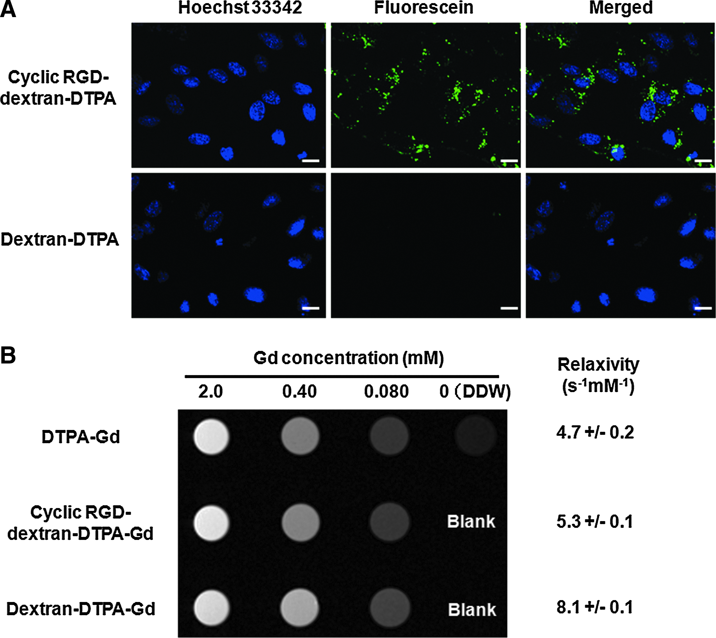
Figure 2A shows fluorescent microscopic images for HUVEC after culturing with the cyclic RGD-dextran-DTPA or the dextran-DTPA. Cyclic RGD-dextran-DTPA was internalized into HUVEC to a greater extent than dextran-DTPA. T1-weighted images and T1 relaxivities for DTPA-Gd, dextran-DTPA-Gd, and cyclic RGD-dextran-DTPA-Gd with different concentrations are shown in Figure 2B. All contrast agents had lower T1 relaxation times than DDW. The relaxivities increased in the order of DTPA-Gd, cyclic RGD-dextran-DTPA-Gd, and dextran-DTPA-Gd.
Evaluation of animal model
Figure 3A shows the blood perfusion image of mice hindlimb scanned by the laser-Doppler image analyzer. At 1 day after ischemia, blood perfusion of mice hindlimb ligated was drastically reduced. Partial recovery of blood perfusion was observed after 7 days. When the blood perfusion was analyzed quantitatively, it was found that the LDII gradually increased without any treatment (Fig. 3B). At 7 days after ischemia, angiogenesis was confirmed in the ischemic-angiogenic tissue of mice hindlimb, while less angiogenesis was observed in the normal region (Fig. 3C).
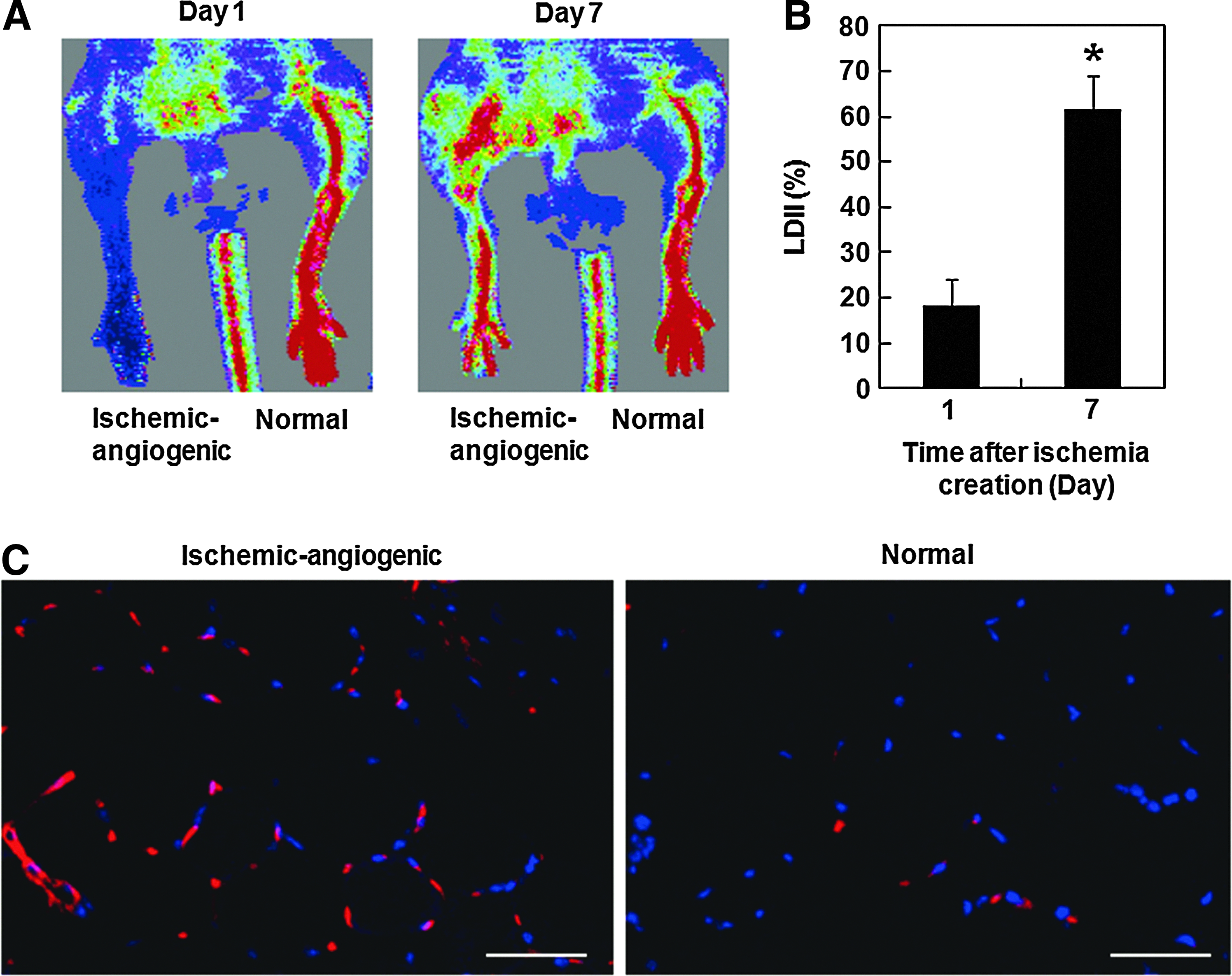
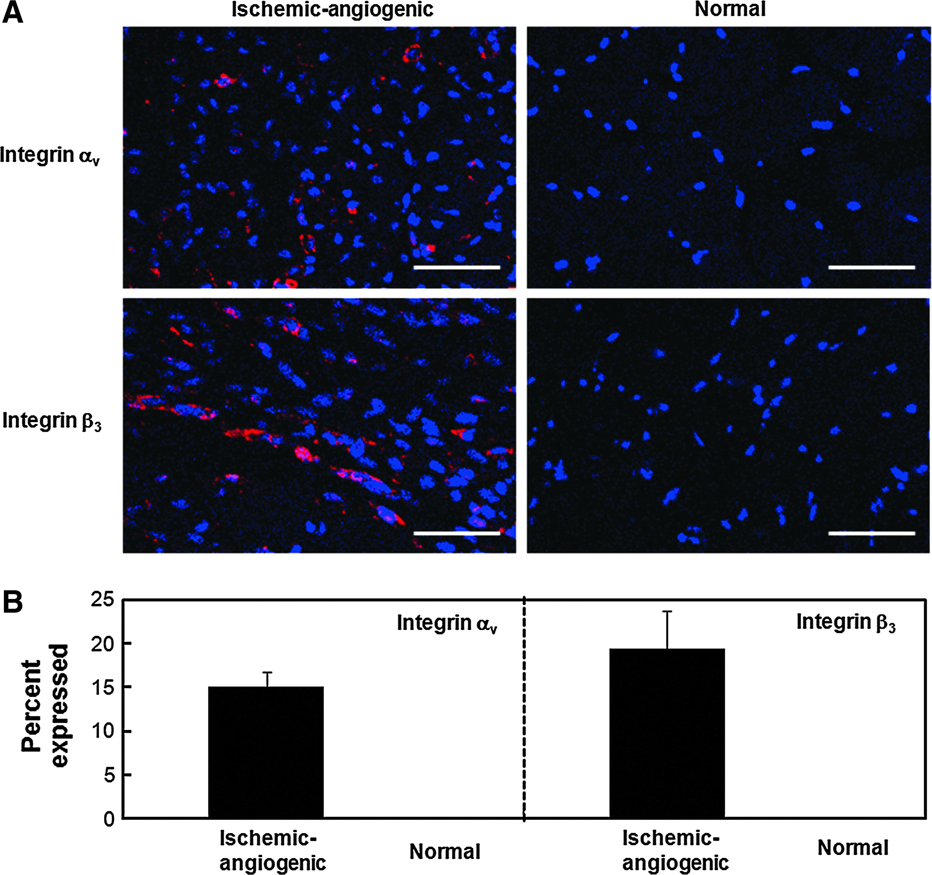
Figure 4 shows the expression images of integrin αvβ3 in the ischemic-angiogenic and normal tissues of mice with hindlimb ischemia created 7 days earlier. Both integrins αv and β3 were clearly detected in the ischemic-angiogenic region, while no signal was observed in the normal region.
In vivo MR imaging of ischemic-angiogenic region with cyclic RGD-dextran-DTPA-Gd
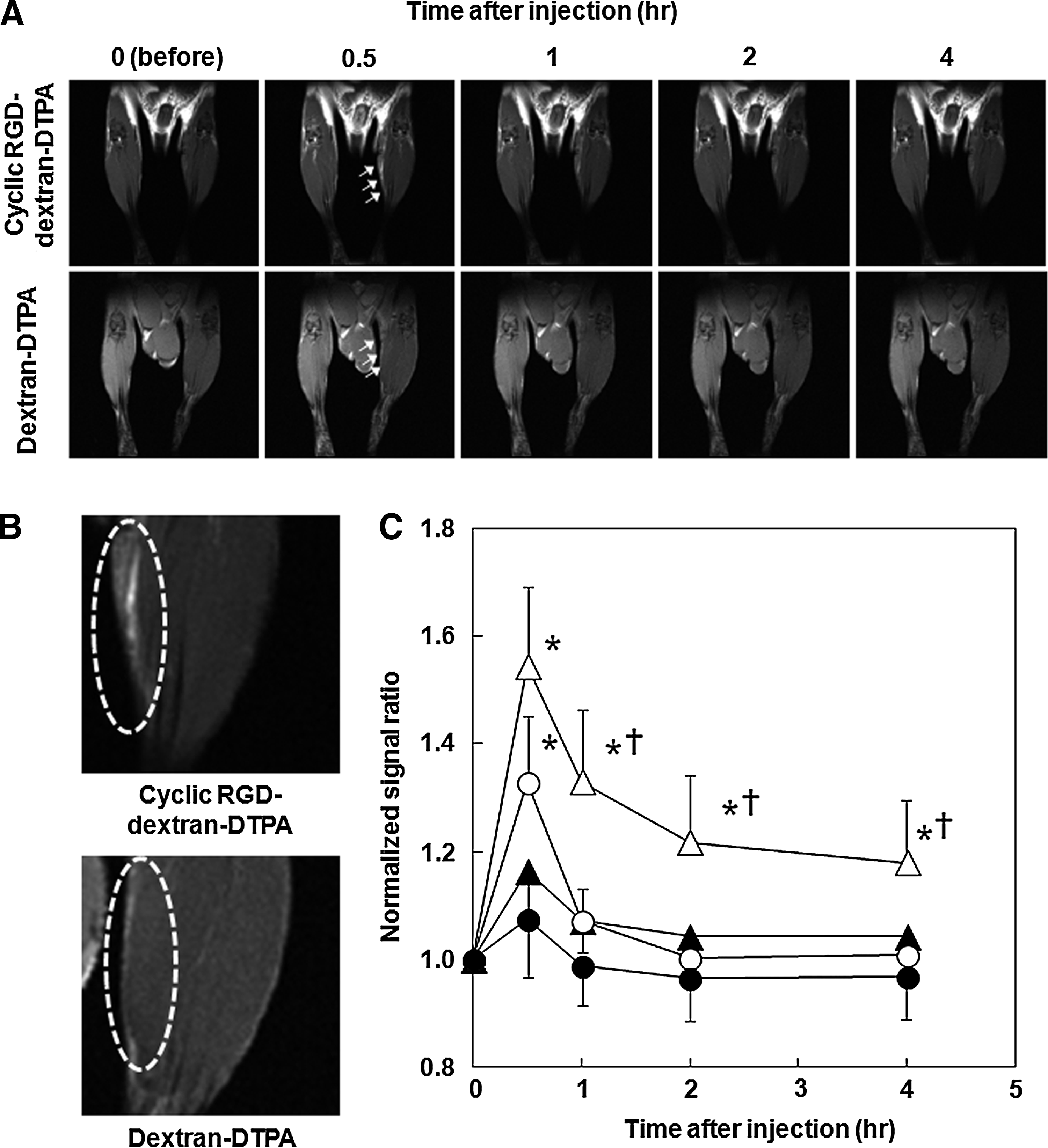
Figures 5A and B show MR images of mice hindlimb region before or after injection of dextran-DTPA-Gd or cyclic RGD-dextran-DTPA-Gd. From these pictures, the T1 signal intensity ratios in the ischemic-angiogenic region or normal region were plotted as a function of time after the injection (Fig. 5C). Cyclic RGD-dextran-DTPA-Gd showed high signal intensity in the ischemic-angiogenic region. On the other hand, the signal intensity ratio in the ischemic-angiogenic region for dextran-DTPA-Gd increased slightly and decreased to a normal region with time.
Accumulation of cyclic RGD-dextran-DTPA in ischemic-angiogenic region
Figure 6 shows the distribution percentages of cyclic-RGD-dextran-DTPA or dextran-DTPA in the ischemic-angiogenic or normal region of mice with hindlimb ischemia created 7 days earlier. From the MRI results (Fig. 5C), the distribution was evaluated 4 h after the injection of cyclic-RGD-dextran-DTPA or dextran-DTPA. Irrespective of cyclic RGD introduction, the extents of dextran-DTPAs accumulated in the ischemic-angiogenic region were higher than those in the normal region. Furthermore, the percentage of cyclic RGD-dextran-DTPA distributed in the ischemic-angiogenic region was significantly higher than that of dextran-DTPA.
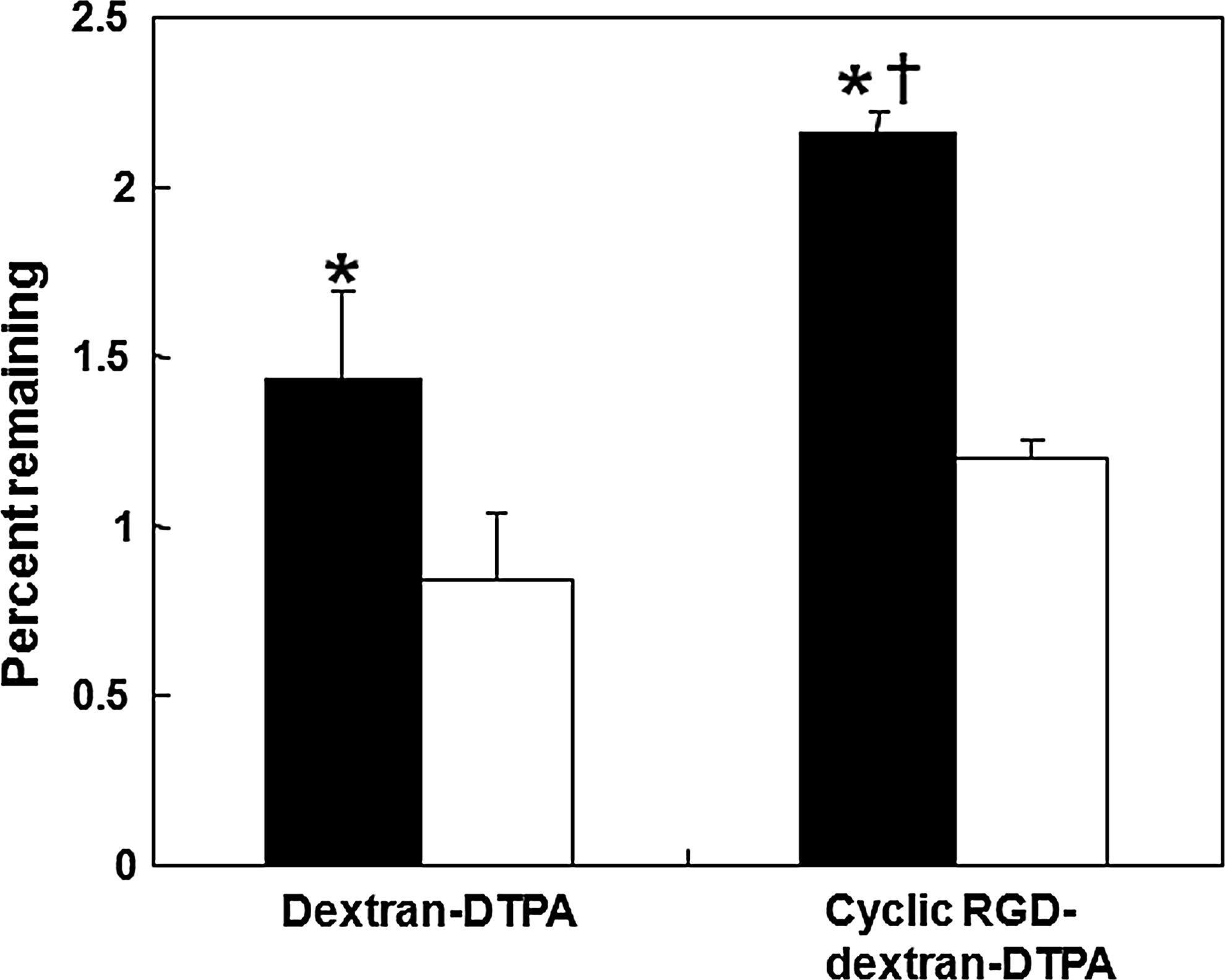
Distribution of 59Fe-labeled dextran-DTPA or cyclic RGD-dextran-DTPA in the ischemic-angiogenic (■) or normal (□) region of mice hindlimb 4 h after intravenous injection. Hindlimb ischemia (three mice each) was created 7 days before the injection. *p<0.05; significant against the percent remaining in normal region after the injection of the corresponding agent. †p<0.05; significant against the percent remaining after the injection of dextran-DTPA-Fe.
Discussion
The goal of this study was to design and prepare a nano-sized contrast agent for molecular imaging of therapeutic angiogenesis. The nano-sized contrast agent prepared in this study was composed of dextran, cyclic RGD, DTPA, and Gd3+. Dextran is a water-soluble and naturally occurring polysaccharide with low immunogenicity and a long history of clinical use as a plasma expander. 49 Cyclic RGD peptide has a high affinity for integrin αvβ3 of vascular endothelial cells expressed during proliferation or migration,43,44 and one type of cyclic RGD (EMD121974, Cilengitide) is used for clinical trial (phase II) as an inhibitor for tumor angiogenesis. 50 The DTPA and Gd3+ are components of Magnevist® of an MRI-positive contrast agent clinically available. Since the dextran has a number of OH groups, the cyclic RGD as a ligand and DTPA as a chelator for imaging probe are expected to be simultaneously introduced to dextran. It has been reported that dextrans with molecular weights of 30,000 or higher are stable in the blood circulation after intravenous injection. 51 Modification with a water-soluble polymer enables the ligands or imaging probes to be stably present in the blood circulation. 52 Therefore, it is highly expected that dextran-based contrast agents will enhance imaging efficacy. From the viewpoint of clinical application, dextran-based contrast agents (cyclic RGD-dextran-DTPA) are feasible, because each component (dextran, cyclic RGD, DTPA, and Gd3+) has been proved to be safe and used clinically, although the safety of cyclic RGD-dextran-DTPA complex needs to be carefully evaluated. Our conceptual study in rodents can easily be upscaled to a preclinical study with larger animals, such as swine.
The cyclic RGD-dextran-DTPA prepared was internalized into HUVEC to a greater extent than dextran-DTPA (Fig. 2A). It is likely that the affinity of cyclic RGD for the αvβ3 integrin expressed on the HUVEC surface enabled the cyclic RGD-dextran-DTPA to strongly interact with the cells, resulting in effective internalization in a receptor-mediated fashion. A lot of research has been conducted on the multimerization53,54 or modification of cyclic RGD with nano-sized carriers55–57 to enhance the affinity for integrin αvβ3. Although multiple cyclic RGD molecules could be introduced to dextran in this study, the cyclic RGD number introduced to dextran was not changed. Further investigations should be performed to optimize the number of cyclic RGD molecules and investigate the effects on the interaction strength with integrin αvβ3.
The conjugation of DTPA enabled dextran to chelate Gd3+ and shortened the relaxation time, while the T1 relaxivities increased in the order of DTPA, cyclic RGD-dextran-DTPA, and dextran-DTPA at the same Gd3+ concentration (Fig. 2B). It is well recognized that the relaxivity is governed by the chemical structure of the compound chelating Gd3+, and the slow and restricted rotation of Gd3+ gives the compound high relaxivity. 58 In the present study, cyclic RGD was chemically introduced to the carboxyl groups of DTPA for the chelation of Gd3+. Therefore, the chelating ability was slightly reduced, which caused a lower number of Gd3+ chelated with cyclic RGD-dextran-DTPA (25 Gd3+) compared with dextran-DTPA (33 Gd3+). It is conceivable that the extent of rotation restriction is increased in the order of DTPA, cyclic RGD-dextran-DTPA, and dextran-DTPA, which corresponds to the order of the relaxivities obtained. It is well known that DTPA has high chelation stability with many metal ions; this characteristic makes it possible to readily introduce other radioactive isotopes for scintigraphy, PET, and SPECT imaging.
The ischemic-angiogenic region could be clearly visualized on MRI after an intravenous injection of cyclic RGD-dextran-DTPA-Gd (Fig. 5A). The signal profiles of dextran-DTPA-Gd and cyclic RGD-dextran-DTPA-Gd mostly agreed with the distribution behaviors conceivable from the result of radioisotope-based tracing analysis (Fig. 6). It was found by immunohistochemical staining that the αvβ3 integrin was expressed in the ischemic-angiogenic region of mice (Fig. 3C). In addition, it was also observed that the fluorescein-labeled cyclic RGD-dextran-DTPA was co-localized in the integrin αvβ3-positive region after an intravenous injection (data not shown). Taken together, it was clearly demonstrated that the affinity of cyclic RGD for the integrin αvβ3 enabled the cyclic RGD-dextran-DTPA to selectively deliver to the ischemic-angiogenic region of mice with hindlimb ischemia. On the other hand, dextran-DTPA without cyclic RGD introduction was also slightly accumulated in the ischemic-angiogenic region. It is well recognized that inflammation is often induced, and the leakiness of blood vessels increases in ischemic-angiogenic regions. In addition, the lack of integrity in blood vessels formed during the initial steps of angiogenesis is involved in the high leakiness. It is, therefore, possible that dextran-DTPA was passively delivered to the ischemic-angiogenic region by extravasation from the blood vessel thereat, resulting in promoted accumulation. The signal ratios of dextran-DTPA and cyclic RGD-dextran-DTPA obtained by the radioisotope-based tracing analysis (Fig. 6) were higher than those by MRI 4 h after injection (Fig. 5C). This may be due to the fact that the sensitivity of 59 Fe (γ counter detection) is higher than that of Gd3+ (MRI detection).
Three points are remaining to be investigated for the practical use of cyclic RGD-dextran-DTPA-Gd. The first point is related to the preparation of multimodal imaging materials. Each imaging modality is based on quite different principles and has advantages and disadvantages. Generally, a single modality does not always correspond to all the requirements for diagnosis imaging. 59 A combinational imaging system composed of different imaging modalities may compensate the deficiencies of single imaging modality. Currently, some prototypes of multimodal imaging system, including MRI-optical, NIRF-SPECT, PET-computed tomography, and SPECT-MRI, have been introduced.60–63 Further design and investigation of dextran-based multimodal imaging probes should be done to realize the idea of multimodal imaging. The second point is regarding the animal model of angiogenesis. In the present study, the natural healing after hindlimb ischemia in mice was used for the animal model of angiogenesis to remove the complicated factors involved in angiogenesis. It has been demonstrated that the controlled release of bioactive substances with gelatin hydrogel enables to promote and enhance the angiogenesis, which is clinically applied for the angiogenic therapy.6,64–66 Therefore, the combination of this technology with the contrast agent prepared in the present study will achieve more practical angiogenesis imaging. The final point is about the imaging timing of ischemic-angiogenic region. The expression of integrin αvβ3 was observed in the ischemic-angiogenic region of mice with hindlimb ischemia created 7 days before (Fig. 3). Several research reports have indicated that the highest integrin αvβ3 expression was observed for mice with ischemia created 7 days earlier.41,67,68 Therefore, the mice with ischemia created 7 days earlier were used in the present study. However, it is practically important not only to detect the maximum angiogenesis but also to trace the integrin αvβ3 expression profiles. Imaging of integrin αvβ3 expression profiles will enable more detailed information to be obtained on the therapeutic effects and planning of additional therapies that are suitable for healing.
Footnotes
Acknowledgments
The authors would like to thank Ms. Sayaka Shibata and Ms. Misao Yoneyama (National Institute of Radiological Sciences, Japan) for their technical assistance. This research was partly supported by a Kakenhi grant from the Japanese Society for the Promotion of Science (JSPS) and the Ministry of Education, Culture, Sports, Science and Technology, Japan.
Disclosure Statement
No competing financial interests exist.