
Abstract
Cadaveric tendon allografts form a readily available and underutilized source of graft material. Because of their material properties, allografts are biomechanically and biologically superior to synthetic scaffolds. However, before clinical use, allografts must undergo decellularization to reduce immunogenicity and oxidation to increase porosity, leaving a nonvital biostatic scaffold. Ex vivo seeding, or revitalization, is thought to hasten graft incorporation and stimulate intrinsic tendon healing, permitting early mobilization and return to function. In this study, we examined physical and biochemical augmentation methods, including scaffold surface scoring (physical) and rehydration of lyophilized scaffolds in serum (biochemical). Scaffolds were divided into four groups: (1) scored scaffolds, (2) lyophilized scaffolds rehydrated in fetal calf serum (FCS), (3) scaffolds both scored and rehydrated in FCS, and (4) control scaffolds. Scaffolds were reseeded with adipose-derived stem cells (ADSCs). Reseeding efficacy was quantified by a live cell and total cell assays and qualified histologically with hematoxylin and eosin, live/dead and SYTO green nucleic acid stains, TUNEL apoptosis stains, procollagen stains, and transmission electron microscopy. Scaffold-seeded cell viability at up to 2 weeks in vitro and up to 4 weeks in vivo was demonstrated with bioluminescent imaging of scaffolds seeded with luciferase-positive ADSCs. The effect of seeding on scaffold biomechanical properties was demonstrated with evaluation of ultimate tensile stress (UTS) and an elastic modulus (EM). We found that scaffold surface scoring led to an increase in live and total cell attachment and penetration (MTS assay, p<0.001 and DNA assay, p=0.003, respectively). Histology confirmed greater total cell number in both construct core and surface in scored compared with unscored constructs. Cells reseeded on scored constructs displayed reduced apoptosis, persistent procollagen production, and had a similar ultrastructural relationship to the surrounding matrix as native tenocytes on transmission electron microscopy. Rehydration of lyophilized scaffolds in serum did not improve reseeding. Seeded constructs demonstrated greater UTS and EM than unseeded constructs. Scaffolds seeded with ADSC-luc2-eGFP demonstrated persistent viability for at least 2 weeks in vitro. In conclusion, tendon surface scoring increases surface and core reseeding in vitro and may be incorporated as a final step in allograft processing before clinical implantation.
Introduction
Tissue-engineered solutions involve the orchestrated combination of candidate cells and supporting scaffolds either with stimulation or with stand-alone application.4,5 Synthetic scaffolds include polyglycolic acid-6,7 and polylactic acid-8,9 based extracellular matrix (ECM) grafts. 4 These materials may be fabricated with adequate porosity to allow cell penetration, but remain inferior in terms of inflammatory response, biocompatibility, biomechanical attributes, and display rapid biomechanical attrition with time.10–19 Biological scaffolds are of mammalian origin (human, porcine, bovine, and equine) and include dermis, pericardium, small intestine submucosa,8,20 and allogenic tendon. 10 These scaffolds are composed of naturally occurring collagen fibers (predominantly type I collagen), and have a surface chemistry and native structure that is bioactive and promotes cell proliferation and tissue ingrowth.4,21
Human allogenic intrasynovial tendons form an underutilized pool of readily available graft material. 22 They are human-derived, nontoxic, and biocompatible. 23 This tissue possesses the ideal characteristics of a tendon scaffold—basic tendon structure, native ECM, and capability of cell seeding. 24 With orthotopic application, they retain their inherent biomechanical advantages and may be selected to match deficient tendons perfectly. In addition, they avoid donor-site morbidity and extended operative times necessary for autograft harvest. 22
Our overall goal is to enhance tendon graft healing. With allografts, remodeling and incorporation are slow because allografts do not harbor live cells.25,26 In addition, certain features must be addressed before an allogenic tendon can be into clinical use. First, the risk of disease transmission and immunogenicity must be addressed. While an immune response may be less noticeable in an immune-privileged location (such as the knee) in well-vascularized locations, we have shown that the rapid immune response quickly compromises tendon scaffold biomechanics. 27 Second, a surface optimized for gliding may not be ideal to facilitate tissue integration. 26 Lubricin, a lubricating glycoprotein present on the surface of an intrasynovial tendon, is known to inhibit cell adhesion. 28 Roughening the surface increases the surface area, 29 thereby improving cell adhesion, which in turn is thought to improve graft incorporation. 26 Third, digital flexor tendons display a close-knit tendon weave and compact superstructure, conferring great biomechanical strength, but making cellular ingrowth extremely challenging.
We have previously discussed processing of human intrasynovial tendon with decellularizing agents to reduce immunogenicity 30 and the use of a porositizing agent to enhance cell penetration. 10 Following treatment, cells are seeded into these biostatic (nonvital) scaffolds, a process described as revitalization.23,31 In spite of prior processing, graft core cellularity following reseeding still remains inferior to that of the freshly harvested tendon.
In this study, we examined the effect of mechanical treatment (surface scoring) and biochemical treatment (lyophilized graft rehydration in fetal calf serum [FCS]) on adipoderived stem cell (ADSC) attachment and penetration.
With surface scoring, it is hypothesized that the surface area and irregularity are increased, facilitating cellular attachment and penetration. With rehydration in serum, we hypothesized that following removal of tendon water, scaffold core rehydrationin serum would create a growth factor-rich internal milieu attractive to cell penetration, proliferation, and survival. We previously demonstrated that the addition of growth factors in combination was beneficial to cell growth.27,32 However, routine use of a carefully titrated recipe of recombinant human growth factors may be less appealing economically and logistically when upscaled for clinical application. As such, we sought a readily available growth factor-rich alternative—FCS. As pooled, deidentified ADSCs were used, bovine serum served as a surrogate for human (recipient) serum. FCS has been shown to be as efficient as human serum in supporting the proliferation and differentiation of human stem cells. 33
Potential seed-cell candidates include mesenchymal stem cells and tendon fibroblasts (tenocytes and tenoblasts). We chose mesenchymal stem cells because tendon fibroblasts make poor seed-cell candidates as they experience phenotypic drift after two to three passages in culture, and cell yield at biopsy is low relative to the amount of tendon material obtained.5,10,34
Methods
Scaffold harvest
Human flexor digitorum profundus (FDP), flexor digitorum superficialis (FDS), and flexor pollicis longus (FPL) were harvested from fresh–frozen cadaveric forearms (Science Care). Epitenon and synovial sheath were meticulously removed. Proximally, tendons were transected at the musculotendinous junction. Distally, FDS tendons were transected 2 cm proximal to the chiasma, and FDP and FPL tendons were transected 1 cm proximal to the osteotendinous junction. Care was taken to ensure that each tendon pair (8–10 cm long) was of the same length. All tendons were frozen at −70°C in phosphate-buffered saline (PBS, Sigma) until decellularization.
Tendon scaffold decellularization
Tendon scaffolds were decellularized according to an established protocol. 30 Briefly, scaffolds were treated with 0.1% ethylenediaminetetraacetic acid (EDTA) for 4 h followed by 0.1% sodium dodecyl sulfate in 0.1% EDTA for 24 h at room temperature with constant agitation. Scaffolds were washed in PBS and stored at −70°C.
Increasing porosity (porositization) with peracetic acid
A solution of buffered 5% peracetic acid (PAA) was created by diluting stock PAA solution (32%, Sigma) with PBS and adjusting pH to 7. Decellularized tendon scaffolds were treated with 5% PAA for 4 h with agitation according to a previously established protocol. 10 Treated scaffolds were then washed with deionized water (DI) and frozen to −70°C.
Cell culture and scaffold reseeding
Commercially obtained ADSCs (up to passage 4, Poietics PT-5006 cryopreserved ADSCs, Lonza) were cultured in the F12 medium (Gibco, Invitrogen) augmented with 10% FCS (Gibco). Cells were grown to confluence at 37°C in a humidified tissue culture chamber with 5% carbon dioxide.
Experimental setup
Scaffolds were divided into four groups (Table 1).
FCS, fetal calf serum.
Treatment groups
Mechanical treatment (surface scoring)
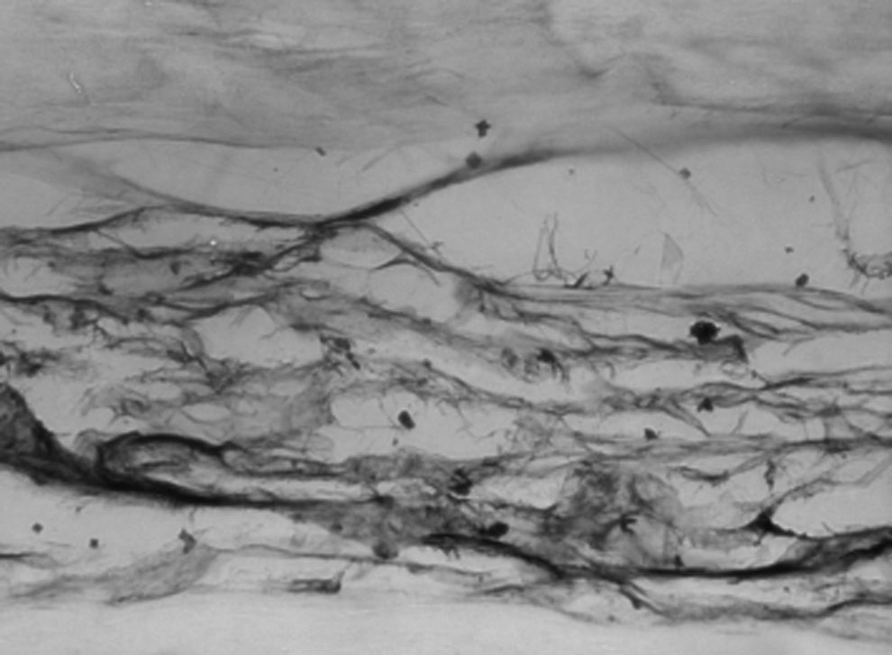
Groups S (surface scoring only) and Group S+F (scoring and 100%FCS hydration) were treated with surface scoring. Using the sharp edge of a 15-blade, both surfaces of each scaffold were gently scored ten times along its length from end to end (Fig. 1) by a single investigator.
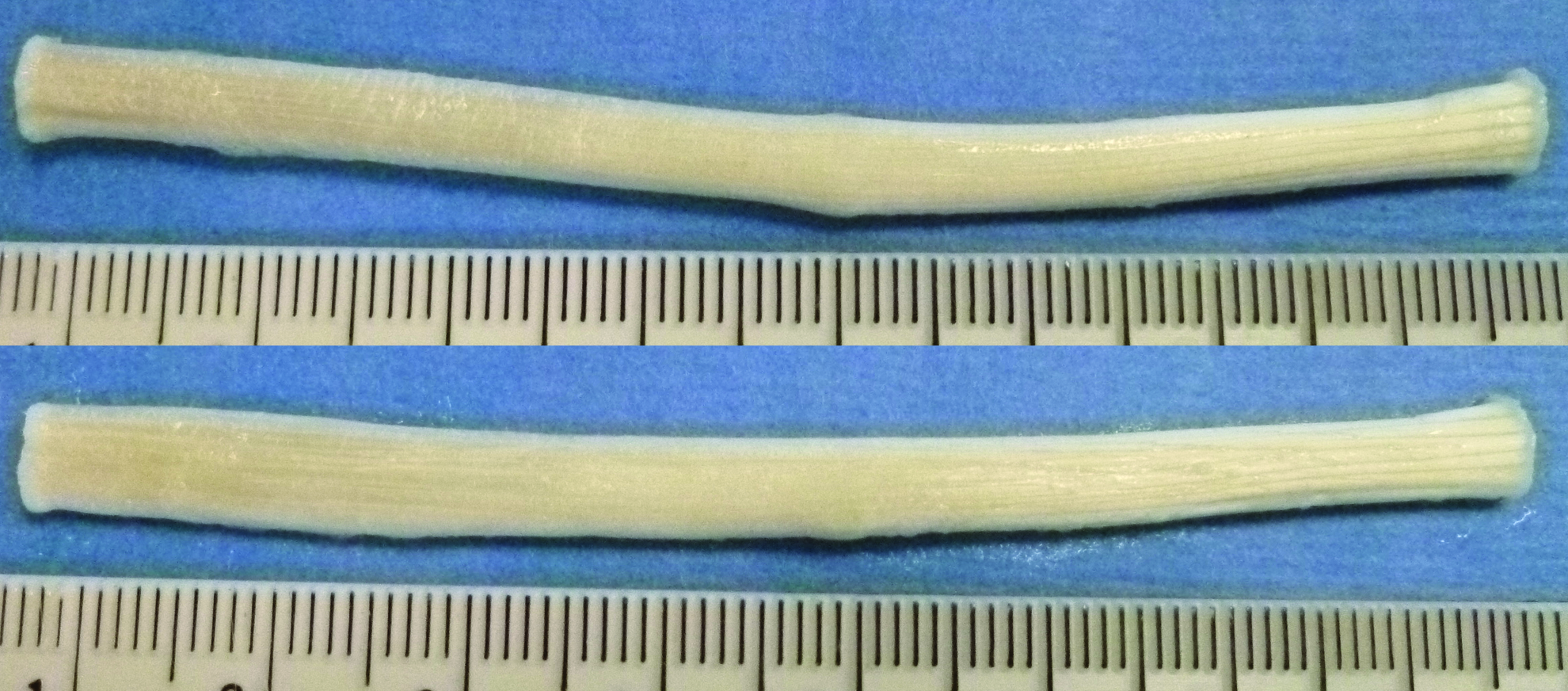
Human flexor tendon before (above) and after (below) surface scoring. Color images available online at www.liebertpub.com/tea
Biochemical treatment (rehydration in 100% FCS)
All four groups were placed in lyophilization flasks connected to the four-port manifold of a lyophilizer (FreeZone 1L Benchtop, Labconco) for 36 h at 0.03 mbar at −46°C. Complete lyophilization was confirmed by observation of complete sublimation of ice and dehydration of tendon scaffolds. Group S+F (scoring and 100% FCS rehydration) and Group F (100% FCS rehydration alone) scaffolds were then rehydrated in 100% FCS (Gibco) overnight (16 h) before reseeding. In contrast, Group S (scoring only) and Group CON (control) scaffolds were rehydrated in F12 media enriched with 10% FCS overnight (16 h) before reseeding.
Reseeding
From pilot experiments, we had determined that ideal parameters involved 6 h of attachment in sealed tubes using 1–2×10⁶ cells/mL for 1-cm tendon scaffolds at 37°C on a rotating mixer (Mix-All; RPI Corp.). Using similar methodology, long flexor tendons (8–10 cm) were reseeded with 1–2×10⁶cells/mL/cm length of scaffold for 6 h. Constructs were then placed in an enriched F12 medium for 2 weeks with medium changes every other day.
Quantification of reseeding success
Live cells
Constructs were divided into 1-cm segments and scaffold dimensions were recorded with digital calipers. As we were only concerned with tangential scaffold reseeding, scaffold ends (0.5 cm) were discarded and only central segments were used. This step eliminates spuriously high readings from cells attached to transected scaffold ends.
Viable cell reseeding success was quantified with the CellTiter 96 Cell Proliferation Assay (Promega). Dye solution was prepared at 1:5 dilution of stock to the F12 medium. Reseeded construct segments were then divided lengthwise to maximize the surface area exposed to a dye solution. The dye solution was then added to divided constructs in 24-well plates and incubated for 1.5 h at 37°C during which bioreduction of the tetrazolium compound (MTS) occurred. Bioreduced dye was transferred to 96-well assay plates and absorbance at 490 nm was read by a spectrophotometer (Epoch, Biotek). Absorbance data were normalized to dry weight.
Total cells
Total cell reseeding success was measured with a DNA assay (DNeasy, Qiagen, Inc.). DNA concentration was determined by its absorbance at 260 nm using a spectrophotometer (Epoch, Biotek) and normalized to dry weight.
From the above tests across four groups, we found that scoring had a greater effect on reseeding than scaffold rehydration in 100% FCS. Further, rehydration in 100% FCS was not superior to rehydration in F12 media augmented with 10% FCS, our current standard. Subsequent investigations (below) were performed on scored reseeded (original Group S) versus unscored (reseeded) constructs (original Group CON, control) rehydrated in F12 media enriched with 10% FCS before reseeding. In subsequent analysis, rehydration in 100% FCS was excluded (original Group F and Group S+F).
Qualification of reseeding success
Nucleic acid staining
Whole tissue mounts were divided longitudinally and immersed in SYTO green (Invitrogen) nucleic acid stainat 1:2000 dilution in DI water for 20 min. Segments were then placed on coverslips and imaged. Separate images were obtained for the outer convex scaffold surface and the inner cut (core) surface.
Hematoxylin and eosin staining
Formalin-fixed, paraffin-embedded (FFPE) sections (5 μm) were stained with hematoxylin and eosin (H&E, Sigma) for assessment of seeding efficacy and scaffold morphology.
Transmission electron microscopy
Tendon segments were fixed with 2% glutaraldehyde in a 0.1 M cacodylate buffer for 24 h at room temperature and postfixed in 2% OsO4 for 2 h at room temperature. Samples were stained en bloc with 4% uranyl acetate for 6 days at 4°C, dehydrated, infiltrated with a 1:1 mixture of propylene oxide and eponate overnight, and embedded in 100% eponate for 2 days at 60°C. Ultrathin sections (60 nm) were cut and collected on uncoated copper 200 grids. Serial sections were placed on formvar-coated copper 1000-μm slot grids, stabilized with carbon film. All sections were stained with uranyl acetate lead citrate and examined with a transmission electron microscope (Jeol1230; JEOL Ltd).
Apoptosis assay
To further assess the viability of reseeded cells, we assessed DNA fragmentation as a marker of apoptosis using the DeadEnd fluorometric TUNEL (TdT-mediated dUTP nick end labeling) assay (Promega) according to the manufacturer's instructions. Briefly, FFPE sections were deparaffinized, permeabilized with proteinase K (20 μg/mL) for 10 min. Sections were then reacted with TdT and fluroscein-labeled nucleotides and the reaction terminated with 2× SSC solution. The Vectashield (Vector Labs) mounting medium with propidium iodide (PI) was added and the slides visualized with a fluorescent microscope.
Procollagen staining
Procollagen staining was performed to evaluate synthetic ability of seeded cells. FFPE sections were treated with 0.6% hydrogen peroxide to neutralize endogenous peroxidase and permeabilized with 0.1% saponin. An anti-human pro-COL1A1 (N-17) primary antibody (Santa Cruz) at 1:200 dilution was added for 1 h and washed. A secondary biotin-conjugated anti-goat antibody was then added (Vectastain ABC, Vector Labs) for 30 min and washed. Peroxidase substrate Vector SG (Vector Labs) was added for 10 min and washed before mounting.
Biomechanical testing
Pair-matched designs were used to compare across two groups during materials testing:
The effect of scoring
To determine if scoring had adverse effects on material properties, scored (unseeded) scaffolds were compared with unscored control (unseeded) scaffolds (contralateral limb, corresponding tendon).
The effect of lyophilization
To determine if lyophilization had an adverse effect on material properties, lyophilized and rehydrated scaffolds were compared with pair-matched nonlyophilized scaffolds.
The effect of scoring+reseeding
Comparing reseeded scaffolds, scored (reseeded) scaffolds were compared with unscored control (reseeded) pair-matched scaffolds.
Tendon ends were fastened between sandpaper sheets using cyanoacrylate glue and attached to the crossheads of a servo-controlled hydraulic materials testing system (MTS 858; MTS Inc.). 35 The glue-sandpaper assembly served to increase the surface contact area and the frictional coefficient of contact surfaces.
Orthogonal photographs together with a known scale were obtained by a digital camera (Nikon 100) tripod-mounted at a fixed distance. Using the formula for an ellipse, the axial cross-sectional area could be calculated at the site of failure using ImageJ software (NIH).10,35
The biomechanical testing protocol consisted of 10 preconditioning cycles at 10 mm/min to 100N with distraction to failure on the 11th cycle. Ultimate tensile stress (UTS) was calculated by dividing force by the cross-sectional area. Elastic modulus (EM) was calculated using the gradient of the stress–strain curve during the linear phase of deformation.
Lentivirus production
To determine ex vivo cell viability, a cell line of luciferase (+) ADSC was created to allow regular follow-up of luciferase activity of cells on seeded constructs. The relationship between luciferase enzyme concentration and the peak height of emitted light in vitro is linear up to 7 to 8 orders of magnitude.36,37 Using standardized quantification techniques, changes in measured radiance could be correlated to the amount of viable cells present.
First, pUbi-luc2-eGFP lentiviral vectors were constructed by Gambhir lab. 38 High-titer lentiviral vectors were produced using a modified version of the protocol presented in Zhang et al. 39
Creation of luciferase (+), GFP(+) mesenchymal stem cell line
A subpopulation of ADSCs were transduced with lentivirus at a multiplicity of infection of 50. The genetically modified ADSC underwent two rounds of FACS sorting (FACSAria III, Becton Dickinson) before use. The stable cell line is referred to as ADSC-luc2-eGFP.
Bioluminescence imaging: ex vivo cell viability
Cell viability of ADSC-luc2-eGFP-seeded scaffolds was monitored using Bioluminescence imaging (BLI) using the Xenogen Spectrum imaging system (Caliper Life Sciences). Short scored constructs (1 cm) seeded with 1×10⁶ cells/mL, 2×10⁶ cells/mL, and 4×10⁶cells/mL were transferred to wells containing D-luciferin (Stanford Center for Innovation in In-Vivo Imaging, 150 μL in 1 mL DPBS). Serial images were taken using Living Image software (Caliper Life Sciences) on alternate days. Sequential images were acquired at 1-min intervals for 20 min. Field of view was set to image all wells at once. Identical settings were used to acquire each image. To quantify measured light, regions of interest were drawn over the seeded scaffolds and the maximum radiance (photons/sec/cm2/steradian) was obtained, as validated previously. 40 Steady-state peak radiance readings were compared. Both superior and inferior surfaces were imaged and the readings averaged.
BLI findings were verified by fluorescent microscopy showing the presence of GFP(+) cellson scored (seeded) samples.
Bioluminescence imaging: in vivo cell viability
To establish proof of concept that seeded cells would survive in vivo, the following experiment was performed using a human-to-immunodeficient rat xenograft model as a surrogate for a human-to-human allograft model. All procedures were performed in accordance with the Guide for the Care and Use of Laboratory Animals.
First, decellularized pair-matched tendons were divided into 4-cm segments, incubated overnight in F12 media (Gibco) containing an antibiotic–antimycotic (Anti-Anti, Gibco–Invitrogen), and divided into two groups: reseeded versus unseeded control (decellularized only). Scaffolds (3 cm long) in the reseeded group were seeded with ADSC-luc2-eGFP at 1×10⁶ cells/mL/cm scaffold for 6 h in the manner previously described.
Next, 12-week-old male nude rats (RNU rats, ∼300 g) were obtained from Charles River and housed at our institutional veterinary medical unit. All procedures were performed in accordance with our institutional review board and within an approved animal protocol. Under general inhalational anesthesia, the dorsal surfaces were shaved and cleansed with povidone iodine. Short longitudinal incisions were made over each flank and subcutaneous pockets dissected out. Reseeded constructs were placed in the left pocket, and control unseeded scaffolds placed in the right pocket. Scaffold ends were anchored to nearby fascia using prolene 4-0 (Ethicon) to prevent construct movement and migration. Incisions were closed with vicryl 4-0 (Ethicon).
Rats were given an intraperitoneal injection of the reporter substrate D-Luciferin (375 mg/kg body weight) and imaged after 30 min. Imaging was performed twice a week for 4 weeks.
Statistical analysis
Data were reported as mean±standard deviation (SD). Comparisons across groups were performed using the Students t-test (two groups) and one-way analysis of variance (ANOVA) (greater than two groups), with post hoc analysis using the least significant difference test. The significance level was set at p<0.05. All analyses were performed using IBM PASW 18 (SPSS, Inc.).
Results
Quantification of reseeding success: live and total cells
The MTS assay showed that live cell content was greatest in the scored groups, in particular, Group S (scoring only, A490nm 0.0041±0.0015, p<0.001, one-way ANOVA, Fig. 2). Group S displayed greater live cell seeding than Group S+F (scoring and FCS, A490nm 0.0037±0.0014, p=0.002), Group F (FCS only, A490nm 0.0018±0.0006, p<0.001), and Group CON (control, A490nm 0.0024±0.0007, p<0.001).
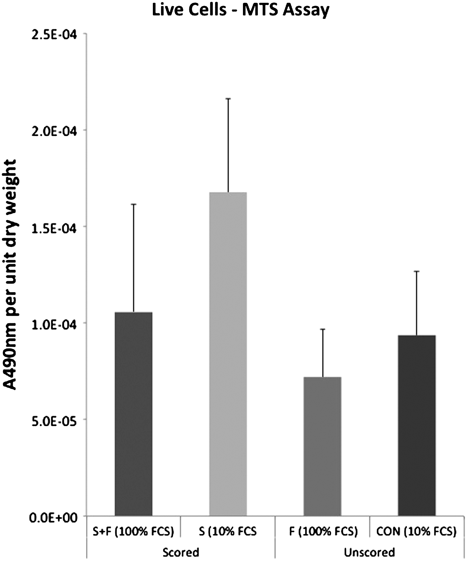
Live cell content (MTS assay). (100%, rehydration in 100% serum; 10%, rehydration in media augmented with 10% serum).
The DNA assay revealed greatest total DNA content in scored constructs (p=0.003, one-way ANOVA, Fig. 3). In particular, Group S (scoring only, DNA content 106.5±39.3 ng/mg dry weight) displayed greater total cell seeding than Group F (FCS only, DNA content 31.1±10.8 ng/mg dry weight, p<0.001) and Group CON (control, DNA content 56.8±28.8 ng/mg dry weight, p=0.02). Also, Group S+F (scoring and FCS) demonstrated greater reseeding (DNA content 106.3±71.0 ng/mg dry weight), than Group F (FCS only, p=0.02).
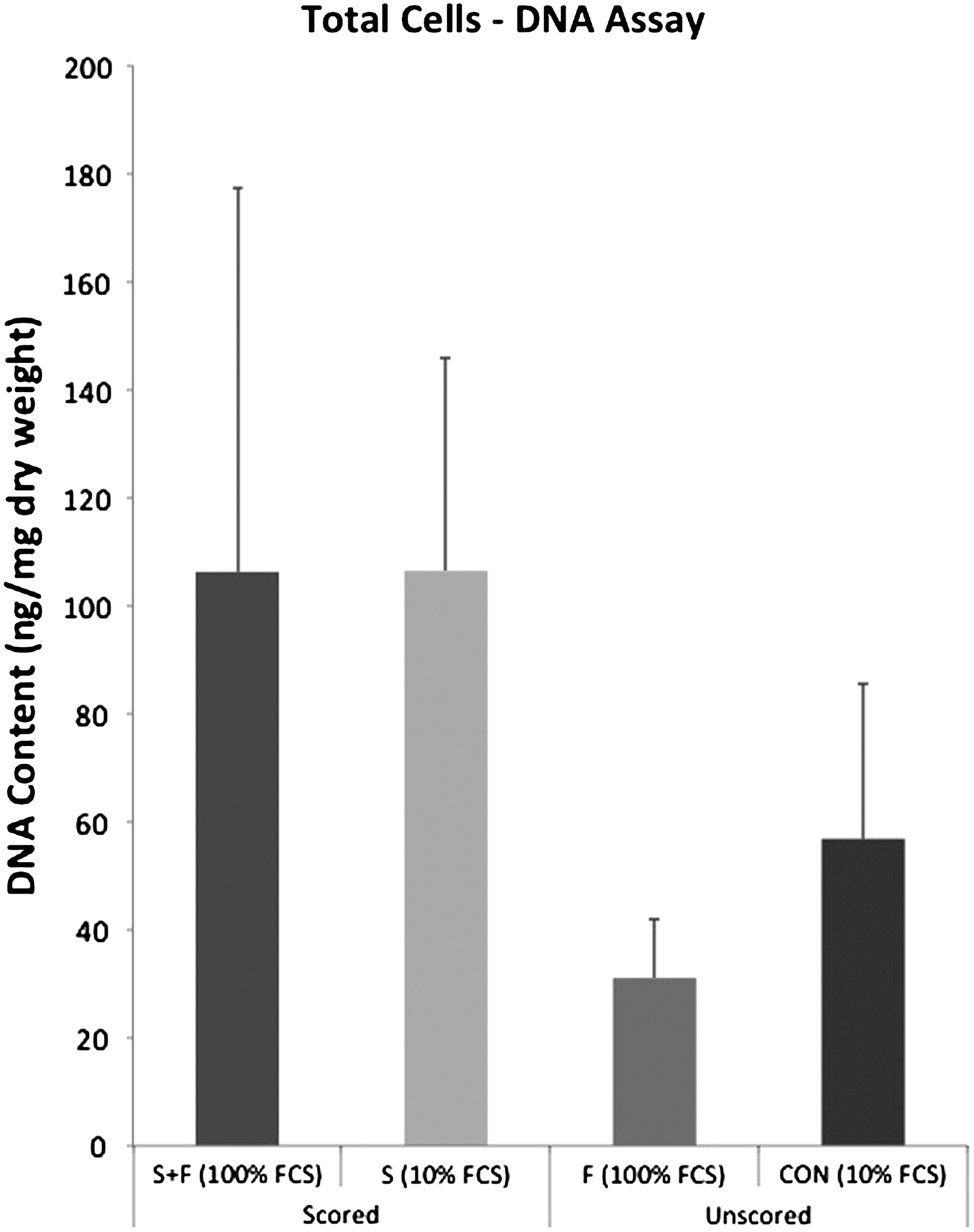
Total cell content (DNA assay). (100%, rehydration in 100% serum; 10%, rehydration in media augmented with 10% serum).
These findings indicated that scoring augmented seeding more than rehydration in 100% FCS. Subsequent qualitative analysis was performed on scored versus unscored (control) scaffolds.
Qualification of reseeding success
Nucleic acid staining
Whole mount SYTO green fluorescent nucleic acid staining revealed greater density of seeded cells on scored scaffolds compared with unscored, control scaffolds (Fig. 4). This was true both for the surface as well as the cut surface (core).
SYTO green nucleic acid staining of whole-tissue mounts showing greater surface (left) and core (right) reseeding in scored
H&E staining
H&E staining confirmed greater scaffold seeding with scored scaffolds (Fig. 5). This difference was most pronounced in the scaffold core.

Hematoxylin and eosin staining showing enhanced core (right) penetration in scored
Transmission electron microscopy
At the ultrastructural level, seeded cells adopted the appearance of native tenocytes, displaying integration and attachment to collagen fibrils (Fig. 6, black arrows). There was no morphological difference between cell–matrix relationship, between reseeded scored and unscored constructs.

Reseeded cells demonstrate good integration with collagen fibrils and similar morphology to native tenocytes (transmission electron microscopy, 300×). (Black arrows, attached cells)
Apoptosis assay
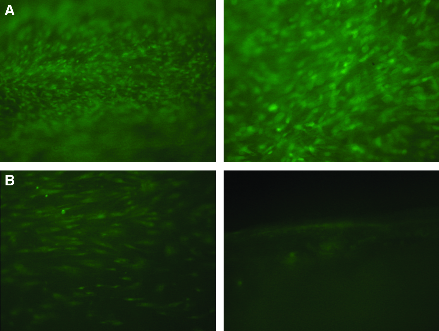
The TUNEL assay revealed a greater ratio of fluorescein-labeled (green) cells and PI-labeled (red) cells in unscored, control scaffolds (Fig. 7C, D), implying a greater percentage of apoptotic cells in this group. In contrast, scored scaffolds demonstrated a smaller proportion of apoptotic (green) to total (red) cells (Fig. 7A, B), suggesting a smaller percentage of apoptotic cells in this group.
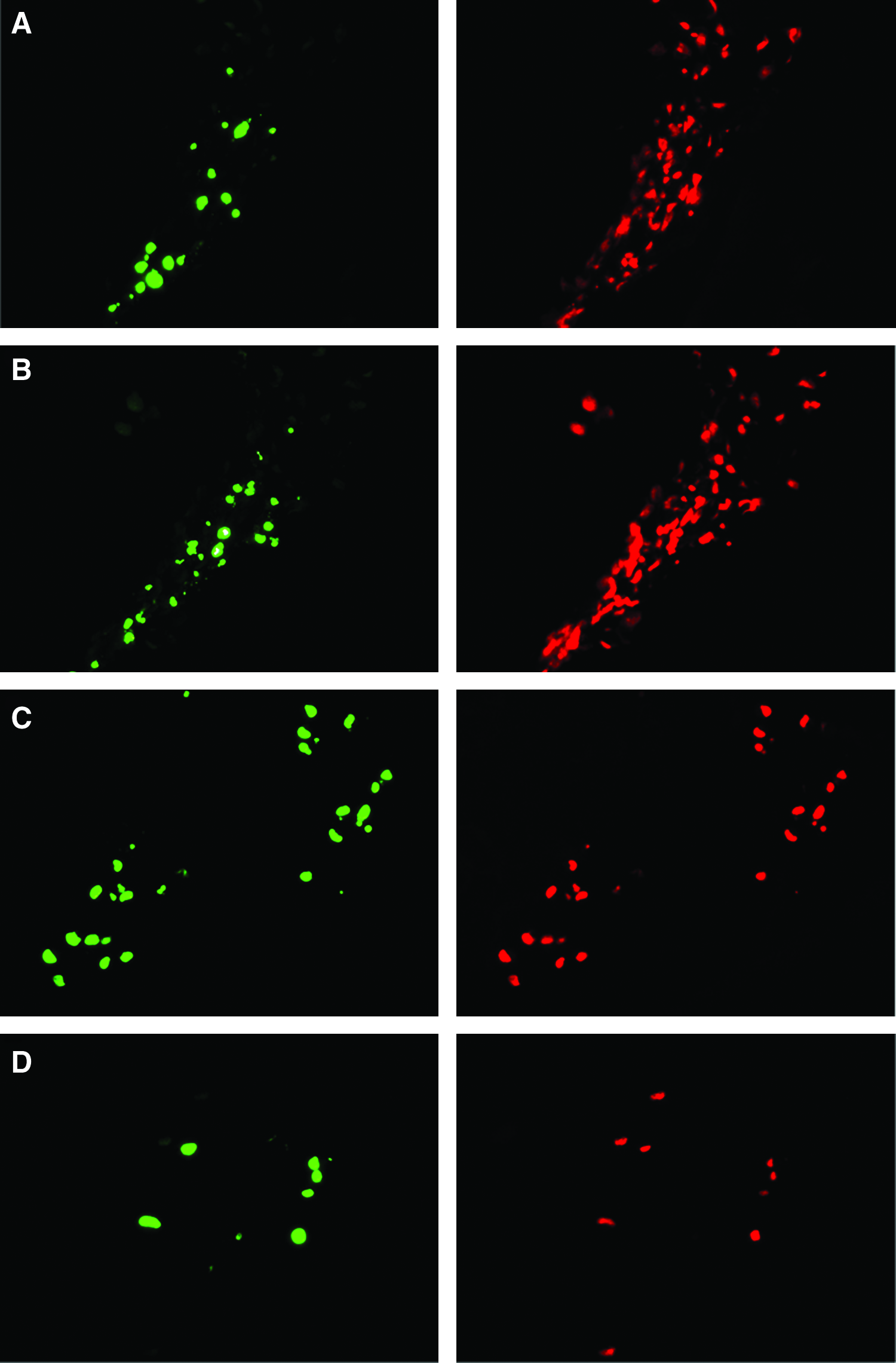
TUNEL assay (paraffin sections) demonstrated greater percentage of apoptotic cells in unscored constructs
Procollagen staining
Procollagen staining revealed active procollagen synthesis following seeding (Fig. 8). Although there were more apoptotic cells in unscored constructs, there was no visible difference in procollagen staining between scored and unscored groups. We believe that this is because dead cells were actively synthesizing procollagen at the time of cell death.
Procollagen staining demonstrates continued synthetic activity in seeded cells (paraffin section, 20×).
Biomechanical testing
The effect of scoring
There was no difference in UTS and EM between scored and unscored pair-matched decellularized scaffolds (p=0.48 and p=0.32, paired t-test, n=10 pairs).
The effect of lyophilization
Decellularized (nonlyophilized) scaffolds were compared with their pair-matched counterparts that were decellularized, lyophilized, and rehydrated in PBS. There was no difference in the UTS and EM (p=0.50 and p=0.18, respectively, paired t-test, n=10 pairs).
The effect of scoring+reseeding on material properties
Finally, with reseeded constructs (n=6 pairs), we found that scored (reseeded) constructs had greater UTS (46.6±10.9 MPa) compared with unscored (reseeded) constructs (37.3±9.7 MPa, p=0.002, Fig. 9A). Scored (reseeded) constructs also displayed greater EM (902.7±389.7 MPa) compared with unscored (reseeded) constructs (722.1±259.4 MPa, p=0.04, Fig. 9B). This suggests that improved reseeding (scored scaffolds) leads to enhanced biomechanical properties. This characteristic of reseeded constructs has been previously demonstrated. 41
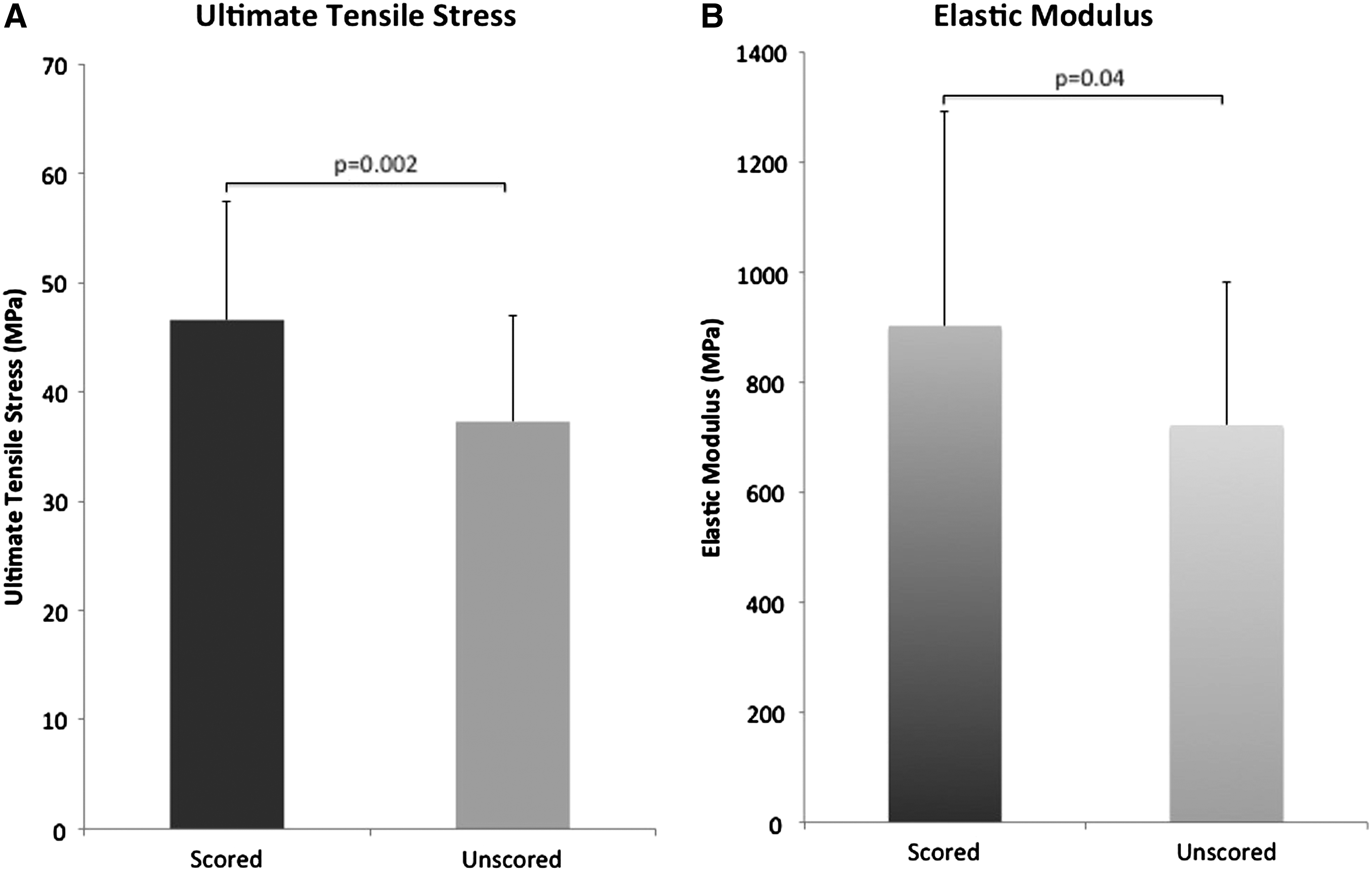
Ultimate tensile stress
Bioluminescence imaging: ex vivo cell viability
BLI confirmed the presence of viable ADSC-luc2-eGFP cells on both surfaces of scaffolds (Fig. 10A) at up to 2 weeks, with negative bioluminescent on the unseeded control scaffold. There was gradual reduction in BLI over time. At 4 days following seeding, radiance was 3.21×10¹⁰, 7.19×10¹⁰, and 1.06×10¹¹ photons/s/cm2/sr for constructs seeded with 1×10⁶, 2×10⁶, and 4×10⁶ ADSC-luc2-eGFP cells/mL, respectively. At 14 days, radiance was 5.37×10⁹, 6.94×10⁹, 7.38×10⁹ photons/s/cm2/sr corresponding to 83%, 90%, and 93% reduction from day 4 readings, respectively, suggesting gradual cell death over the course of 2 weeks in an ex vivo environment. Findings were verified with fluorescent microscopy at the conclusion of the experiment, demonstrating the presence of GFP(+) cells over seeded scaffolds (Fig. 10B) with scaffolds seeded with 1×10⁶, 2×10⁶, and 4×10⁶ cells/mL displaying increasing density of adherent cells.
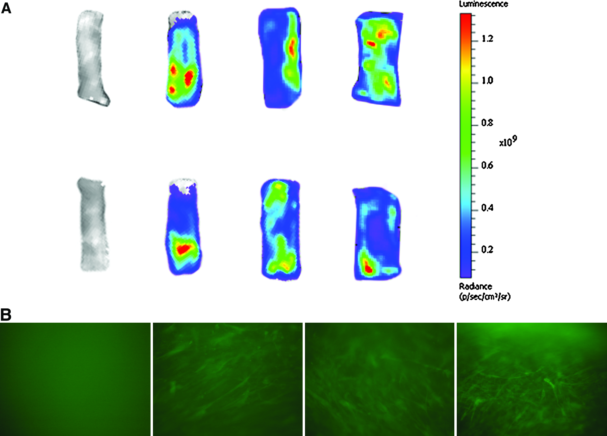
Ex vivo bioluminescent images of ADSC-luc2-eGFP-seeded constructs at 14 days postseeding with no cells (control, far left), 1×10⁶ cells/mL (center left), 2×10⁶ cells/mL (center right), and 4×10⁶cells/mL (far right) demonstrate viable cells distributed over the construct surface
Bioluminescence imaging: in vivo cell viability
BLI revealed persistent cell survival at up to 4 weeks. However, there was 84.2%±12.1% reduction in radiance for week 4 readings compared with week 1. This suggests cell attrition over time. 42
Discussion
The concept of revitalization of biostatic allografts is not new and reseeded constructs have already seen clinical use. Decellularized and subsequently reseeded human trachea has been successfully implanted in a young woman with end-stage bronchomalacia.43,44
Is the ex vivo revitalization of a decellularized tendon graft truly necessary? After implantation, a biostatic allograft function undergoes degradation and remodeling, with gradual substitution by ECM synthesized by recipient cells. However, this process is not always complete due to a lack of reaction by recipient cells.23,26 One notable example is incomplete remodeling of massive distal femoral bone allografts. Without recipient cells and the synthesis of new tissue, the mechanical features of the graft will be adversely affected. 23 With structural, load-bearing grafts, such as bone (compression) and tendon (tension), the restoration of mechanical features is particularly important.
Furthermore, tendons are innately hypocellular and hypovascular, and possess a close-knit tendon weave and compact superstructure, making remodeling slow and inefficient. 45 To facilitate early engraftment, we believe that tissue-engineered tendon constructs should be prepopulated with recipient cells to initiate the repair process and encourage intrinsic rather than extrinsic tendon healing. 46
Mesenchymal stem cells are attractive candidate seed-cells and have been used by other investigators for similar purposes. We chose ADSC instead of bone marrow stem cells because of the high cell yield per unit volume of fat compared with marrow. Zuk et al. noted that bone marrow yields ∼1 mesenchymal stem cell per 105 stromal cells, while fat yields 2–6×10⁸ stem cells per 300cc of fat. 47 Cell harvest and expansion is thus easily achieved with a small volume of lipoaspirate obtained during the initial surgery.
In this article, we examined the role of additional biochemical and physical intervention steps in optimizing ex vivo reseeding. We found that rehydration in growth factor-rich serum was insufficient to drive cell migration into the tendon core. We also noted that scaffolds rehydrated in 100% serum (Group F and Group S+F) displayed reduced seeding compared to their counterparts hydrated in 10% serum (Group CON and Group S, respectively). This may be explained by the potential toxic effect of serum when applied in concentrations greater than 40%.48,49 Thus, the routine use of serum rehydration over the current standard (media enriched with 10% serum) cannot be justified.
On the other hand, we demonstrated that surface scoring increases reseeding, leading to an increase in live and total cell content (Figs. 2–5). This is believed to be because introducing cells to an uninterrupted external tendon face (e.g., remnant tendon sheath, epitenon, and compact superstructure) leaves superficially adherent cells that have tenuous hold and slough off easily. Scoring disrupts the tendon surface, creating a foothold for cell attachment and penetration.
The role of surface intervention in ex vivo reseeding is not new and has been explored by other authors frustrated by difficulty in achieving cell attachment and penetration in the face of uninterrupted tendon surfaces. Cartmell and Dunn performed partial-thickness lacerations in patella tendons intended for ACL reconstruction. 50 Tischer et al. investigated injection reseeding of rabbit semitendinosus tendons. 51 Using intrasynovial tendons, Hashimoto et al. investigated physical abrasion with sandpaper and chemical abrasion with trypsin 26 and reported findings similar to ours that surface disruption resulted in greater cell attachment.
We also demonstrated that seeded cells demonstrate an ultrastructural relationship to the surrounding matrix reminiscent of native tendon (Fig. 6) and these cells display a persistent metabolic activity and procollagen synthesis (Fig. 8), suggesting adoption of a phenotype concordant with the host environment. Furthermore, cell viability is enhanced, as evidenced by reduced apoptosis (Fig. 7). Seeded ADSCs also demonstrate prolonged ex vivo survival for at least 2 weeks on BLI (Fig. 10), although with noticeable attrition. This has implications for future upscalable application, as graft preparation and ex vivo seeding may occur long before clinical implantation.
Increased cellularity leads to increased synthetic and contractile ability, increasing construct strength and stiffness (Fig. 10). The contribution of cells to a construct's biomechanical properties had previously been demonstrated by Androjna et al., 52 who hypothesized that the attached cells generated contractile forces, resisting tension via their cytoskeleton. 41
Finally, as proof of concept, we further demonstrated that revitalized constructs remain viable for up to 4 weeks in vivo in a nude animal model. From this, we can conclude that: (1) the seed-cells survive beyond the critical 2-week strength nadir of a healing tendon repair, (2) derive independent nutrition (neovascularization) necessary for prolonged survival, and (3) are not targeted by immune cells for elimination early in the remodeling process. The progressive reduction in radiance suggests normal seed-cell attrition with time as host cells increasingly populate the scaffold, which is expected in remodeling tissue.
Physical intervention may be less readily accepted with digital flexor tendons, where the benefits of strength and early engraftment can only be fully appreciated in the presence of restored gliding. Many authors have emphasized the role of the tendon sheath, with some authors emphasizing en bloc grafting with the tendon sheath. 53 This may be less important with allografts, as an attached cellular sheath layer may incite an inflammatory reaction and act as a barrier to repopulation by recipient cells. In addition, the tendon sheath forms a physical barrier to chemical agents, leading to incomplete decellularization and the attendant potential complications.
To minimize the risk of compromised gliding after surface disruption, scoring is performed in line with tendon fibers, rather than across the tendon weave. Furthermore, we hypothesize that a scored external tendon face may allow for enough in vitro or in vivo cell attachment and hyaluronic acid synthesis sufficient to recreate a gliding surface. These hypotheses remain to be tested.
There are some limitations to this study. Because of the novelty of this manual technique, it is impossible to standardize the manual scoring technique with regard to scoring depth and location. It is thus impossible to quantify the increase in scaffold surface area. Machine-scored scaffolds may offer a more standardized alternative for upscaled application.
Some questions remain unanswered. First, the optimal seeding density for any given length of a natural scaffold remains unknown. Because only a small percentage of suspended cells finally adhere to the scaffold, intuition would suggest that increased cell density leads to increased reseeding. However, this theory ignores cell attrition from metabolic stressors as density increases. Second, while there was an appreciable increase in cell attachment and penetration with scoring, cell populations appeared in clusters, presumably coincident to the location of surface troughs. Homogenous and uniform cellularity, akin to native tendon, remains unattainable by current techniques. Finally, if surface scoring and other mechanical disruption techniques lead to greater cell penetration and reseeding by host (recipient) cells, then ex vivo reseeding may not be necessary.
Beyond this study, we intend to explore the following: (1) establish normative values for seeded cell survival—to determine how long-seeded grafts remain viable in vivo beyond the current 4-week period, (2) to determine how cell concentration affects graft outcome and if there is an optimal concentration threshold, (3) to determine if other seeded tissue constructs (e.g., bone and cartilage) display similar outcomes, and (4) to determine if complex tissues, such as composite multitissue constructs, can be similarly seeded with favorable outcomes. Pluripotent candidate seed-cells (ADSCs) are ideally suited for the purpose of en bloc reseeding of composite multitissue scaffolds.
In conclusion, natural tendon scaffolds form the ideal graft for flexor tendon reconstruction, as they possess desirable biomechanical characteristics that make them well-suited for the role. Reseeded tendons may allow for faster graft incorporation and remodeling, permitting early motion to mitigate adhesion formation. Tendon surface scoring is one method that leads to greater reseeding in vitro and may be incorporated as a final step in allograft processing before clinical implantation.
Footnotes
Disclosure Statement
No competing financial interests exist.