
Abstract
The culture surface can affect the in vitro differentiation of stem cells. In this study, we investigated whether modifying the culture surface with 3,4-dihydroxy-
Introduction
There have been attempts to modulate stem cell differentiation with cell-adhesive substrates.13–20 Stem cell differentiation can be controlled by changing the chemical functional groups of culture substrates, such as methyl, carboxyl acid, hydroxyl, and amine groups.15,16,21 However, certain nanomaterials were found to enhance stem cell differentiation.22–27 For example, carbon nanotubes have been demonstrated to enhance the osteogenic differentiation of stem cells. 27 The interaction of these nanomaterials with the cell membranes or with proteins in the cytoplasm activated the mitogen-activated protein kinase (Mapk) signaling pathway, which regulated the expression of genes, inducing osteogenic transcription. 28
In this study, 3,4-dihydroxy-
Materials and Methods
DOPA coated on the culture surface
For DOPA coated on culture plates, DOPA (Sigma, St. Louis, MO) was dissolved in the Tris buffer (10 mM, pH 8.5) at 2 mg/mL and placed in a normal cell culture plate (Corning, Corning, NY) at 25°C for 16 h. After incubation, the plate was washed with phosphate-buffered saline (PBS) three times. 31 To confirm DOPA coated, X-ray photoelectron spectroscopy (XPS) and an ultrahigh vacuum multipurpose surface analysis system (Sigma Probe, Thermo, Essex, United Kingdom) at base pressures of <10−10 mbar were used. The photoelectron spectra were excited using an Al Kα (1486.6 eV) anode operating at a constant power of 100 W (15 kV and 10 mA). During the spectra acquisition, the constant analyzer energy mode was employed at a pass energy of 30 eV and a step of 0.1 eV. The binding energy scale was calibrated from the hydrocarbon contamination using the C 1s peak at 285 eV. The hydrophilicity/hydrophobicity of the surface was measured with a water contact angle analyzer (DSA 100; Krüss, Hamburg, Germany).
Cell culture
For this study, hBMMSCs were purchased from Lonza (Walkersville, MD) and cultured on a noncoated plate or a DOPA-coated plate in a growth medium composed of the Dulbecco's modified Eagle's medium (DMEM; Gibco BRL, Gaithersburg, MD) containing 10% (v/v) fetal bovine serum (FBS; Gibco BRL) and 1% (v/v) penicillin/streptomycin (Gibco BRL). To induce the osteogenic differentiation of hBMMSCs, the cells were cultured in an osteogenic medium composed of the DMEM (high glucose) supplemented with 10% FBS, 0.1 mM dexamethasone (Sigma), 50 μM ascorbate-2-phosphate (Sigma), and 10 mM b-glycerophosphate (Sigma). The medium was changed every 2 days, and hBMMSCs at four passages were used for experiments.
Cell adhesion and spreading
The hBMMSCs were cultured in the growth medium. The cell adhesion ratio was calculated by dividing the number of adherent cells at various time points by the initial plating cell number (n=5). Trypsinized cells were counted using a hemocytometer and plated on culture plates. The initial cell plating number was determined so that the cells reach confluency at 2×10⁶/100 cm2 after 1 week of culture. 36 To evaluate cell spreading, hBMMSCs were stained with 1,1′-dioctadecyl-3,3,3′,3′-testramethylindocarbocyanine perchlorate (DiI; Sigma). A stock solution of DiI in dimethylsulfoxide (Sigma) (0.25 μg/μL) was added to the cell culture medium at 1 μg of DiI/mL of medium, and hBMMSCs were incubated at 37°C for 2 h, washed three times with PBS, stained with 4,6-diamidino-2-phenylindole (DAPI; Vector Laboratories, Burlingame, CA), and mounted. Thirty different images were randomly selected from each group for the calculation of the DiI-labeled cell adhesion area. DiI-positive cell areas were digitally quantified using Image Pro Plus software (Media Cybernetics, Silver Springs, MD).
Cell apoptosis and viability
The terminal deoxynucleotidyl transferase dUTP nick end labeling assay was performed to determine the apoptotic activity of cultured hBMMSCs in the growth medium using an ApopTag Red in situ apoptosis detection kit (Chemicon, Temecula, CA) according to the manufacturer's instructions. The viability of the cells cultured on DOPA-coated surfaces in the growth medium was evaluated with a Neutral Red assay as previously described. 37 The results are expressed as a percentage of the positive control (cells cultured on noncoated plates, n=5 for each group).
Alkaline phosphate activity
The alkaline phosphate (ALP) activity of hBMMSCs was determined with p-nitrophenol phosphate (Anaspec, San Jose, CA) as the substrate. Specifically, hBMMSCs were rinsed twice with PBS and lysed in the alkaline lysis buffer followed by three freeze–thaw cycles at −70°C and 37°C. The aliquots were incubated in the glycine buffer containing 2 mg/mL of p-nitrophenol phosphate. After 30 min, 3 N sodium hydroxide was added to stop the reaction. The absorbance of p-nitrophenol was measured at 405 nm.
Calcium assay
For von Kossa staining, the cells were fixed with 4% (w/v) paraformaldehyde for 10 min at room temperature, washed with deionized water, incubated in a 2% (w/v) silver nitrate (Sigma) solution with exposure to a 60-W lamp for 1 h, and subsequently treated with a 5% sodium thiosulfate (Sigma) solution for 5 min. To determine the amount of deposited calcium, the cells were rinsed twice with deionized water and incubated with 0.6 N hydrochloric acid for 4 h. Calcium was extracted by shaking the sample for 4 h at 4°C. The lysate was subsequently centrifuged at 1000 g for 5 min, and the supernatant was used to determine the calcium content. The calcium concentration in the lysates was quantified spectrophotometrically with cresolphthalein complexone (Sigma). Three minutes after the addition of reagents, the samples were read at 575 nm using a microplate reader (PowerWave ×340; Bio-Tek Instruments, Winooski, VT). The calcium concentration was calculated from a standard curve generated from the serial dilution of a standard calcium solution (Sigma).
Real-time polymerase chain reaction
For the relative quantification of the mRNA expression of runt-related transcription factor 2 (RUNX2), ALP, and osteocalcin (OC), real-time reverse transcriptase polymerase chain reaction (qRT-PCR) was performed. Using a Light Cycler 480 (Roche, Basel, Switzerland) with SYBR Green I (Takara, Otsu, Shiga, Japan), qRT-PCRs were performed. After 5 min of preincubation, 35 amplification cycles were performed. Each cycle consisted of the following three steps: 30 s at 94°C, 45 s at 60°C, and 45 s at 72°C. The primer sequences for qRT-PCR are shown in Table 1. All of the data were analyzed with the 2−ΔΔCt method.
RUNX2, runt-related transcription factor 2; ALP, alkaline phosphate; OC, osteocalcin.
hBMMSC transplantation and in vivo bone formation
Poly(lactic-co-glycolic acid) sponge (PLGA) scaffolds (5×5×2 mm3, 95% porosity) were fabricated with the solvent-casting and particle-leaching method. 1 Female athymic mice (BALB/c-nu, 6 weeks old; Orient Bio, Sungnam, Korea) were anesthetized with xylazine (20 mg/kg) and ketamine (100 mg/kg). hBMMSCs were cultured in the osteogenic media 3 weeks. Osteogenically differentiated hBMMSCs were seeded on PLGA scaffolds and implanted into dorsal subcutaneous spaces of athymic mice (n=8 implants per group). Eight weeks after the implantation, the mice were euthanized, and the implants were retrieved and fixed in a 4% (w/v) paraformaldehyde solution. Bone formation was evaluated with computed tomography (CT) scans (n=3 per group) and histological analysis (n=5 per group). CT images were obtained with a micro-CT system (SkyScan-1172; Skyscan, Kontich, Belgium) and further processed using Skyscan CT analyzer software (Skyscan) to visualize the signal as red for explanted tissue and white for the mineralization region. The mineralization volume was calculated with CT analyzer software. Histological specimens were fixed in paraformaldehyde, embedded in paraffin, sectioned transversely at a thickness of 4 μm, and examined with Goldner's trichrome staining and immunohistochemical staining. The tissue sections were immunohistochemically stained with antibodies against Runx2 (Abcam, Cambridge, United Kingdom) and Col 1 (Abcam). The staining signal was visualized with rhodamine isothiocyanate-conjugated secondary antibodies (Jackson ImmunoResearch Laboratories). The slides were counterstained with DAPI (Vector Laboratories) to stain the nuclei of cells and photographed with a fluorescence microscope (IX71 inverted microscope; Olympus, Tokyo, Japan). The animal study was approved by the Institutional Animal Care and Use Committee of Seoul National University (SNU-110121-4).
Statistical analysis
Quantitative data are expressed as the means±standard deviations. The number n refers to n independent experiments. The statistical analysis was performed with a one-way analysis of variance with the Tukey's post hoc test using SPSS software (SPSS Inc., Chicago, IL). A value of p<0.05 was considered to be statistically significant.
Results
To confirm DOPA coated on the culture substrate, the surfaces of noncoated and DOPA-coated plates were analyzed using XPS. The chemical composition of the noncoated plate was polystyrene (Fig. 1E). Nitrogen peaks of the DOPA-coated group appeared at 400 eV (Fig. 1B). The oxygen peak decreased in the DOPA-coated group at 532 eV compared with the noncoated coating group (Fig. 1A, B). The carbon peak increased in the DOPA-coated group at 285 eV compared with the noncoated group. The atomic quantification showed increased carbon and nitrogen and decreased oxygen amounts in the DOPA-coated group (Fig. 1C). The surface hydrophilicity was also investigated with contact angle measurements (Fig. 2). DOPA coated decreased the water contact angle (42.0±9.3 versus 19.2±8.2; no-coating versus DOPA coated), indicating that DOPA coated increased surface hydrophilicity.

X-ray photoelectron spectroscopy (XPS) analyses of culture plate surfaces that were
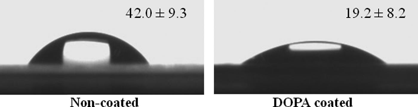
Water contact angle of culture surfaces that were uncoated or coated with DOPA.
The cell adhesion and spreading were evaluated. The DOPA-coated surface showed significantly higher cell adhesion after 24 h compared with the noncoated surface (Fig. 3). Additionally, the DOPA-coated surface showed significantly higher cell spreading compared with the noncoated surface (Fig. 4). Both the noncoated and DOPA-coated surfaces showed negligible hBMMSC apoptosis (Fig. 5). The cell viability was not different between the groups (Fig. 6).
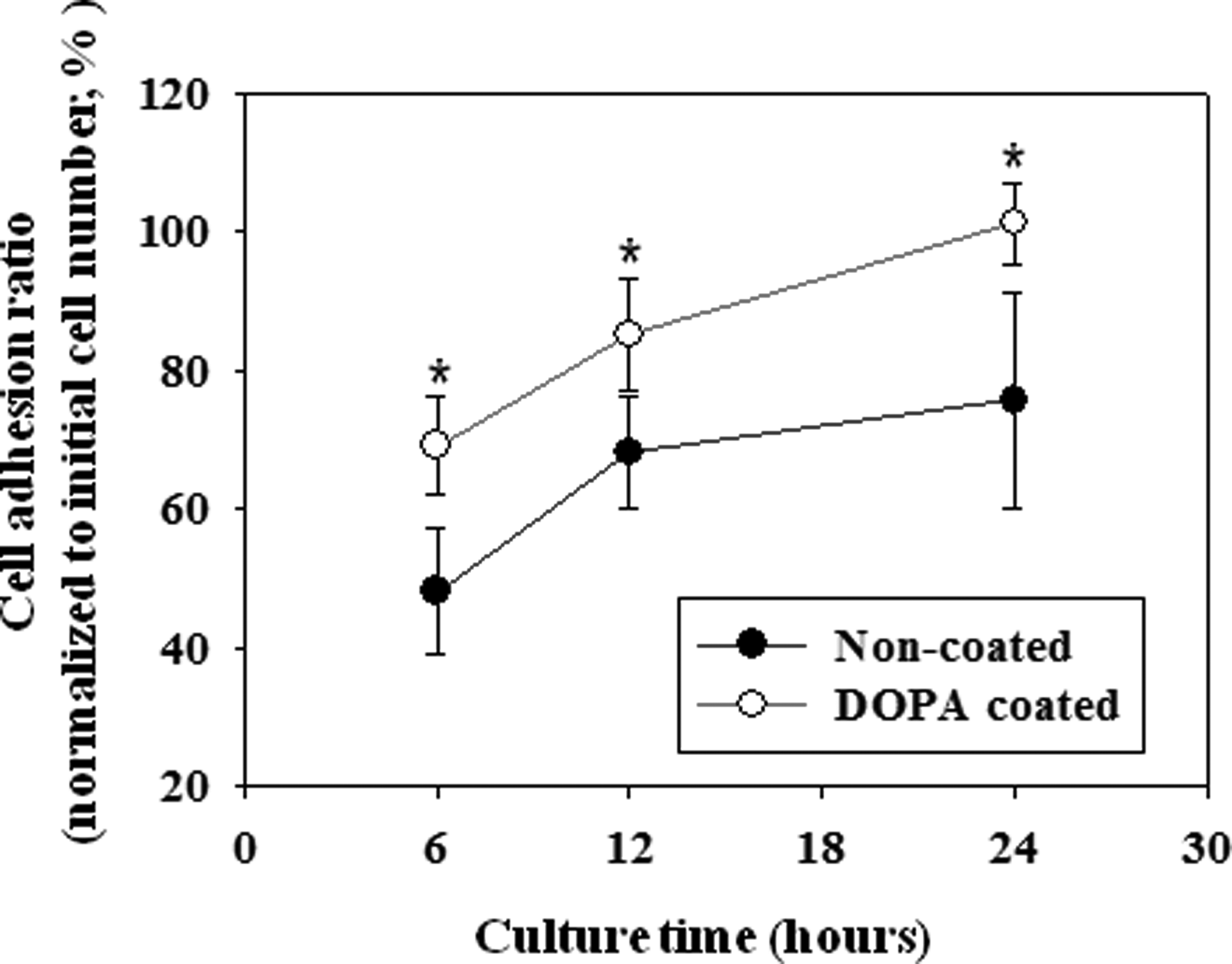
Cell adhesion ratio of human bone marrow-derived mesenchymal stem cells cultured on a DOPA-coated surface or a noncoated surface. The numbers of adherent cells were determined by counting adherent cells using a hemocytometer after trypsinization at various time points. *p<0.05 between the two groups.
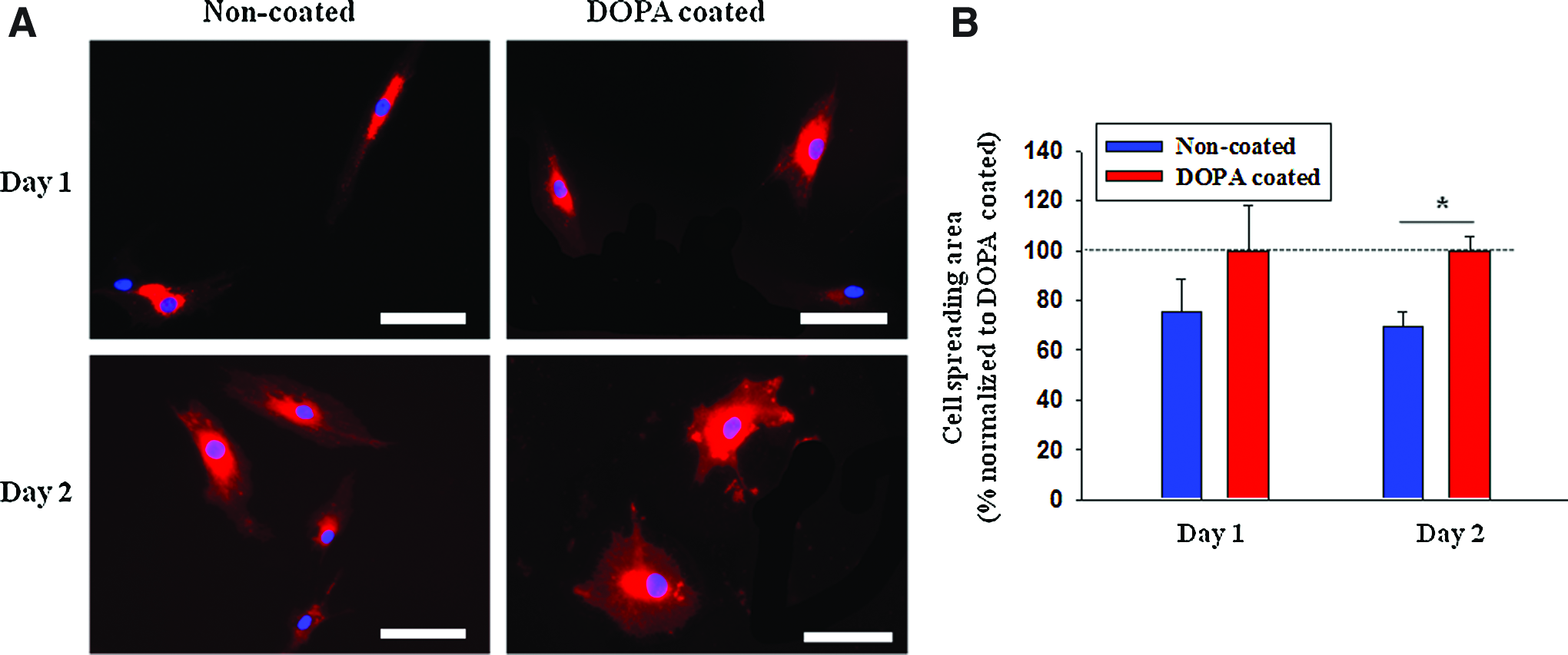
Enhanced human bone marrow-derived mesenchymal stem cell (hBMMSC) spreading after culture on DOPA-coated surfaces.

Apoptosis and viability of hBMMSCs cultured on a DOPA-coated surface or a noncoated surface.

The viability of hBMMSCs as determined with the neutral red assay. Color images available online at www.liebertpub.com/tea
The effects of DOPA coated on the osteogenic differentiation of hBMMSCs were examined. The ALP activity was significantly higher in the DOPA-coated group compared with the no-coating group (Fig. 7). Culture in the growth medium (negative control) showed a negligible ALP activity. Calcium deposition was observed in both the DOPA-coated group and the no-coating group 1 week after culture in the differentiation medium (Fig. 8). The calcium deposition was significantly higher in the DOPA-coated group than the no-coating group at 2 and 3 weeks. Culture in the growth medium (negative control) showed negligible calcium deposition during 3 weeks.

The osteogenic differentiation of hBMMSCs cultured on either noncoated or DOPA-coated surfaces, as evaluated with the alkaline phosphate (ALP) assay. ●; noncoated+growth medium. ○; noncoated+osteogenic medium. ▼; DOPA coated+osteogenic medium (n=3 per group; *p<0.01 compared with noncoated+osteogenic medium).

The osteogenic differentiation of hBMMSCs cultured on either noncoated or DOPA-coated surfaces as evaluated with a calcium assay.
The mRNA expression of osteogenic markers was examined using qRT-PCR analyses (Fig. 9). The mRNA expression of RUNX2, ALP, and OC showed no difference between the noncoated and DOPA-coated group at 1 week. The mRNA expression of RUNX2, an osteogenic transcription factor, was significantly higher in the DOPA-coated group at 2 weeks compared with the no-coating group. The mRNA of ALP, an early osteogenic marker, was also expressed at a significantly higher level in the DOPA-coated group at 2 and 3 weeks. The mRNA expression of OC, a late osteogenic maker, was only significantly higher in the DOPA-coated group at 3 weeks.

The osteogenic differentiation of hBMMSCs that were cultured on either noncoated or DOPA-coated surfaces, as evaluated with real-time polymerase chain reaction analysis to detect the mRNA expression of runt-related transcription factor 2 (RUNX2), ALP, and osteocalcin (OC). Blue bars indicate noncoated+osteogenic medium, and red bars indicate DOPA coated+osteogenic medium (n=5 per group; *p<0.05 compared with the other group at the same time point). Color images available online at www.liebertpub.com/tea
Finally, the in vivo bone formation efficacy of hBMMSCs cultured on the two different surfaces was examined. Eight weeks after implantation, hBMMSCs cultured on the DOPA-coated surface showed a significantly higher calcium deposition, as detected with micro-CT, compared with hBMMSCs cultured on a noncoated surface (Fig. 10). The mineralization volume was also significantly greater in the DOPA-coated group. The enhanced bone formation was also confirmed with histological and immunohistological analyses (Fig. 11). The DOPA-coated group showed bone tissue with lacuna structures, whereas only fibrous tissues were observed in the no-coating group. A higher degree of positive staining for type 1 collagen in the DOPA group suggested increased bone formation. A higher expression of Runx2 in the DOPA-coated group indicated a larger number of osteoblastic cells.

Evaluation of the in vivo bone formation ability of hBMMSCs cultured on either noncoated or DOPA-coated plates and implanted subcutaneously into athymic mice for 8 weeks.

Histological or immunohistological analysis of implants retrieved 8 weeks after implantation.
Discussion
In this study, tissue plates were coated with DOPA to enhance the in vitro osteogenic differentiation and in vivo bone formation capacity of hBMMSCs. With its high adhesion strength in holding mussels in place in a rough marine environment, DOPA can be used for modifying the cell culture surface.29–34 Additionally, DOPA can increase the hydrophilicity of the cell culture surface, due to its functional groups, such as catechol and amine.33–35 We hypothesized that DOPA coated on the culture surface may affect cell adhesion properties and the hydrophilicity of the surface to enhance the in vitro osteogenic differentiation of culture stem cells.
The osteogenic differentiation of mesenchymal stem cells can be controlled by regulating Runx2, an osteogenic transcription factor.13–15 For the upregulation of Runx2, MAPK kinase (MEK) upregulation is required through two pathways.12–14 First, soluble osteogenic factors, such as dexamethasone and bone morphogenetic protein-2, directly upregulate the MEK pathway.13,27 Second, indirect upregulation through increased interactions between integrins and the cytoplasm upregulates ROCK1 and RhoA. 13 RhoA can replace soluble factor signaling, whereas ROCK1 promotes downstream cell adhesion and spreading.13,15,16 In this study, the same soluble factor (i.e., dexamethasone) was used for both the no-coating group and the DOPA-coated group. Thus, we focused on the second pathway to explain the difference in osteogenic differentiation between the two groups.
The enhanced osteogenic differentiation of hBMMSCs cultured on DOPA-coated plates could be at least partly due to fibronectin adsorbed onto DOPA. Cell adhesion proteins contained in the serum, such as fibronectin and vitronectin, adsorb onto cell culture plates, and cells adhere to the proteins when they are cultured in a serum-containing medium. Fibronectin plays important roles in BMMSC behavior. It promotes not only cell adhesion and spreading, but also promotes the osteogenic differentiation of BMMSCs. 38 However, these proteins can be denatured upon adsorption to normal culture plates. 31 DOPA could prevent the denaturation of cell adhesion proteins when they are adsorbed onto DOPA-coated plates. 31
Another factor that could explain the enhanced osteogenic differentiation of hBMMSCs by DOPA is its chemical structure.29–35 Previous studies have shown that MSC behaviors can be affected by chemical functional groups of the culture surface. 21 MSCs cultured on methyl groups showed an undifferentiated phenotype. 21 However, MSCs cultured on alcohol or carboxyl acid showed chondrogenic differentiation. 21 MSCs cultured on amines showed osteogenic differentiation. 21 The amine group can promote cell adhesion35,38 and spreading.31,33,35,36 Compared with noncoated culture plates composed of polystyrene, DOPA contains much more amine groups.29–34 The amine group of DOPA may play a critical role in the enhanced osteogenic differentiation of hBMMSCs.
The more extensive in vivo bone formation of the DOPA-coated group (Figs. 10 and 11) compared with the no-coating group may be due to the implantation of more osteogenically differentiated cells in the DOPA group. Previous studies have shown that the implantation of osteogenically differentiated cells results in more bone formation in vivo than osteogenically undifferentiated cells.11,12 Thus, culture on DOPA-coated culture plates may enhance the in vivo bone formation efficacy of hBMMSCs.
Conclusions
The culture of hBMMSCs on DOPA-coated culture plates significantly enhanced osteogenic differentiation compared with noncoated conventional culture plates. The enhanced osteogenic differentiation was mediated at least partly by RhoA signaling. hBMMSCs cultured on DOPA-coated plates exerted an enhanced ability of in vivo bone formation compared with those cultured on conventional plates. DOPA coated on culture plates may be useful in enhancing the therapeutic efficacy of hBMMSCs for bone regeneration.
Footnotes
Acknowledgments
This study was supported by the Korea Health 21 R&D Project, Ministry of Health and Welfare (A100443), and the National Research Foundation of Korea (2009-0092213, 2010-0020352).
Disclosure Statement
No competing financial interests exist.