
Abstract
Physical barriers are commonly used to reduce peritendinous adhesion after injury. However, the inflammatory response to surgery cannot be prevented. This study was designed to evaluate the ability of ibuprofen-loaded poly(l-lactic acid)-polyethylene glycol (PELA) diblock copolymer fibrous membranes in preventing adhesion formation and reduce inflammation. Electrospun PELA fibrous membranes underwent mechanical testing and were characterized by morphology, surface wettability, drug release, and degradation. Results of an in vitro drug release study showed that a burst release was followed by sustained release from fibrous membranes with high initial ibuprofen content. Fewer L929 mouse fibroblasts adhered to and proliferated on the ibuprofen-loaded PELA fibrous membrane compared with tissue culture plates or PELA fibrous membrane without ibuprofen. In a chicken model of flexor digitorum profundus tendon surgery, the ibuprofen-loaded PELA fibrous membranes prevented tissue adhesion and significantly reduced inflammation. Taken together, these results demonstrate that ibuprofen-loaded PELA fibrous membranes prevent peritendinous adhesion formation better than membranes that do not contain ibuprofen, through anti-adhesion and anti-inflammatory actions.
Introduction
Recently, various physical membranes have been used as barriers to minimize extrinsic healing, an important part of adhesion formation. 4 Electrospun fibrous membranes are effective for tissue separation and drug delivery because of their large surface area-to-volume ratio, high porosity, and very small pore size.5,6 The microporous structure allows the passage of nutrients from outside the tendon sheath to promote intrinsic healing. 4 Of these electrospun fibrous membranes, electrospun poly(l-lactic acid)-polyethylene glycol (PELA) diblock copolymer fibrous membranes are more flexible and easier to use than commonly used anti-adhesion films (e.g., poly-D-L-lactic acid and Seprafilm® films) in the prevention of abdominal wall adhesions.7,8 With better flexibility and hydrophilicity than poly(l-lactic acid) (PLLA), PELA is also effective for drug delivery applications. By varying the ratio of polyethylene glycol (PEG), PELA electrospun fibrous membranes can be optimized to produce favorable hydrophilic properties and degradation patterns and prevent cell attachment and proliferation. 9 However, PELA fibrous membranes cannot block the inflammatory response to surgery. Moreover, PELA electrospun fibrous membranes have not yet been used to prevent peritendinous adhesion formation.
Acute inflammatory response to surgery is a risk factor for tendon adhesion formation. 10 Despite the initial promise shown by barrier materials in clinical applications, absorbable membranes often degrade rapidly, sometimes inducing a severe foreign-body reaction. 11 Inflammatory cells and fibroblasts migrate to the site of injury and new capillaries form during the process of adhesion formation. 4 Nonsteroidal anti-inflammatory drugs (NSAIDs) such as ibuprofen target the inflammatory phase of tendon healing to reduce adhesions. However, NSAIDs alone cannot completely prevent adhesion formation because of their rapid clearance and potential for severe side effects when taken orally. 4
Ibuprofen inhibits both cyclooxygenase (COX)-1 and COX-2, and therefore inhibits adhesion formation after flexor tendon repair better than COX-2 selective NSAIDs. 12 In this study, ibuprofen-loaded PELA diblock copolymer fibrous membranes prepared by electrospinning were tested for the ability to minimize adhesion formation and inflammation caused by surgery and material degradation. The electrospun ibuprofen-loaded PELA fibrous membranes were characterized and evaluated in vitro and in vivo for their ability to prevent tissue adhesion through synergistic anti-adhesion and anti-inflammatory effects.
Materials and Methods
Materials
PELA (Mw=40 kDa, E/L=10/90, Mw/Mn=1.56), ibuprofen, 3-(4,5-dimethyl-2-thia-zolyl)-2,5-diphenyl-2H-tetrazolium bromide (MTT), dimethyl sulfoxide, acridine orange, and propidium iodide were purchased from Sigma-Aldrich and used without further purification. Dulbecco's modified Eagle's medium (DMEM) and fetal bovine serum were supplied by Gibco. All other chemicals and solvents were of reagent grade or better and purchased from GuoYao Regents Company, unless otherwise indicated.
Electrospinning of nanofibrous membranes
Electrospinning was carried out according to our previous report. 9 In brief, to prepare ibuprofen-loaded PELA electrospun fibrous membranes, 1 g PELA and ibuprofen were completely dissolved in a solvent containing 2.5 g dichloromethane and 1.5 g acetone. The solutions containing 2.0%, 6%, or 10% (w/w) ibuprofen were drawn into 2.0-mL glass syringes fitted with 0.7-mm-diameter needles to prepare the drug-loaded fibers (PELA-2%, PELA-6%, and PELA-10%, respectively). PELA fibrous membranes were fabricated by electrospinning at an applied voltage of 15 kV using a high-voltage power supplier. A syringe pump was used to feed the polymeric solution into the needle tip (feeding rate 3.0 mL/h). The fibrous membranes were collected on grounded aluminum foil. The distance between the needle tip and the collector was 15 cm. The fibrous membranes were then vacuum-dried at room temperature for 1 day before the investigation.
Characterization of the electrospun fibrous membranes
The thickness of the fibrous membranes was measured with a micrometer, and their apparent density and porosity were calculated according to previously published methods. 13 Morphology was observed by scanning electron microscopy (SEM, FEI Quanta 200). At least five images of each sample (10,000× magnification) were obtained. The mean diameter of the fibers was determined with Photoshop 8.0 using at least 20 different fibers and 200 different segments from each image selected randomly. 14
To test mechanical properties, the dry fibrous membranes were punched into small strips (70.0×7.0×0.6 mm). Uniaxial tensile tests were performed using an all-purpose mechanical testing machine (Instron 5567, Norwood, MA). The stress–strain curves of the fibrous membranes were constructed from the load deformation curves recorded at a stretching speed of 0.5 mm/s (n=5). Young's modulus, tensile strength, and elongation at break of the fibrous membranes were derived from the stress–strain curves.
Surface wettability of the PELA fibrous membranes was evaluated using the water contact angle method at room temperature. The water contact angles of different fibrous membranes were determined with a Krüss GmbH DSA 100 Mk 2 goniometer, followed by image processing of sessile drop profiles with DSA 1.8 software. 9
Drug release study
The ibuprofen-loaded PELA fibrous membranes were punched into small squares (20×20 mm; total mass ∼50 mg) and immersed in 20 mL phosphate buffered saline (PBS, 154 mM, pH 7.4) containing 0.02% sodium azide as a bacteriostatic agent. The suspension was maintained in a thermostated shaking water bath (Taichang Medical Apparatus Co.) at 37°C and 100 cycles/min. At predetermined time intervals, 5.0 mL release buffer was removed for analysis and replaced with 5.0 mL fresh PBS.
The structure integrity and amount of released ibuprofen was detected by ultraviolet–visible spectrophotometry (UV-2550, Shimadzu, Japan). A linear correlation (γ2=0.9992) was determined between absorption and ibuprofen concentration of standard samples (0, 10, 20, 30, 40, and 50 μg/mL). The percentage of ibuprofen released from triplicate samples was then determined based on the initial weight of the drug incorporated into the electrospun fibrous membrane.
In vitro degradation of fibrous membranes
The degree of degradation was estimated from the decrease in molecular weight and loss of mass. Ibuprofen-loaded PELA fibrous membranes were accurately preweighed (∼100 mg each) and added to 50 mL PBS (pH 7.4) containing 0.02% sodium azide as a bacteriostatic agent. At predetermined time intervals, a sample of each group (containing 2%, 6%, or 10% ibuprofen) was retrieved, rinsed with distilled water to remove residual buffer salts, and dried to a constant weight in a vacuum desiccator. Mass loss was determined gravimetrically by comparing the initial weight with the dry weight remaining at a specific time. Molecular weight was determined by gel permeation chromatography (Waters 2695 and 2414 refractive index detectors; Waters Corporation) using polystyrene as a standard and a Styragel HT 4 column (7.8×300 mm; Waters Corporation). The mobile phase consisted of tetrahydrofuran as the elution solvent at a flow rate of 1.0 mL/min.
In vitro cell culture
L929 mouse fibroblasts were used to evaluate adhesion and proliferation on PELA electrospun fibrous membrane surfaces with or without ibuprofen. The cells were incubated in DMEM supplemented with 10% fetal bovine serum, 100 U/mL penicillin, and 100 μg/mL streptomycin at 37°C in a humidified atmosphere with 5% CO2. The culture medium was changed every 3 days. After the cells reached confluence, they were harvested with 0.25% trypsin. After sterilizing by immersion in 75% ethanol for 1.5 h, the electrospun matrices were washed repeatedly with PBS to remove residual ethanol. The samples were then placed in a 24-well plate.
Cell viability assay
Cells (1×10⁵ cells/mL) were seeded into each well (100 μL/well). After incubation for 1, 4, or 7 days, MTT solution (5 mg/mL) was added to each sample (100 μL/well), and the cells were incubated for an additional 4 h at 37°C. The medium was then removed, and the samples were washed with PBS. The MTT formazan crystals were dissolved in 1 mL dimethyl sulfoxide on a shaking platform for 10 min, and 200 μL of the solution was transferred into a 96-well plate. Absorbance at 490 nm was determined using a spectrophotometer (Synergy 2; BioTek).
Fluorescent staining and observation
The presence of L929 cells on the electrospun fibrous membranes with or without ibuprofen was evaluated by fluorescent microscopy. Cells (1×10⁵ cells/mL) were seeded into each well (50 μL/well). These cultures containing electrospun fibrous membranes were stained with acridine orange and propidium iodide on day 1 and day 4. Thirty minutes later, the cells were observed under a fluorescence microscope (LEICA DM 4000 B). After light excitation, the nuclei of living cells on the surface of the fibrous membranes were stained bright green, and dead cells were stained red.
Preliminary animal study
Leghorn chickens were obtained from the Laboratory Animal Center of Shanghai Institute for Biological Science (Shanghai, China; certificate number SCXK 2003-0003). Animal experiments were carried out in accordance with the policies of Shanghai Jiao Tong University School of Medicine and the National Institutes of Health.
The chickens (n=90, 1.5–2 kg each) were anesthetized by intramuscular injection of ketamine hydrochloride (50 mg/kg). After sterile skin preparation, an elastic tourniquet was applied. A 1.5-cm linear skin incision was made on the lateral aspect of the proximal phalanx of the third toe of the right foot. The flexor tendon sheath was incised, isolating the flexor digitorum profundus (FDP). The tendon was transversely incised and then repaired using a modified Kessler tendon repair with 6-0 prolene suture (Ethicon Ltd.). 5 The animals were randomly assigned to treatment group, and a 1×1 cm piece of PELA fibrous membrane (with or without ibuprofen) was wrapped around the repair site of the FDP. The control group received no treatment before closure. The skin was closed with 4-0 silk sutures, and the extremity was immobilized in a weight-bearing splint (Fig. 1).

Macroscopic evaluation
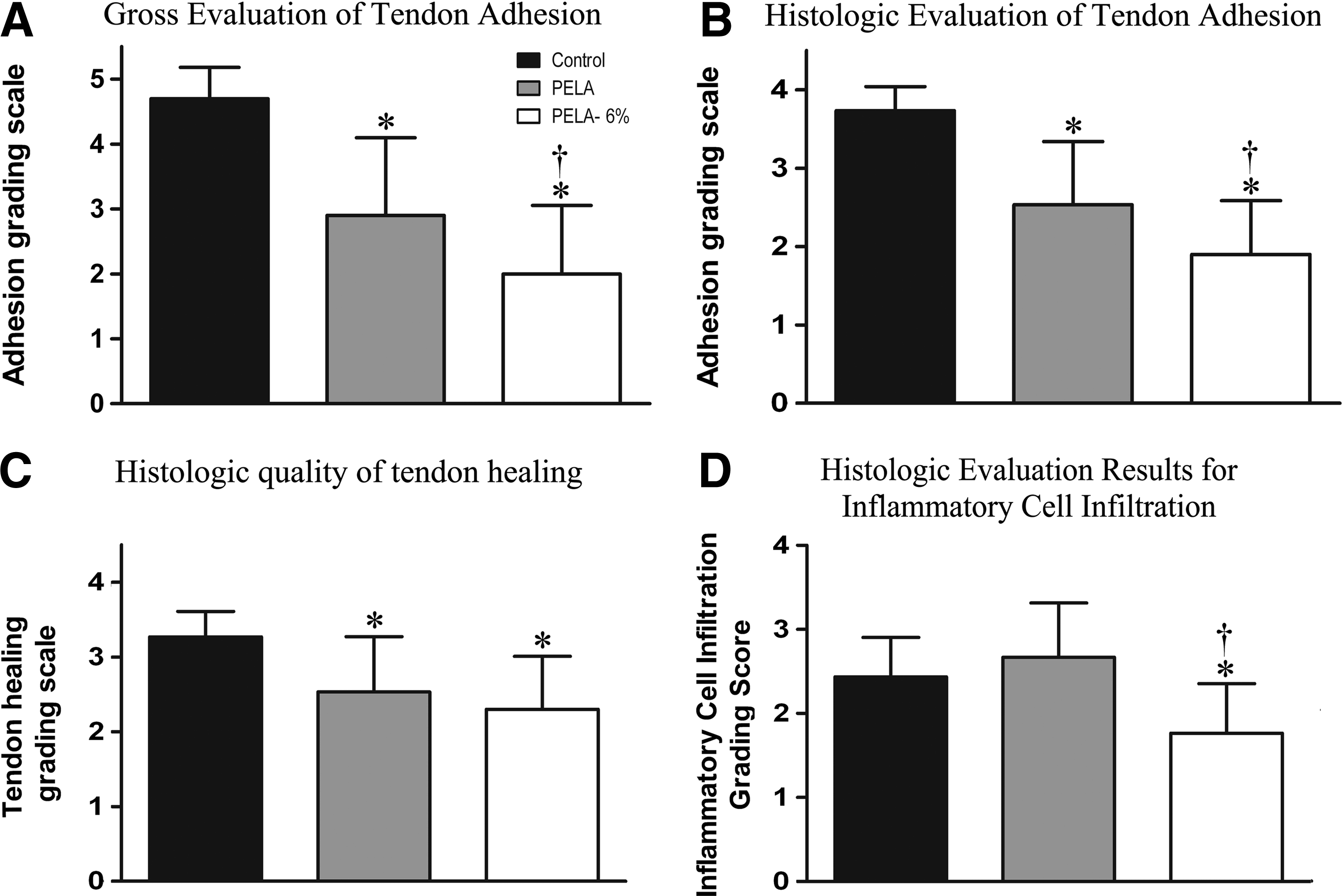
Before sacrificing the animals, the skin incision was visually checked for signs of inflammation or ulcer. To evaluate peritendinous adhesions, a semiquantitative grading scale was used to categorize the extent and severity of adhesion at the repair site on the basis of the macro views: grade 1, no adhesions; grade 2, filmy (separable from surrounding tissue); grade 3, mild (not separable from surrounding tissue); grade 4, moderate (35%–60% of the injured area); grade 5, severe (>60% of the injured area). 5 Adhesions were evaluated by two independent investigators blinded to treatment.
Histological evaluation
The third toes were fixed overnight in 4% paraformaldehyde buffered with PBS (pH 7.4). The specimens were decalcified for 1 month in 10% EDTA buffered with PBS (pH 7.4) at room temperature. After dehydration with increasing concentrations of ethanol and paraffin embedment, 4-mm-thick transverse sections were stained by hematoxylin and eosin (HE) and Masson. Histological assessments of adhesions and tendon healing were then performed.15,16 Adhesions were classified as follows: grade 1, no adhesions; grade 2, mild (<33% of the tendon surface); grade 3, moderate (33%–66% of the tendon surface); or grade 4, severe (>66% of the tendon surface). 15 Tendon healing was classified as follows: grade 1, excellent (good tendon continuity and smooth epitenon surface); grade 2, good (intratendinous collagen bundles exhibited good repair, but the epitenon was interrupted by adhesions); grade 3, fair (irregularly arranged and partly broken intratendinous collagen bundles); grade 4, poor (failed healing or massive overgrowth of granulation tissue). 16 For each specimen, histology slides in three comparable layers (transverse sections: distal, middle, and proximal layers) were evaluated under light microscopy (LEICA DM 4000 B) by two independent investigators blinded to treatment.
Histopathologic examination of inflammation
To evaluate the potential of anti-adhesion properties of the ibuprofen-loaded fibrous membrane, inflammatory cell infiltration was classified as follows: grade 1, a single inflammatory cell detected; grade 2, mild infiltration; grade 3, moderate infiltration; or grade 4, severe infiltration. These examinations were performed by two independent investigators blinded to treatment.
Biomechanical evaluation
The FDP tendon of the third toe was exposed at the ankle for the biomechanical test. Flexion and maximum tensile strength were measured using a rheometer (Instron 5548; Instron). The proximal end of the FDP tendon was fixed to a force gauge, and the proximal phalanx of the toe was attached to a self-made device with the distal interdigital joint fixed by stainless steel rods. To evaluate work of flexion, load (Newton) and displacement (mm) were measured when the FDP tendon was pulled at 10 mm/min until the range of proximal interdigital joint was 40°. Work of flexion was then obtained by calculating the area under the curve. Maximum tensile strength represents the force required to pull the FDP tendon out of the tendon sheath.
Statistical analysis
Results are expressed as mean±standard deviation. Statistical software SPSS 10.0 was used to analyze the data by one-way analysis of variance; p<0.05 considered significant.
Results
Characterization of electrospun ibuprofen-loaded PELA fibrous membranes
SEM micrographs of the PELA fibers are shown in Figure 2A–D. The mean diameter of the fibers decreased as the ibuprofen concentration increased: 1.45±0.71 μm (without ibuprofen), 1.40±0.52 μm (2% ibuprofen), 1.32±0.67 μm (6% ibuprofen), and 1.25±0.59 μm (10% ibuprofen). The porosity of the fibrous membranes decreased as the amount of ibuprofen increased (range 61.6%–71.3%) (Table 1). As shown in Figure 2, the PELA fibers from each group were round, bead-free, randomly arrayed, and very porous. Because ibuprofen has excellent solubility in dichloromethane, the polymer/drug solution was stable and homogeneous, and the electrospinning was successful. The surfaces of the fibers loaded with ibuprofen were smooth, and no drug crystals were apparent (Fig. 2B–D). The water contact angles were 118.6±2.1° (unloaded PELA fibers), 119.5±3.1° (PELA-2%), 121.9±3.2° (PELA-6%), and 123.7±2.6° (PELA-10%) (Table 1). These findings indicate that addition of a small molecule drug (IBU) led to formation of smaller diameter fibers. The possible reason for this improvement is that addition of drugs disturbed the polymer solution, lowered the surface tension, and thus enhanced the bending instability. 5
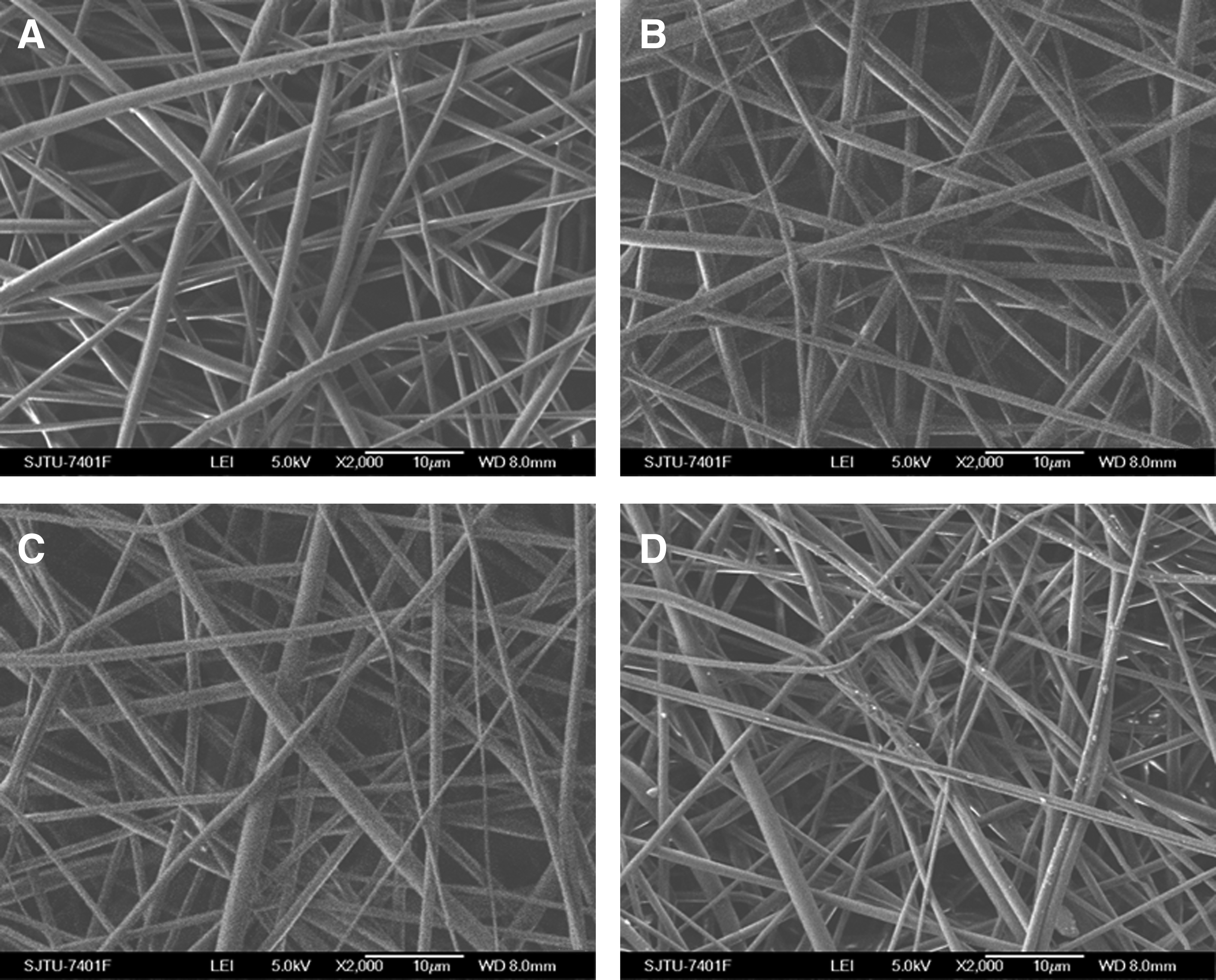
SEM images of PELA electrospun fibers containing
PELA, poly(l-lactic acid)-polyethylene glycol.
To determine the effects of ibuprofen on the mechanical properties of PELA fibrous membranes, the strain–stress response was tested (Table 2). Our results showed higher modulus but lower tensile strength and elongation at break for ibuprofen-loaded PELA fibers compared with unloaded PELA fibers. As the ibuprofen concentration increased, the maximum mechanical tensile strength and maximum elongation decreased, and maximum modulus of elasticity increased.
In vitro drug release and membrane degradation
The drug release profiles of the ibuprofen-loaded PELA fibrous membranes are shown in Figure 3. As the ibuprofen concentration is increased, drug molecules tend to aggregate on the fiber surface, leading to a larger initial burst release. During the first 2 days, the ibuprofen burst release from the electrospun fibrous membranes was 38% (PELA-2%), 47% (PELA-6%), and 62% (PELA-10%), followed by a sustained-release stage during the subsequent 10 days. Because drug release behavior depends primarily on polymer matrix degradation and drug diffusion, the rate of drug release increases as the ibuprofen content increases (Fig. 4).

Cumulative ibuprofen release from electrospun fibers containing 2% ibuprofen (PELA-2%), 6% ibuprofen (PELA-6%), or 10% ibuprofen (PELA-10%) after incubating in phosphate buffered saline (PBS) at 37°C. Color images available online at www.liebertpub.com/tea

Degradation of the fibrous membrane during incubation was determined by gravimetric analysis (Fig. 4A). The mass loss in the early stage of degradation may result from diffusion of relatively low-molecular-weight fibers and drug molecules on the surface dissolving into the medium. In the middle stage, a slight loss of mass may be caused by drug diffusion and polymer degradation. The mass loss from the PELA fibers was 45% (PELA-2%), 60% (PELA-6%), and 72% (PELA-10%,). As shown in Figure 5B, the molecular weight of the fibrous membrane decreased gradually over time, with approximately 30% to 50% lost after a 6-week incubation. This findings of IBU releasing and PELA fiber degradation were expected because the IBU distribution, pores of drug diffusion, high specific surface area, and hydrophilic PEG content of the fibers should result in difference drug-releasing and polymer degradation speed.5,17,18

MTT analysis of fibroblasts on the surface of PELA fibrous membranes with or without ibuprofen (each group, n=3).
In vitro cell adhesion and proliferation
The viability of L929 cells on the surface of PELA fibers (with or without ibuprofen) was compared after 1, 4, and 7 days in culture (Fig. 5). Our results showed that cells grew on the surfaces of all PELA fibers, but grew less well on ibuprofen-loaded fibers. Fluorescent micrographs show the adherence of L929 mouse fibroblasts to ibuprofen-loaded PELA fibrous membranes after 1 and 4 days of incubation. Fewer cells adhered to the ibuprofen-loaded PELA fibers than to the unloaded fibers or tissue culture plate (Fig. 6A–F). In addition, the number of cells decreased as the ibuprofen concentration increased. These fluorescent observations were consistent with the cell viability assay results.
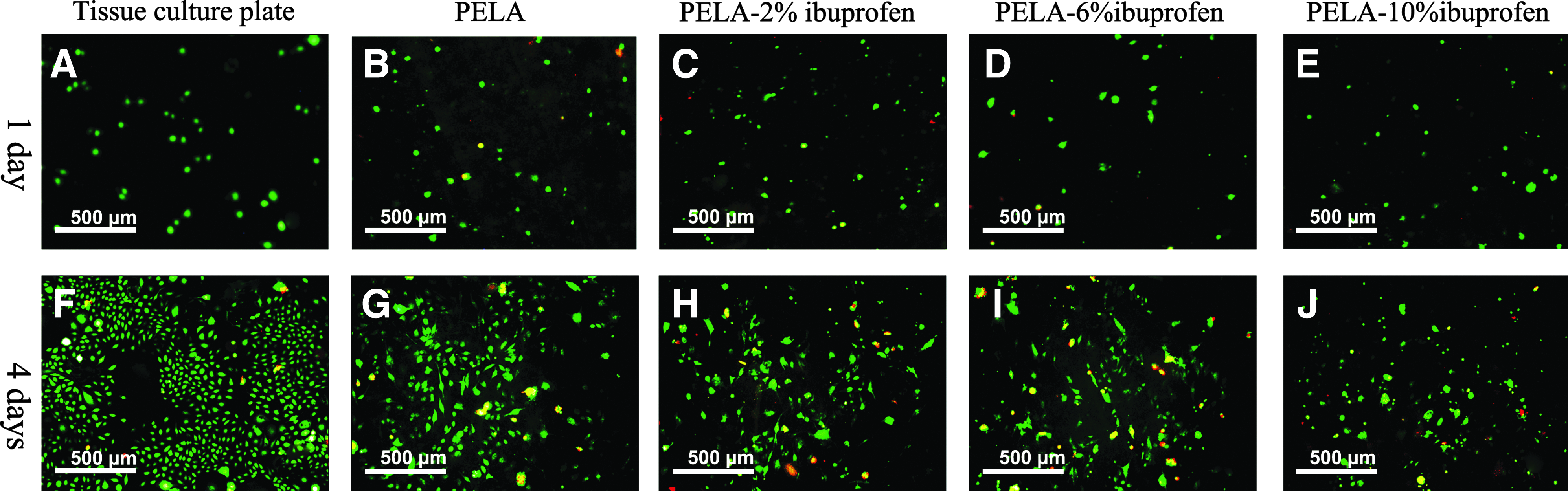
Fluorescent micrographs of L929 mouse fibroblasts after 1 and 4 days of incubation
Animal implant study
After 21 days, the peritendinous adhesions and tendon healing were evaluated by direct observation after the surgical dissection of the repair sites (Fig. 7). In the untreated control group, severe peritendinous adhesions were observed at the repair site, and these were difficult to explore even by sharp dissection (Fig. 7A). Use of PELA fibrous membranes without ibuprofen resulted in scar tissue bridging between the tendon and surrounding tissue (Fig. 7B), but ibuprofen-loaded PELA fibrous membranes demonstrated improved anti-adhesion properties (Fig. 7C).

Gross evaluation of a chicken model of flexor digitorum profundus tendon surgery after 21 days.
In the control group, histological examination of the sutured tendon revealed a dense layer of connective tissue. In most specimens, the peritendinous space was obliterated (Fig. 8A, D). Histological examination of tendons wrapped with the unloaded PELA fibrous membrane revealed cord-like fibrotic tissues in some specimens and general preservation of the peritendinous space (Fig. 8B, E). In contrast, no peritendinous adhesions were detected in most tendons treated with the ibuprofen-loaded PELA fibrous membrane (Fig. 8C, F). Compared with the ibuprofen-loaded PELA fibrous membrane group, inflammatory cell infiltration was increased in tissues surrounding the repair site in the control and unloaded PELA fibrous membrane group. Evaluation of tissue adhesion in the three treatment groups is summarized in Figure 9 (Supplementary Figs. S1–S4; Supplementary Data are available online at www.liebertpub.com/tea).
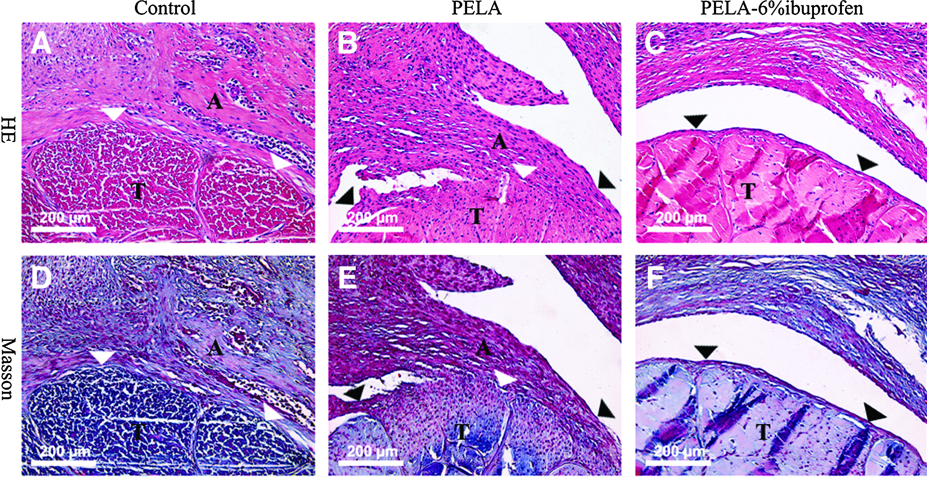
HE and Masson staining of sections cut transversely.
The repaired flexor digitorum profundus tendon was evaluated 21 days after the operation by
Biomechanical analysis
To evaluate tendon healing and peritendinous adhesions, work of flexion and maximum tensile strength were measured using a rheometer. Work of flexion differed significantly between groups treated with the PELA fibrous membrane (with or without ibuprofen) and the untreated control group, and between the groups treated with unloaded membrane and the ibuprofen-loaded membrane (Fig. 10). However, maximum tensile strength did not differ significantly among the three groups.

Tendon repair and peritendinous adhesions were evaluated by determining work of flexion
Discussion
In this study we tested the ability of ibuprofen-loaded PELA fibrous membranes prepared by electrospinning to prevent peritendinous adhesion and reduce inflammatory reaction after surgery. Incorporation of PEG increases the flexibility and hydrophilicity of PLLA. The improved flexibility allows the anti-adhesion fibrous membranes to be more easily handled by surgeons. Cells more readily adhere to and proliferate on moderately hydrophilic substrates than on hydrophobic or very hydrophilic substrates. 19 Therefore, cell adhesion can be prevented by incorporating PEG. During the electrospinning process, air entrapment between fiber interfaces increases surface hydrophobicity. As a result, the water contact angle of the PELA copolymer containing 10% PEG is 118.6±2.1°, which is considerably higher than that of the casting film (70.1±2.3°). 20 With higher PEG contents (40% or 50%), closure of the interconnecting pores may inhibit passage of cytokines and growth factors from outside the tendon sheath, which are needed to promote intrinsic healing.20,21 PELA fibers containing 10% PEG can more effectively block cell adhesion/proliferation, and was therefore chosen for this study.
Systemic delivery of NSAIDs requires adequate blood supply to the injured area; however, surgery often prevents delivery of NSAIDs by reducing blood flow. 22 In addition, the rapid clearance of NSAIDS and gastrointestinal and renal side effects limit the safety and effectiveness of this method of NSAID delivery. A previous study reported higher rates of drug release from electrospun fibrous membranes than from solvent-casting polymer film, 17 suggesting that the electrospun fibrous membrane is more effective as a delivery system. The superiority of PELA membranes for drug delivery applications is due to the hydrophilic PEG chains and hydrophobic PLLA chains. Strong hydrophobic interactions between PLLA chains inhibit water from entering and dissolving the drug before the membrane degrades. However, water conducted into the fibrous membrane by PEG chains allow the drug to be released from the fibrous membrane after hydration. Electrospinning produces a large surface area-to-volume ratio and high porosity with a very small pore size, enabling the continuous release of the drug from the inner part of the membrane after diffusion of drug molecules from the outer layer. 17 In the present study, ibuprofen release from the drug-loaded PELA membranes was 38% (2% ibuprofen), 47% (6% ibuprofen), and 62% (10% ibuprofen) during the first 2 days of incubation. This initial burst release was followed by a sustained release stage during in the subsequent 10 days. Our results showed that ibuprofen influenced the characteristics of the electrospun fibers. Introduction of ibuprofen into the polymer matrix resulted in straighter and more uniform electrospun fibers. This effect may be due to lowered surface tension of the polymer solution, which enhanced the bending instability. Ibuprofen rendered the PELA electrospun fiber matrix stiffer and less plastic; therefore, 6% ibuprofen was chosen for the in vivo study and the in vitro biomechanical tests.
NSAIDs including ibuprofen have long been used to prevent adhesion formation. Ibuprofen can reduce the inflammatory response to trauma and degradation products, prevent mass migration of inflammatory cells, and reduce capillary development in the injured area, thus reducing tissue adhesion.11,23 Compared with COX-2 selective NSAIDs such as rofecoxib, ibuprofen is more effective at limiting adhesion formation after flexor tendon repair because it inhibits both COX-1 and COX-2. 12 Furthermore, NSAIDs block the biosynthesis of inflammatory prostaglandins, especially prostaglandin E2.24,25 Therefore, ibuprofen can also alleviate perioperative pain to some extent, but further study is needed. When incorporated into PELA nanofibers, ibuprofen can be stably released to decrease inflammation during tendon healing, thus reducing tendon adhesion with few side effects. In this study, histology results demonstrated that inflammatory cell infiltration was significantly reduced by the ibuprofen-loaded PELA fibrous membrane. In addition, peritendinous adhesions were reduced by the PELA fibrous membranes, especially by ibuprofen-loaded membranes.
Previous studies have reported that ibuprofen-loaded alginate gel,11,26 solvent-casting PELA film, 11 and hyaluronic acid gel 27 used as physical barriers and drug delivery systems reduce adhesions and inhibit inflammation. However, either rapid degradation of these products or their primary applications for use on peritoneal defects limit their use in preventing peritendinous adhesions. The present study focuses on the anti-adhesion properties of ibuprofen-loaded PELA fibrous membranes. Our histologic and biomechanical findings demonstrated that the ibuprofen-loaded PELA fibrous membrane not only acts as a physical barrier to prevent peritendinous adhesions but also inhibits inflammation without interfering with tendon healing.
Conclusions
Electrospun PELA fibrous membranes containing ibuprofen are effective for the prevention of peritendinous adhesions. The PELA membranes inhibited cell adhesion and proliferation and allowed the controlled release of ibuprofen to inhibit inflammation. In addition, the membranes could be easily wrapped around the tendon in leghorn chickens to reduce peritendinous adhesion formation. Taken together, our results demonstrate that the electrospun ibuprofen-loaded PELA fibrous membrane shows promise as a multifunctional barrier for clinical applications.
Footnotes
Acknowledgments
This work was supported by the Nano-tech Foundation of Shanghai (1052nm0570045 and 11nm0503100
Disclosure Statement
No competing financial interests exist.
References
Supplementary Material
Please find the following supplemental material available below.
For Open Access articles published under a Creative Commons License, all supplemental material carries the same license as the article it is associated with.
For non-Open Access articles published, all supplemental material carries a non-exclusive license, and permission requests for re-use of supplemental material or any part of supplemental material shall be sent directly to the copyright owner as specified in the copyright notice associated with the article.