
Abstract
Fibrous encapsulation can impair implant osseointegration and cause implant failure but currently there are limited strategies to address this problem. Since bisphosphonates (BPs), a class of drugs widely used to treat bone diseases, was recently found to induce fibroblast apoptosis, we hypothesize that by loading BPs on titanium (Ti) implant surface, fibrous encapsulation may be inhibited with simultaneous enhancement of implant osseointegration. This strategy of local administration can also be expected to minimize the adverse side effects of BPs, which are associated with intravenous injections. To verify this hypothesis, alendronate was loaded on Ti surface via a hydroxyapatite (CaP) coating, and the effects of the loaded alendronate on fibroblast proliferation and apoptosis, and osteoblast proliferation, alkaline phosphatase (ALP) activity, and apoptosis were investigated in vitro. With a surface density of loaded alendronate 0.046 mg/cm2 or higher, fibroblast proliferation was suppressed due to increased apoptosis, while osteoblast proliferation and ALP activity increased with minimal apoptosis. In a coculture of fibroblasts and osteoblasts in a 1:1 ratio, ∼60% of the cells on these alendronate-loaded substrates were osteoblasts 1 day after cell seeding. The percentage of osteoblasts increased to about 75% 4 days after cell seeding. These results suggest that fibroblasts and osteoblasts respond differently toward the alendronate-modified substrates, and this phenomenon can potentially be capitalized to reduce fibrous encapsulation.
Introduction
Although fibrous encapsulation can impair osseointegration and the success of orthopedic implants, it receives insufficient attention in scientific and clinical research. To the best of our knowledge, there is hardly any development of techniques to reduce fibrous encapsulation on implant surfaces. Decreasing fibroblast proliferation on implant surfaces may lead to less fibrous encapsulation, but this is difficult to accomplish since fibroblasts possess great heterogeneity,8,9 and receive stimuli via direct crosstalk with different cells such as lymphocytes and epithelial cells.10,11 These characteristics of fibroblasts make it difficult to control their proliferation by a single or several cytokines.
Bisphosphonates (BPs) are common drugs to treat bone diseases such as osteoporosis, bony metastases, and Paget's disease. Although millions of patients receive BP therapy worldwide, 12 the mechanism is not completely understood. At tissue level, administration of BPs results in promotion of bone mineralization due to decreased bone turnover.13,14 At cellular level, it is widely accepted that BPs can inhibit the activity of osteoclasts, induce their apoptosis, and affect osteoclastic bone resorption via the mevalonate pathway. 15 Further, BPs can enhance osteoblast functions and prevent its apoptosis.16–18 In addition to the effects of BPs on osteoclasts and osteoblasts, recent studies indicated that BPs, especially the highly potent nitrogen-containing BPs, can inhibit fibroblast proliferation and increase their apoptosis.19–22 Thus, treatment with BPs may help to reduce fibrous encapsulation and enhance implant osseointegration. However, since a number of recent studies have shown that systematic administration of BPs especially by intravenous injection may be associated with a severe bone disease, osteonecrosis of the jaws (ONJ),23–25 development of strategies for local BP administration may be necessary to prevent this adverse effect.
Herein, we hypothesize that loading BPs on titanium (Ti) surfaces can reduce fibroblast proliferation while simultaneously enhancing osteoblast activities as a result of BP-induced fibroblast apoptosis and the positive effects of BPs toward osteoblasts. To verify this hypothesis, alendronate (Alen), an FDA-approved BP, was loaded on Ti surface via a precoated hydroxyapatite (CaP) layer. The effects of the loaded alendronate on the proliferation and apoptosis of fibroblasts and osteoblasts were investigated. To simulate in vivo conditions, a coculture of fibroblasts and osteoblasts was used to investigate the effects of the loaded alendronate.
Materials and Methods
Materials
Ti foils (0.52 mm thick) were purchased from Goodfellow, Inc. Alendronate sodium trihydrate (97%) were purchased from Sigma-Aldrich Chemical Co. 3T3 fibroblasts, and osteoblasts (MC3T3-E1 subclone 14) were obtained from American Type Culture Collection (ATCC). All other chemicals, if not specified, were purchased from Sigma-Aldrich Chemical Co.
Preparation of substrates
Ti foils were cut into squares of 1×1 cm2, and then ultrasonically cleaned for 10 min in Kroll's reagent (4.0% HF, 7.2% HNO3, 88.8% water). 26 Ti-CaP substrate was prepared according to the method reported in an earlier work 27 : Ti surface was activated via immersion in a 5 M NaOH aqueous solution for 24 h at 60°C, followed by heating at 600°C for 3 h. After cooling to room temperature, the activated Ti substrates were immersed in a simplified simulated body fluid (SSBF) solution for 1 day, followed by five washes with distilled water, and drying in a stream of nitrogen to obtain the Ti-CaP substrate. The SSBF solution was prepared at 37°C with the sequential addition of NaHCO3 (0.3525 g/L), K2HPO4 (0.2305 g/L), CaCl2 (0.2890 g/L), and tris-hydroxymethyl aminomethane (TRIS, 6.118 g/L). The pH value was adjusted to 7.4 by using 1 M HCl. The Ti-CaP substrates were immersed in an aqueous solution of alendronate (of concentration of 0.2, 0.5, or 1 mg/mL) for 1 h at room temperature followed by washing thrice with distilled water, and drying in a stream of nitrogen, to prepare the Ti-CaP-Alen0.2, Ti-CaP-Alen0.5, and Ti-CaP-Alen1 substrates. The washing solution was collected and the surface density of loaded alendronate was calculated as described below.
Surface characterization and alendronate release test
The chemical composition of the surfaces was analyzed by X-ray photoelectron spectroscopy (XPS) on a Kratos AXIS UltraDLD spectrometer (Kratos Analytical Ltd.) with a monochromatized Al Kα X-ray source (1486.7 eV photons). All binding energies were referenced to the C 1s (C-C bond) peak at 284.5 eV. Static contact angles of the different surfaces were measured at room temperature by the sessile drop method using a 3 μL water droplet in a telescopic goniometer (Rame-Hart). For each sample, at least 10 measurements from different regions of the surface were taken. The surface density of alendronate on the substrates was calculated from the difference between the amount of alendronate in solution before and after loading. The alendronate concentration in the washing solution combined with the remaining loading solution was determined using the method reported earlier 28 : 0.3 mL of the analyte solution was added into 0.7 mL of a mixture containing one part of ascorbic acid aqueous solution (10%) and six parts of ammonium molybdate solution (0.42% in 0.5M H2SO4), and incubated for 1 h at 37°C. The optical density of the solution at 820 nm was then measured, and the alendronate concentrations were obtained using a standard curve.
Tests to monitor the possible release of alendronate from the substrates were conducted by immersing the substrates in 1 mL of distilled water over 5 days. At the time points of 5, 10, 24, 48, 72, 96, and 120 h, the alendronate concentration in the water was measured as described above.
Cell culture
3T3 fibroblasts and MC3T3-E1 osteoblasts were cultured in Dulbecco's modified Eagle's medium supplemented with 10% fetal bovine serum (HyClone Ltd.), 100 unit/mL penicillin, and 100 μg/mL streptomycin (Invitrogen, Inc.). Cell cultures were maintained at 37°C under a humidified atmosphere of 5% CO2 in air. The medium was changed every 2 days, and the cells were passaged at least once a week. The cells were detached from the culture flask by using 1×Trypsin-EDTA solution (PAA Laboratories), and the cell suspension was transferred to a 15 mL Falcon tube and centrifuged at 210 g for 5 min. After removal of the trypsin solution, the remaining cell pellets were resuspended in fresh medium for subsequent experiments.
Cell attachment and proliferation
The pristine and modified Ti substrates were placed into a 24-well microplate. For cell attachment assay, 0.1 mL of cell suspension containing 50,000 cells was carefully seeded on each substrate to avoid cell adhesion on the well. After incubation for 6 h at 37°C, the substrates were washed thrice with phosphate-buffered saline (PBS). The cells on the substrates were then detached using trypsin and counted with a hemocytometer. For the proliferation assay, 0.1 mL of cell suspension containing 5000 cells was first carefully seeded on the substrates, and 0.9 mL of medium was then added after 6 h of incubation. After 1, 4, and 7 days of culture, cell proliferation was measured using the thiazolyl blue tetrazolium bromide (MTT) assay. 29 The cells on the substrates were observed using scanning electron microscopy (SEM) (Jeol; JSM-5600LV) after fixation with 3.7% paraformaldehyde in PBS for 10 min and dehydration with serial ethanol (10 min for each step).
Alkaline phosphatase activity assay
The alkaline phosphatase (ALP) activity assay was conducted according to the procedure described in our earlier work 30 : osteoblasts were seeded at a density of 5000 cells/cm2, and cultured in growth medium supplemented with 50 μg/mL ascorbic acid and 10 mM sodium β-glycerophosphate. After 2 weeks of culture, the substrates were washed with PBS and the cells were lysed by 3 cycles of freezing and thawing in water for the analysis of ALP activity and total protein level. ALP activity was determined by measuring the amount of p-nitrophenol released from p-nitrophenylphosphate substrate (Sigma). About 100 μL of the p-nitrophenylphosphate substrate was added to 100 μL of the cell lysate and incubated at 37°C for 30 min. Then, 50 μL of 1 M NaOH was added to stop the reaction. Optical intensity was measured at 405 nm with a microplate reader. The amount of p-nitrophenol produced was quantified using a standard curve obtained from known concentrations of p-nitrophenol. The micro BCA protein assay kit (Pierce Chemical) was used to determine the protein concentration with bovine serum albumin as the standard. ALP activity was expressed as μM of p-nitrophenol formation per minute per milligram of total proteins.
Apoptosis assay
Apoptosis assay was performed using the TdT-mediated dUTP nick end labeling (TUNEL) method. 31 After the cells were cultured on the substrates at a seeding density of 10,000 cells/cm2 for 24 h, they were fixed with 3.7% paraformaldehyde in PBS for 10 min, and permeabilized with PBS containing 0.1% Triton X-100 and 0.1% sodium citrate for 2 min on ice. TUNEL staining was then conducted with the TACS®2 TdT-Fluor in situ apoptosis detection kit (Trevigen, Inc.) as per the manufacturer's instructions. The positive control was prepared by treating the cells on pristine Ti with TACS-Nuclease™ (provided in the kit to induce cell apoptosis). After the TUNEL staining, the cells on the substrates were stained using the mounting medium with DAPI (Fluoroshield; Sigma), and examined under a Nikon Eclipse Ti inverted microscope system with C-HGFIE Intensilight fiber illuminator. At least five separate fields were acquired at×200 for each substrate. All TUNEL-positive cells (bright blue fluorescence in the nucleus) were counted for each field and expressed as a percentage of the total number of nuclei in that field.
Coculture of fibroblasts and osteoblasts
Fibroblasts and osteoblasts were labeled with the Qtracker® 525 and Qtracker 655 cell labeling kits (Invitrogen) respectively, according to the manufacturer's protocol. The labeled cells were mixed together at a ratio of 1:1, and then seeded on the substrates at a total density of 10,000 cells/cm2. A Nikon Eclipse Ti inverted microscope system with C-HGFIE Intensilight fiber illuminator was used to observe the osteoblasts (labeled with red fluorescence) and fibroblasts (labeled with green fluorescence) after 1 and 4 days of coculture. For qualitative observation, the cells on the substrates were fixed with 3.7% paraformaldehyde in PBS for 10 min, followed by staining of the nuclei with mounting medium containing DAPI. To quantify the distribution of the two types of cells on the substrates, the Qtracker-labeled cells on the substrates were detached using trypsin after 1 or 4 days of coculture and reseeded on a 24-well microplate at a density of ∼10,000 cells per well. Six hours after reseeding, the cells were fixed and stained with DAPI as described above and the number of fibroblasts and osteoblasts were counted under the fluorescence microscope. At least five separate fields were acquired at×200 for each substrate.
Statistical analysis
At least five samples per time point for each experimental condition were used. One-way analysis of variance (ANOVA) with Tukey post hoc test was used to assess the data. The results reported herein are expressed as mean±standard deviation. Statistical significance was accepted at p<0.05.
Results and Discussion
Surface characterization and alendronate release test
The surface elemental compositions of the pristine Ti, Ti-CaP, Ti-CaP-Alen0.2, Ti-CaP-Alen0.5, and Ti-CaP-Alen1 substrates, and the XPS wide-scan spectra of the pristine Ti, Ti-CaP, and Ti-CaP-Alen1 substrates are shown in Table 1 and Figure 1, respectively. The spectra of the Ti-CaP-Alen0.2 and Ti-CaP-Alen0.5 substrates are similar to that of the Ti-CaP-Alen1 substrate and hence not shown here. Carbon is typically present in the wide-scan spectrum of pristine Ti due to unavoidable hydrocarbon contamination, and it is widely used as an internal reference at 284.5 eV for peak position calibration. A small amount of N may be attributed to adventitious contamination. As shown in Figure 1, the successful deposition of CaP on the pristine Ti substrate is indicated by the presence of Ca 2s, Ca 2p, and P 2p signals. 27 The presence of sodium as indicated by the Na KLL Auger signal is due to the use of NaOH for activating the Ti surface prior to the CaP deposition. Comparing the wide-scan spectrum of the Ti-CaP-Alen1 substrate with that of the Ti-CaP substrate, the enhanced P 2p signal and decreased Ca 2p signal indicate the successful loading of alendronate. The N 1s signal can also serve to confirm the successful loading of alendronate since each alendronate molecule has one amine group. 32 It can be seen that the surface N content increased with higher alendronate concentration in the loading solution (Table 1). Contact angle measurement can also provide supporting evidence that the Ti surface has been successfully modified (Table 1).33,34 The pristine Ti substrate is relatively hydrophobic (with a contact angle of 61°±1.2°), but after CaP coating and alendronate loading, the contact angle decreases due to the hydrophilicity of CaP and alendronate.33,34
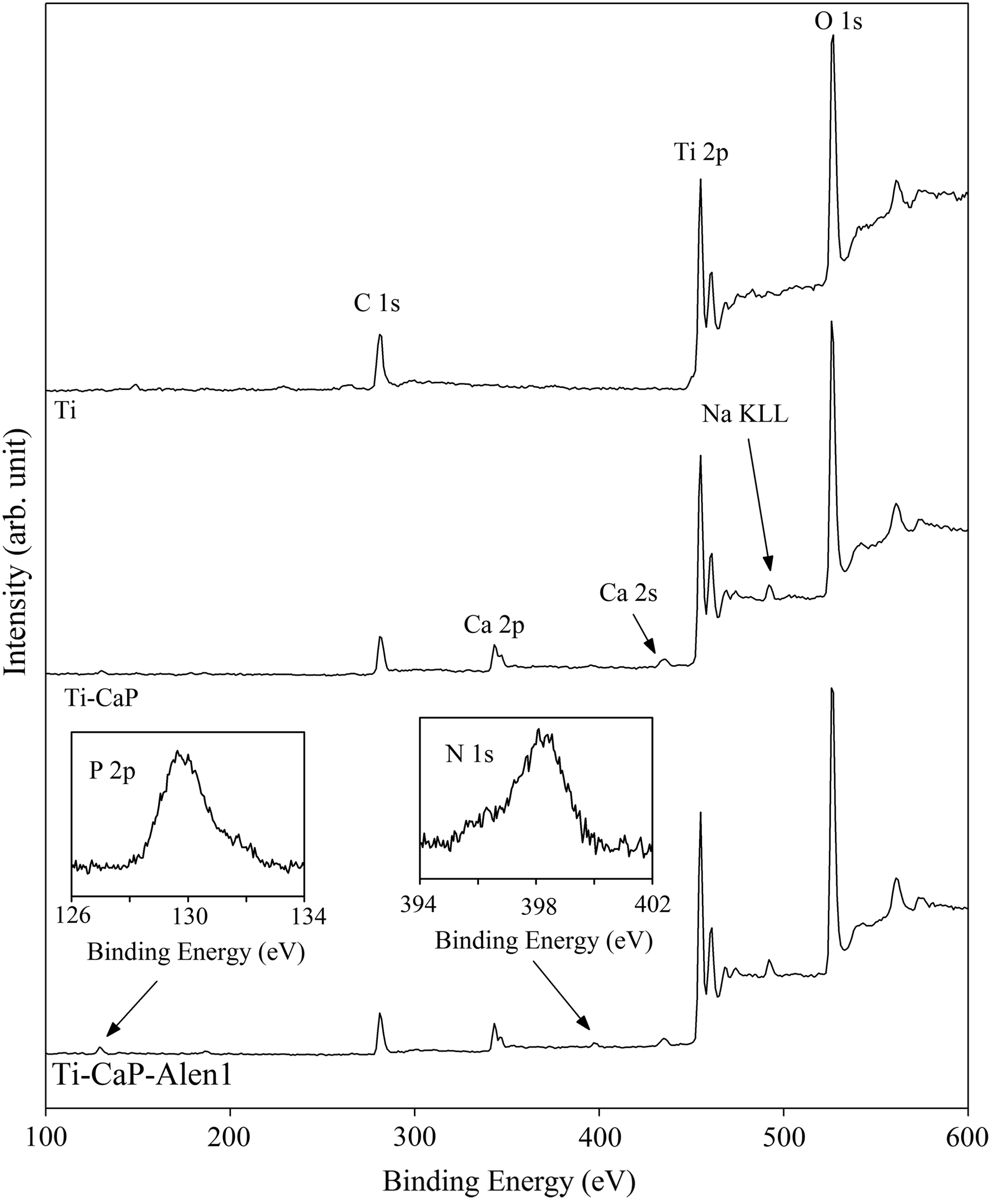
X-ray photoelectron spectroscopy wide-scan spectra of the pristine Ti, Ti-CaP, and Ti-CaP-Alen1 substrates. The insets show the P 2p and N 1s core-level spectra of the Ti-CaP-Alen1 substrate. Alen, alendronate; Ti, titanium; CaP, hydroxyapatite.
Alen, alendronate; XPS, X-ray photoelectron spectroscopy; CaP, hydroxyapatite; Ti, titanium.
The amount of alendronate bound to the Ti-CaP substrates is also shown in Table 1. When the alendronate concentration in the loading solution increased from 0.2 to 1 mg/mL, the surface density of loaded alendronate linearly increased from 0.019±0.01 mg/cm2 (Ti-CaP-Alen0.2) to 0.11±0.02 mg/cm2 (Ti-CaP-Alen1). The loading efficiency (defined as the percentage of alendronate in the loading solution that was loaded on the Ti-CaP substrate) was about 10%.
Release tests were conducted to investigate whether alendronate can be released from the CaP coatings. Figure 2 shows that there was no measurable alendronate release from the Ti-CaP-Alen0.2 and Ti-CaP-Alen0.5 substrates over 5 days of immersion in water. For the Ti-CaP-Alen1 substrate, a slow release was observed, and ∼80% of the initial loaded alendronate remained on the surface after 5 days. These results suggest that most of the loaded alendronate was strongly bound to the CaP layer. Alendronate was reported to have a zwitterionic character, with the nitrogen in the amine group bearing a positive charge and the two phosphonate groups bearing negative charges. The overall charge of this zwitterion is −1, which enables alendronate to form 2:1 complexes with Ca2+ ions in the lattice of CaP. 35 It was also reported that the hydroxyl group linked to the carbon atom in alendronate can coordinate to calcium, which may further enhance its chemisorption on CaP. 36
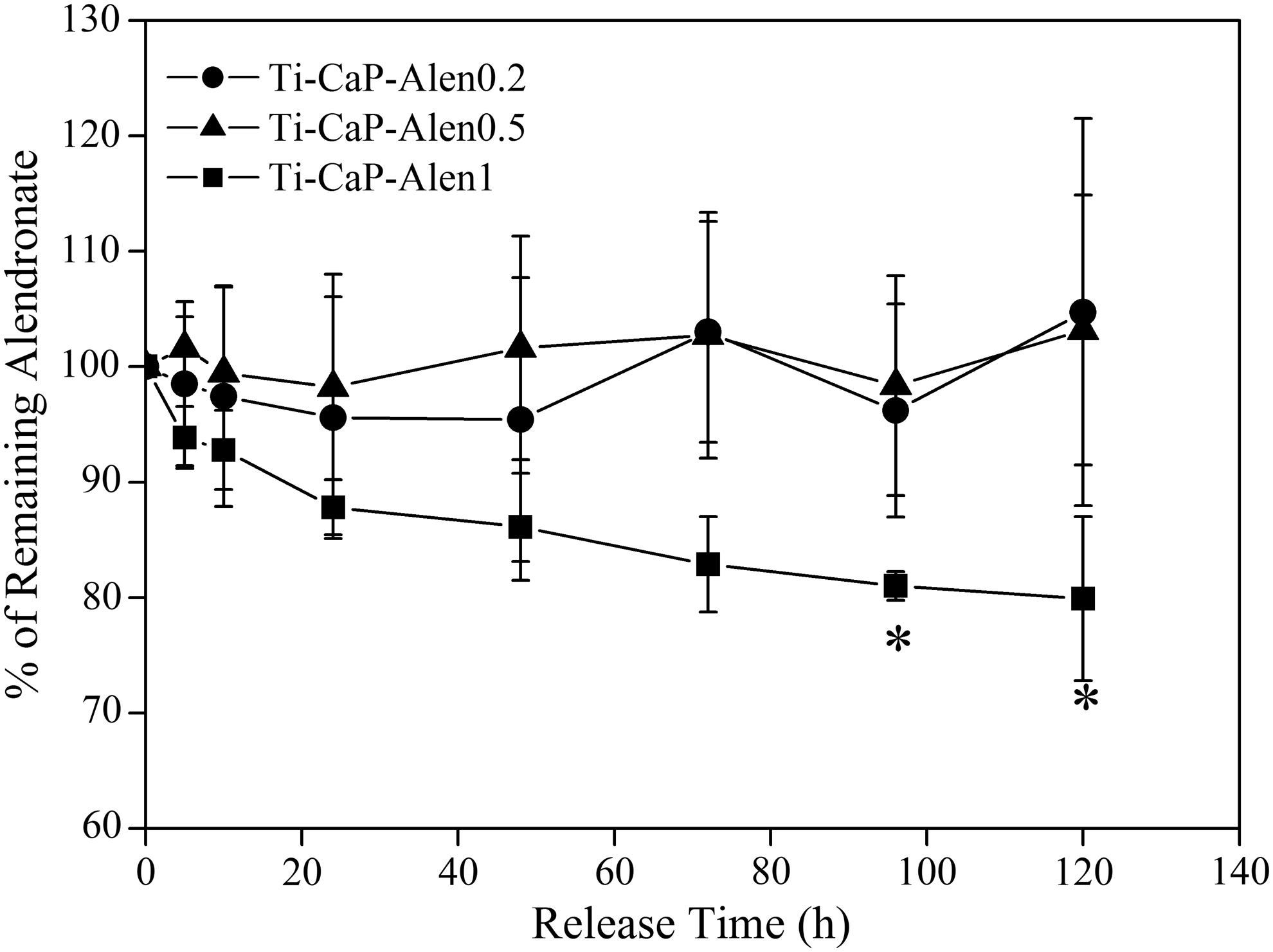
Alendronate release profiles for the Ti-CaP-Alen0.2, Ti-CaP-Alen0.5, and Ti-CaP-Alen1 substrates. * denotes significant difference (p<0.05) compared with that after 5 h.
Fibroblast attachment, proliferation, and apoptosis
A similar number of adherent fibroblasts were observed on the pristine and functionalized Ti substrates 6 h after cell seeding (Supplementary Fig. S1; Supplementary Data are available online at www.liebertpub.com/tea), indicating that the loaded alendronate had no significant effect on fibroblast attachment. Fibroblast proliferation on the different substrates after 1, 4, and 7 days of culture are shown in Figure 3. On the pristine Ti, Ti-CaP, and Ti-CaP-Alen0.2 substrates, cell proliferation steadily progressed over 7 days of culture and the proliferation rates on the these substrates were not significantly different. However, on the Ti-CaP-Alen0.5 and Ti-CaP-Alen1 substrates, the proliferation was ∼50% lower compared to that on the pristine Ti substrate after 4 days of culture. Representative SEM images of fibroblasts on the pristine Ti and Ti-CaP-Alen0.5 substrates on day 1, 4, and 7 are shown in Figure 4. The observed decrease in cell number on the Ti-CaP-Alen0.5 substrate (Fig. 4d, f) compared with that on the pristine Ti substrate (Fig. 4c, e) is consistent with the MTT assay (Fig. 3). The SEM images of fibroblasts on the Ti-CaP and Ti-CaP-Alen0.2 substrates are similar to that on the pristine Ti substrate, while the SEM images of fibroblasts on the Ti-CaP-Alen1 substrate are similar to that on the Ti-CaP-Alen0.5 substrate.

Fibroblast proliferation on the pristine and functionalized Ti substrates as determined from the MTT assay. * denotes significant difference (p<0.05) compared with that on the pristine Ti substrate.
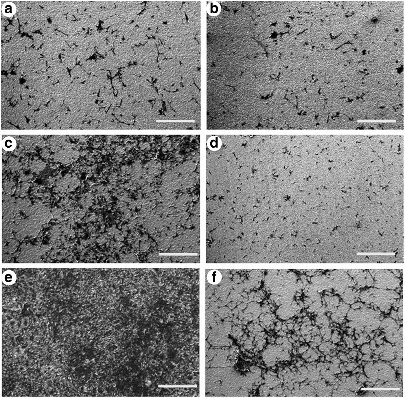
SEM images of fibroblasts on the pristine Ti
TUNEL assay is a commonly-used method to identify DNA fragmentation that results from apoptotic signaling cascades. 37 The fluorescence microscopy images of TUNEL-stained fibroblasts on the different substrates 1 day after cell seeding are shown in Figure 5. A positive control experiment was carried out by the addition of TACS-Nuclease to the cells on pristine Ti to induce their apoptosis, and the results are shown in Supplementary Fig. S2. These images indicate that fibroblasts on the Ti, Ti-CaP, and Ti-CaP-Alen0.2 substrates suffered minimal apoptosis, whereas significant apoptosis occurred on the Ti-CaP-Alen0.5 and Ti-CaP-Alen1 substrates. The quantitative assessment of TUNEL-positive cells is shown in Figure 5f, and it can be observed that there is more than a 6-fold increase in apoptotic fibroblasts on the Ti-CaP-Alen0.5 and Ti-CaP-Alen1 substrates when compared with that on the pristine Ti substrate. As a result of apoptosis, by day 2 the number of fibroblasts on the Ti-CaP-Alen0.5 and Ti-CaP-Alen1 substrates was less than on pristine Ti (Supplementary Fig. S3). Earlier, in vitro studies indicated that the effective concentration of soluble BPs to induce significant apoptosis and suppress fibroblast proliferation is in the range of 10–100 μM,20,22,38 but the relevant data for immobilized BPs are not available. Our results show that with a surface density of loaded alendronate of only 0.14±0.08 μmol/cm2 (0.046±0.03 mg/cm2) (i.e., Ti-CaP-Alen0.5 substrate), a significant decrease in fibroblast proliferation was attained.
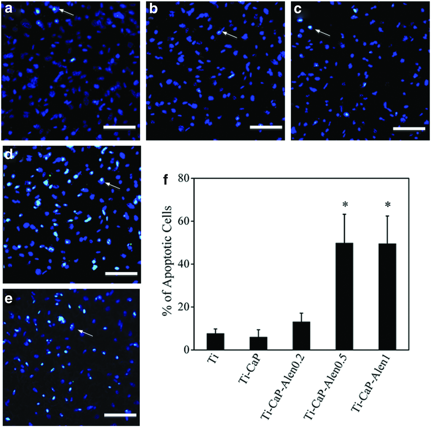
Fluorescence microscopy images of fibroblasts labeled by TUNEL staining on the Ti
The mechanism by which BPs decrease fibroblast proliferation and induce the apoptosis is still unclear. Scheper et al. reported that for BP-treated fibroblasts, the expression of some apoptotic inhibitor proteins such as B-cell lymphoma 2 (BCL2) and BCL2/adenovirus E1B 19 kDa protein-interacting protein 3 was downregulated, and the cleavage and activation of caspase 3 and 9 (which act to induce apoptosis) increased. 21 BP has also been reported to impair prenylation of small GTPases, which could result in fibroblast apoptosis. 39 Cornish et al. loaded BP on bone slices, and found that the bound BP inhibited proliferation of adjacent nonbone cells. They proved that such inhibition was not due to the BP released into the culture medium, but that direct contact of the cultured cells with the BP-loaded bone was necessary. 40 To fully understand the biochemical mechanisms of BP-induced apoptosis in fibroblasts, further studies are required.
Osteoblast attachment, proliferation, differentiation, and apoptosis
As shown in Supplementary Fig. S1, the number of adherent osteoblasts on the Ti-CaP-Alen0.5 and Ti-CaP-Alen1 substrates 6 h after cell seeding was significantly higher than on pristine Ti, indicating the enhancement of osteoblast attachment by these alendronate-modified substrates. The effect of loaded alendronate on the proliferation of osteoblasts on day 1, 4, and 7 are shown in Figure 6. On day 1 and 4, there was a significant increase in osteoblast proliferation on the Ti-CaP-Alen0.5 and Ti-CaP-Alen1 substrates when compared with that on the pristine Ti substrate. The CaP coating on Ti did not significantly affect osteoblast proliferation, which is consistent with earlier literature reports.41,42 On day 7, no significant difference in osteoblast proliferation on the pristine and functionalized Ti substrates was observed. Representative SEM images of osteoblasts on the pristine Ti and Ti-CaP-Alen0.5 substrates on day 1, 4, and 7 are shown in Figure 7. The increase in osteoblast proliferation on the Ti-CaP-Alen0.5 substrate (Fig. 7b, d), compared with that on the pristine Ti substrate (Fig. 7a, c) on day 1 and 4, is consistent with the MTT results. By day 7, the osteoblasts on both the pristine and Ti-CaP-Alen0.5 substrates have almost reached confluence (Fig. 7e, f). The SEM images of osteoblasts on the Ti-CaP and Ti-CaP-Alen0.2 substrates are similar to that on the pristine Ti substrate, while the SEM images of osteoblasts on the Ti-CaP-Alen1 substrate are similar to that on the Ti-CaP-Alen0.5 substrate.
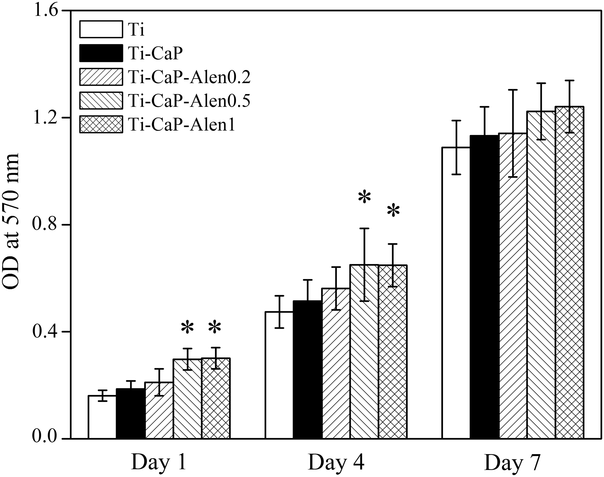
Osteoblast proliferation on the pristine and functionalized Ti substrates as determined from the MTT assay. * denotes significant difference (p<0.05) compared with that on the pristine Ti substrate.
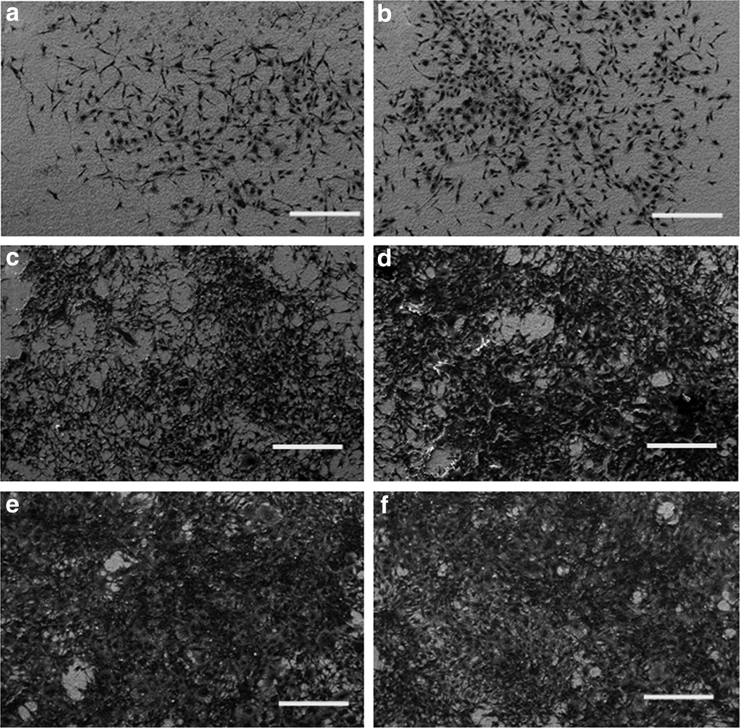
SEM images of osteoblasts on the pristine Ti
The effect of loaded alendronate on ALP activity, an early marker for the differentiation of osteoblast-like cells, was evaluated after the cells had been cultured for 2 weeks on the different substrates. As shown in Figure 8, osteoblasts cultivated on the Ti-CaP and alendronate-modified substrates had a significantly higher ALP activity than those on the pristine Ti. The enhancement of ALP activity was over 1.5-fold and 2-fold for the Ti-CaP and alendronate-modified substrates, respectively. The ALP activity of osteoblasts on the alendronate-loaded substrates was also significantly higher than that on the Ti-CaP substrate. For the alendronate-modified substrates, a 5-fold increase in surface density of loaded alendronate from 0.019±0.01 mg/cm2 to 0.11±0.02 mg/cm2 did not have any significant effect on the ALP activity.
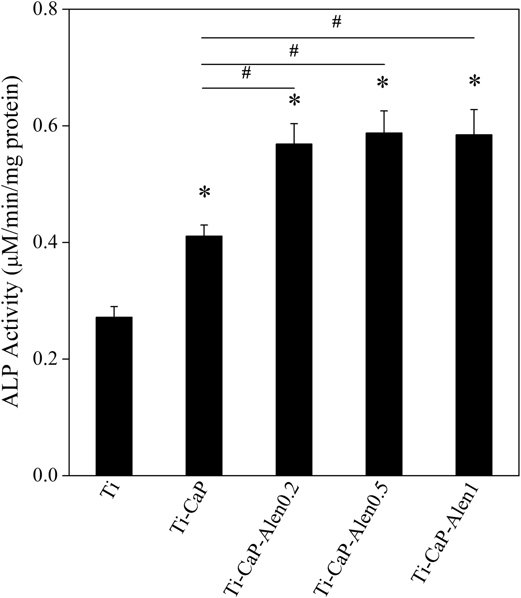
ALP activity of osteoblasts cultured on the pristine and functionalized Ti substrates. * and # denote significant differences (p<0.05) compared with that on the pristine Ti substrate, and between the designated groups, respectively. ALP, alkaline phosphatase.
The reported effects of soluble BPs on the proliferation and ALP activity of osteoblasts are controversial. Some studies indicated a decrease in the proliferation and ALP activity of osteoblasts in cultures containing BPs,38,43,44 whereas other studies demonstrated an opposite effect.18,45,46 Although the reasons for the conflicting results are not known, possible contributing factors are differences in cell types, duration of treatment, the BP analogues, and BP concentrations. Marolt et al. proposed that low BP concentration (10−5 M or lower) can lead to an increase in osteoblast proliferation and ALP activity, whereas high BP concentration (10−4 M or higher) may have the opposite effect. 38 Unlike the reported controversy regarding the effects of soluble BPs, alendronate loaded either on CaP nanocrystal or CaP coating resulted in increased osteoblast proliferation and higher values of differentiation parameters.17,47 In vivo studies also indicated that implantation of BP-loaded biomaterials resulted in a significant increase in relative bone content, and an improvement of its micro-architecture and mechanical fixation.48,49
Our results show that although CaP coating had no significant effect on osteoblast proliferation (Fig. 6), it significantly enhanced ALP activity (Fig. 8), which plays a crucial role in osteoblast differentiation. In vivo studies also indicated that CaP coating on implant surface can enhance osseointegration and fixation.50,51 Thus, the CaP coating may work synergistically with the loaded alendronate in enhancing osteoblast functions while not affecting the alendronate's inhibiting effect on fibroblast proliferation.
TUNEL staining of osteoblasts on the different substrates is shown in Figure 9, and the positive control obtained by the addition of TACS-Nuclease to the cells on pristine Ti to induce their apoptosis is shown in Supplementary Figure S2. The fluorescence microscopy images in Figure 9a–e indicate that there were few apoptotic osteoblasts on all the substrates. The quantitative assessment of the TUNEL-positive cells shown in Figure 9f indicates that osteoblast apoptosis on all the substrates was less than 10%. It has been proposed that BPs can suppress apoptosis in osteoblasts by inducing the rapid phosphorylation of extracellular signal-regulated kinases. 16
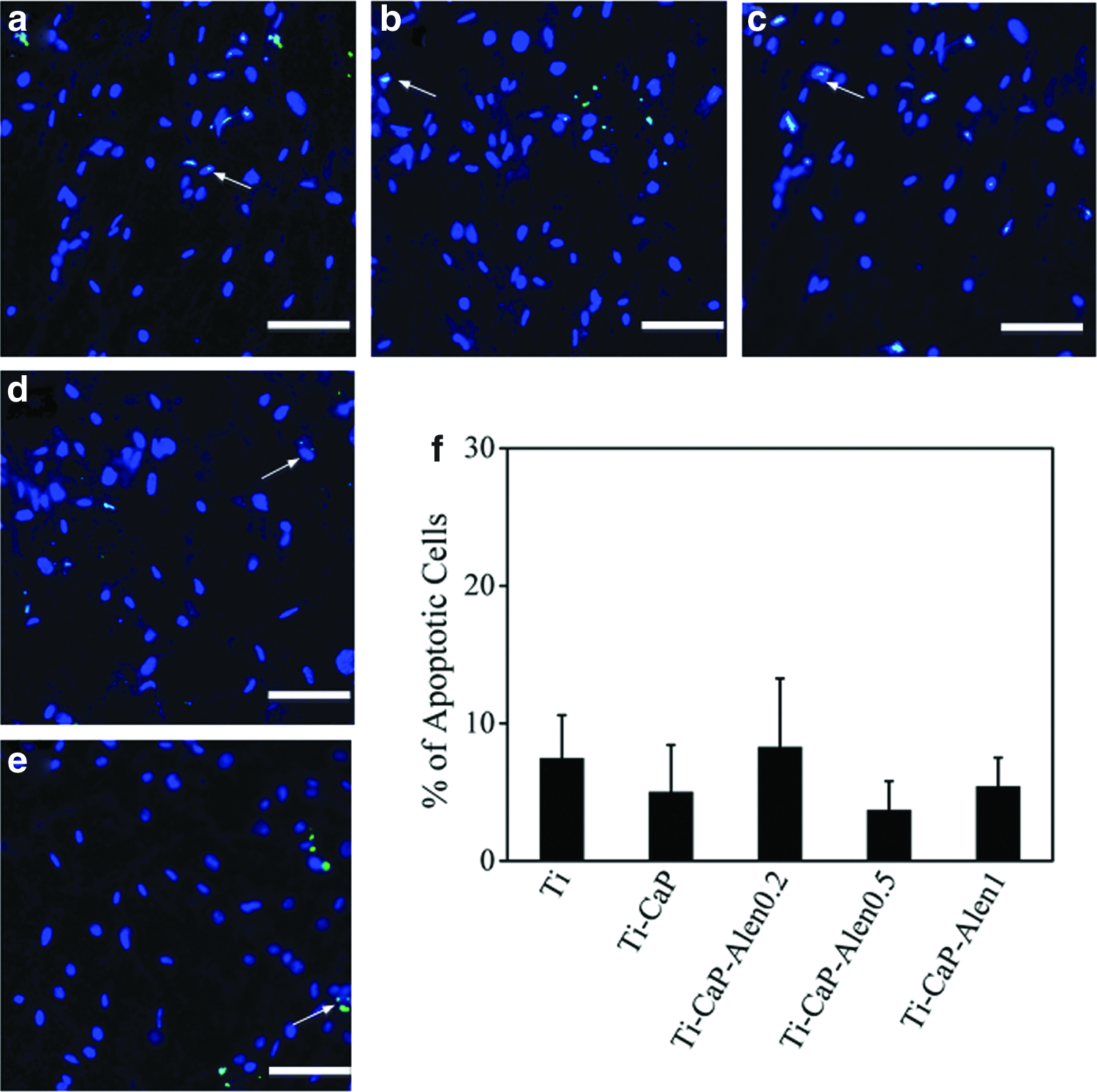
Fluorescence microscopy images of osteoblasts labeled by TUNEL staining on the Ti
Although BPs have an excellent safety profile, there are several negative side effects. 52 Oral BPs may result in upper gastrointestinal irritation, while intravenous BPs may cause acute phase response. Both effects are due to the inhibition of farnesyl pyrophosphate synthase in the relevant cells. 53 ONJ was recently discovered as a serious adverse effect associated with high-dose or long-term intravenous BP therapy. It was reported that cells in soft tissues take up BPs from circulation, and suffer apoptosis or impairment of their functions. 54 In our work, the alendronate was strongly bound to the CaP coating on Ti. Thus, it has the advantage of only directly affecting the area surrounding the implant, which may reduce the above-mentioned adverse side effects of BPs.
Coculture of fibroblasts and osteoblasts
To further evaluate the potential application of the surface-loaded alendronate in preventing implant fibrous encapsulation, fibroblasts and osteoblasts labeled with different fluorescent nanocrystals (i.e., the tracking dyes in the Qtracker labeling kits) were cocultured on the different substrates. The nanocrystals were distributed in the cell cytoplasm, and had no significant effect on fibroblast or osteoblast proliferation as determined by the MTT assay (Supplementary Fig. S4). The nanocrystals inside the cells are expected to be transferred to the daughter cells rather than to the adjacent cells, since the nanocrystals (with a size about 10 nm) are much larger than gap junction channels (with a diameter about 1 nm) that act to interchange cytoplasm between cells. 55 Thus, the nanocrystal labeling technique has been used to differentiate cells in coculture.56,57
Figure 10a–d show the representative fluorescence microscopy images of osteoblasts and fibroblasts cocultured on the pristine Ti and Ti-CaP-Alen0.5 substrates. There were nearly equal numbers of osteoblasts and fibroblasts on the pristine Ti substrate on day 1 and 4, whereas on the Ti-CaP-Alen0.5 substrate, the number of osteoblasts was higher than fibroblasts on day 1 and the cells were predominantly osteoblasts 4 days after coculture. Since it is difficult to accurately count the number of cells on the substrates due to cell clumping (especially on day 4), the cells on the substrates were detached by trypsin followed by reseeding on 24-well microplate at a lower density (∼10,000 cells/well). As shown in Figure 10e, the percentage of osteoblasts on the Ti, Ti-CaP, and Ti-CaP-Alen0.2 substrates 1 and 4 days after cell seeding was close to 50%, that is, in the same ratio as in the seeding suspension, indicating that both fibroblasts and osteoblasts attached and proliferated well on these substrates. On the Ti-CaP-Alen0.5 and Ti-CaP-Alen1 substrates, the percentage of osteoblasts was 60%±7% and 62%±8%, respectively, after 1 day, and after 4 days of coculture, the percentage of osteoblasts on these substrates increased to 74%±10% and 76%±6%, respectively. To ensure there were no specific dye-cell effects, an experiment was performed whereby the trackers for labeling the cells were switched (i.e., staining fibroblasts and osteoblasts with Qtracker 655 and Qtracker 525 cell labeling kits, respectively), and the labeled fibroblasts and osteoblasts were cocultured on Ti in a ratio of 1:1 for 1 day. The percentage of osteoblasts on pristine Ti was 48%±6%, which is not significantly different to the value of 46%±5% shown in Figure 10e.
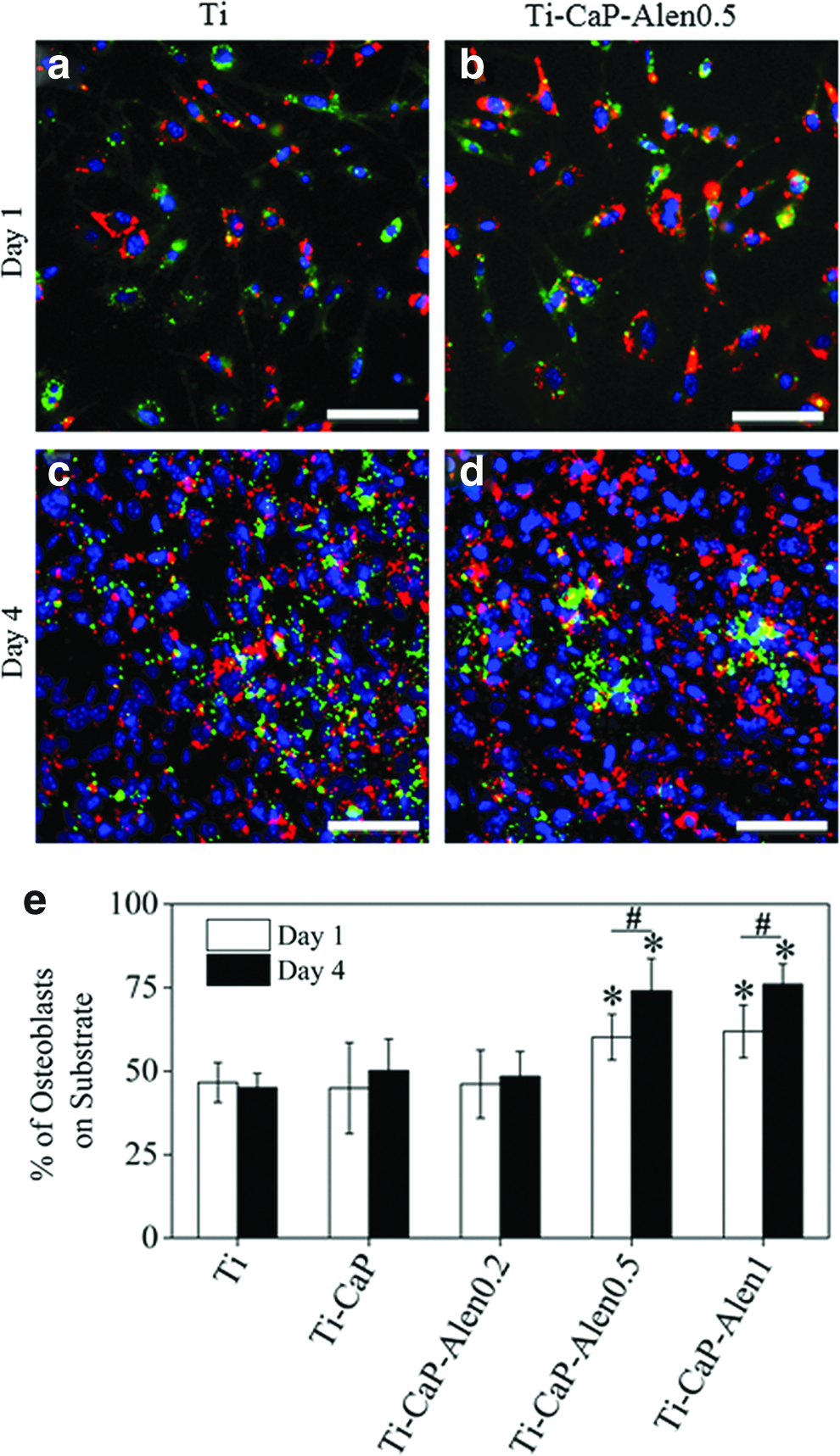
Fluorescence microscopy images of osteoblasts and fibroblasts cocultured on the pristine Ti
The results obtained from the attachment (Supplementary Fig. S1) and proliferation (Figs. 3 and 6) assays carried out with fibroblast and osteoblast monocultures indicate that the loaded alendronate had no significant effect on fibroblast attachment, while the Ti-CaP-Alen0.5 and Ti-CaP-Alen1 substrates enhanced osteoblast attachment. Thus, when fibroblasts and osteoblasts were cocultured on the Ti-CaP-Alen0.5 and Ti-CaP-Alen1 substrates, the percentage of osteoblasts can be expected to be greater than 50% on day 1, which is supported by Figure 10e. With progressive apoptosis of fibroblasts induced by the loaded alendronate (Fig. 3), the cells on the Ti-CaP-Alen0.5 and Ti-CaP-Alen1 substrates were predominantly osteoblasts after 4 days of coculture. This opposing effect of the loaded alendronate on the cocultured fibroblasts and osteoblasts strongly suggests that there may be inherently distinct signaling pathways by which BPs control the life span of these two cell types.
Recently, there have been many studies that focused on the application of BPs in bone tissue engineering and regeneration, as highlighted by the review article by Cattalini et al. 58 Some in vivo studies have indicated that coating BPs on Ti implant surfaces can significantly improve osseointegration.59–61 The mechanism for this enhancement in osseointegration is believed to involve reduction in osteoclast activity and increase in osteoblast activity. 58 Since BPs can suppress fibroblast proliferation, as observed herein as well as in other studies,19,21,22 BP-modified surfaces can potentially reduce fibrous encapsulation and provide another positive aspect for osseointegration. However, since we have used mouse cell lines as the model fibroblast and osteoblast system, we cannot rule out that the corresponding human cells may act differently. Further, in vitro studies cannot reflect the series of biological events occurring at the implant-bone interface, 62 and the very complicated crosstalk among various cells. 63 Thus, in vivo studies will be needed to confirm the ability of surface-loaded BP to reduce fibrous encapsulation of implants.
Conclusion
Alendronate was loaded on Ti substrates precoated with a CaP layer in controlled amounts, and most of the loaded alendronate remained on the substrate even after 5 days of immersion in water. When the surface density of loaded alendronate is 0.046 mg/cm2 or higher, the proliferation of fibroblasts is adversely affected due to an increase in apoptosis, whereas osteoblast proliferation and ALP activity are enhanced. By using a coculture of fibroblasts and osteoblasts on the alendronate-loaded substrates, we have demonstrated the advantage of these substrates in promoting selective surface coverage with osteoblasts. These in vitro results highlight the concept of surface-loaded alendronate on implants for reducing fibrous encapsulation, which may offer promising opportunities in orthopedic applications. However, the findings should be confirmed with animal model studies since fibrous encapsulation is a much more complex process than can be simulated using in vitro coculture.
Footnotes
Acknowledgment
This work was financially supported by the Singapore Stem Cell Consortium Grant SSCC/09/019.
Disclosure Statement
No competing financial interests exist.
References
Supplementary Material
Please find the following supplemental material available below.
For Open Access articles published under a Creative Commons License, all supplemental material carries the same license as the article it is associated with.
For non-Open Access articles published, all supplemental material carries a non-exclusive license, and permission requests for re-use of supplemental material or any part of supplemental material shall be sent directly to the copyright owner as specified in the copyright notice associated with the article.