
Abstract
Objectives:
To investigate whether the intervention of nucleus pulposus (NP) cells or human telomerase reverse transcriptase (hTERT) gene-transfected NP cells can prevent the degeneration process after allograft total disc transplantation.
Methods:
Eighteen canine lumbar intervertebral discs were obtained from five canines and cryopreserved in liquid nitrogen. Canine nucleus pulposus cells were isolated and transduced with recombinant adeno-associated virus (rAAV)-hTERT. The cells were injected into the discs to construct a tissue-engineered allograft disc (group A). NP cells and DMEM/F12 were used for positive control (group B) and blank control (group C). 18 beagle dogs received the three groups of allograft intervertebral disc (IVD) composites implantation, respectively. Radiographic examinations were performed at 4, 8, and 12 weeks postimplantation. At 12 weeks after operation, all dogs were sacrificed and the lumbar spines were harvested for the biomechanical analysis, and then the allografts underwent histological analysis, ectogenic NP cell tracing, and hTERT mRNA analysis.
Results:
Bony fusion between the intervertebral disc allograft and the adjacent host intervertebral body were observed in all animals. The disc height and T2 signal intensity preservation in group A and B was better than group C. Magnetic resonance images (MRI) showed typical degenerative changes in group C. In group A, the normalized grayscale of the transplanted disc on MRI was significant higher compared with the controls at 12 weeks. A biomechanical test showed a poor stability preservation in group C compared to group A and B. PKH-26-positive cells were identified within the allograft discs in group A at 12 weeks, providing evidence of cell survival. Histological analysis showed the NP cell morphology, cell number, and distribution of the allograft discs was better preserved in group A and B compared to group C at a 12-week follow-up.
Conclusion:
The present study demonstrated that NP cells or hTERT-loaded NP cells intervention could effectively resist the degeneration of the allogenic transplanted intervertebral discs in a beagle model. The hTERT-loaded NP cells had a better antidegeneration effect on the transplanted disc than NP cells. This modified disc regeneration technique through NP cell injection or manipulation may have the potential to ensure the long-term function preservation of allograft disc transplantation.
Introduction
When conservative management fails to alleviate the pain and neurologic deficits caused by DDD, the patient is given the option of surgical decompression of the affected intervertebral disc. Discectomy can provide an immediate relief of pressure to the spinal cord and associated nerve roots via partial or complete removal of the diseased disc, and thereby alleviate the suffering of patients. However, removal of the entire disc without further reconstruction of the defect alters the normal structure and alignment of the spine, which might lead to the recurrence of back pain and segmental instability.5,6 To compensate for these mechanical instabilities, discectomies can be followed by a solid fusion between adjacent vertebrae. 7 However, fusion of a relatively mobile spinal functional unit is not an ideal reconstruction and can increase the stress on the discs above and below the fusion and accelerate the degeneration process of the adjacent motion segment.8,9
Biological regeneration of the disc seems to be an ideal treatment for DDD and acquired remarkable achievements in basic research. Regenerative strategies focus on altering the phenotype of cells within the disc or introducing new cell populations. Injection of growth factors, such as the bone morphogenetic protein 7 (BMP-7), the transforming growth factor-β, and others into the disc has been widely reported to stimulate ECM production and cell proliferation.10–12 By delivering disc cells or mesenchymal stem cells into the disc, the obvious antidegeneration effect, including preservation of disc height and water content, enhancement of ECM expression, has been observed.13,14 Another focus in this area is on tissue engineering; nucleus pulposus (NP) tissue engineering has been widely studied and reported to be a promising treatment for DDD, both in vitro and in vivo.15,16 Recently, interest has turned to the AF and to whole disc composite tissues. Although the regeneration proved to be promising in basic research, the translation of such treatments to human application and clinical use is still challenging. The potential success of anabolic growth factors injected directly into the disc might be limited, both owing to the short biological half-life of the factors and their rapid diffusion away from the delivery site. Cell therapy and tissue engineering regeneration can not find a translational pathway that leads to a clinical trial and therapeutic development for lacking of a standard preclinic research animal model.
Our department has reported the treatment of cervical DDD by fresh-frozen endplate-disc-endplate composite allograft transplantations after anterior discectomy. 17 The treatment resulted in a partly viable disc, preserved segmental stability and mobility, and a satisfactory 5-year clinical outcome. These findings suggested that the disc transplantation might be a promising treatment option for DDD. However, late degenerative changes, such as loss of disc height and water content, were found in the transplanted disc, which may limit its long-term clinical outcome and broad clinical application. To ensure the disc cell survival and the long-term function preservation of the transplanted disc, we chose a combination strategy of cell-based NP regeneration and allograft disc transplantation. The allograft disc was used as a natural biological scaffold for intervertebral disc tissue engineering, and exogenous NP cells were injected into the disc to optimize the effect of allograft total disc transplantation. In our previous study, we have successfully extended the lifespan and phenotype preservation of NP cells in vitro by human telomerase reverse transcriptase (hTERT) gene transfection. 18 We hypothesised that this activation of NP cells might upregulate the antidegeneration effect after allograft total disc transplantation.
In the present study, we used a well-established beagle model 16 and tested whether the intervention of hTERT gene-transfected NP cells can prevent the degeneration process after allograft total disc transplantation in a beagle dog model in vivo; we also intended to examine whether the hTERT-activated NP cells can make any difference compare to normal NP cells in the antidegeneration effect.
Materials and Methods
Canine disc harvesting and cryopreservation
The canine lumbar intervertebral disc (IVDs) (L1–5) were obtained from five beagles (weight between 10–12 kg). The process of IVD harvesting and preparation were carried out as previously described. 19 In brief, after discs with endplates harvested with handsaw and trimmed with blade, the absence of growth plates and the thickness of the vertebral endplates (≤2 mm on each side) were checked by X-ray. The endplates surfaces were lavaged with saline solution using the Pulsavac™ wound debridement irrigation system (Zimmer) to remove cutting debris and blood clots. All samples were cryopreserved with a slightly modified standard protocol for disc cryopreservation. 20 Briefly, harvested discs were incubated for 2 h at 4°C with 10% (v/v) dimethyl sulfoxide (Sigma-Aldrich) in DMEM/F12, cooled to −80°C by placing in containers at a −80°C freezer overnight, and stored at −196°C in liquid nitrogen for later use (8–12 weeks). Before injecting NP cells, the discs were thawed quickly at 37°C in a water bath and washed with 4°C Hanks' balanced salt solution.
NP cell isolation and rAAV-hTERT vector transduction
The lumbar spinal column was dissected from a 1-year-old beagle dog under a strict sterilization technique. The intervertebral discs from L1 to L6, together with 1.5–2 mm of the adjacent endplates, were removed with osteotomes. The discs were washed twice in the phosphate-buffered saline (Gibco) solution, and then cut into two pieces transversally in the middle of each disc. The jelly-like nucleus pulposus tissue was collected, pooled together and incubated with 0.5 mg/mL of trypsin (type I; Gibco) for 20 min, and then digested in 10 mL 0.2% collagenase type II (Gibco) at 37°C for 2 h. After removal of tissue debris by filtering through a nylon mesh (100 μm), the cell–collagenase solution was centrifuged at 1000 rpm for 5 min. Cells were seeded in 100-mL culture flasks with Ham's F-12 media containing 10% fetal calf serum (Gibco), streptomycin (100 μg/mL), penicillin G (100 IU/mL), and amphotericin (1 μg/mL) at 37°C in an atmosphere of 5% CO2 and 95% air. Cells were fed twice a week and split routinely when nearing confluence.
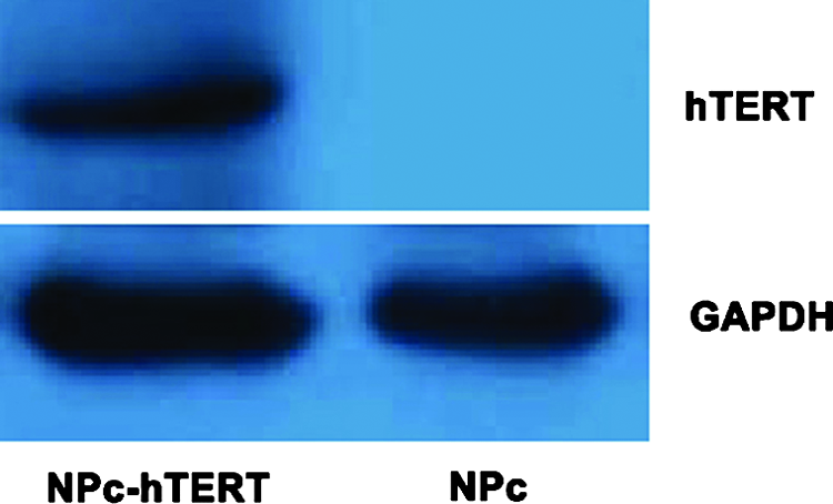
The 2nd passage cells were transduced by the recombinant adeno-associated virus (rAAV)-hTERT vector according to the previously described method. 20 Briefly, the 2nd passage NP cells were placed in monolayer culture in six-well plates (2×10⁵ cells per well), in DMEM/F12 containing 10% fetal bovine serum (FBS). Subconfluent cells were incubated for 1 h at 37°C with the AAV2- hTERT vector at a MOI of 1×10⁵ vector genomes (vg)/cell. The medium was then aspirated and 2 mL of growth medium (DMEM/F12 supplemented with 10% FBS and 30 mM sodium butyrate) was added. Following incubation for 24 h, the growth medium was exchanged for DMEM/F12 supplemented with 10% FBS. Transfected nucleus pulposus cells (NPCs) were cultured according to the routine technique and labeled with PKH-26 (Sigma-Aldrich) using the method we previously described. 16 The efficiency of staining was monitored by fluorescent microscopic analysis (Bio-Rad Radiance 2100™ confocal system in conjunction with a Nikon TE300 microscope). The hTERT mRNA and protein were analyzed by reverse transcriptase polymerase chain reaction (RT-PCR) and Western blot, respectively. Canine glyceraldehyde-3-phosphate dehydrogenase (GAPDH, Gen- Bank accession number: L-23961) was amplified as an internal control for RNA loading. The PCR primer sequences used in RT-PCR analysis were
GAPDH
5′-GAT GCT GGT GCT GAG TAT GT-3′,
5′-CTC CAC GAT GCC GAA G-3′
hTERT
5′-GCC AGC ATC ATC AAA CCC-3′
5′-CCA CGA ACT GTC GCA TGT AC-3′
Construction of cell-allograft IVD composites
Eighteen cryopreserved allograft IVD were randomly divided into three groups.Group A: injected by 20 μL 1×10⁵ transfected NPCs, group B: injected by 20 μL 1×10⁵ NPCs, group C: injected by 20 μL DMEM/F12 without any cells. The injection was carried out through the posterior AF by a microinjector (Hamilton) with a 22G 45°sharp-headed needle at a speed of 20 μL/min. We confined the penetration depth to 6–8 mm to ensure that the cell suspension was totally injected into the NP tissue. No leakage was observed after injection. All discs were cultured in a 50-mL centrifuge tube for 7 days before implantation. The culture medium was changed once every 2 days.
Surgical procedures
The animal experiment protocol was approved by the local animal ethics committee. Eighteen skeletal mature beagle dogs weighing in average 9.8 kg at the time of surgery were divided into three groups randomly and received the three groups of allograft IVD composites implantation, respectively. All the surgeries were performed under general anesthesia (ketamin 0.1 mL/kg and sumianxinzhusheye 0.08 mL/kg intramuscular). After meticulous skin preparation and sterilization, the spine was exposed through an anterior–lateral approach. A standard transplantation of the allogenic IVD was performed at the L4–5 level according to the method of Ruan et al. 17 The stability of the graft was checked by rocking it with a Kocher's clamp with the distraction released. An x-ray examination was used to confirm the position of the graft during surgical procedure. No internal fixation was used. The wound was closed in layers. All animals were given 3 days penicillin intramuscular injection and were handled under the same condition up to 12 weeks. At 12 weeks postoperation, all dogs were sacrificed and the lumbar spines were harvested for the biomechanical analysis and the intervertebral disc allografts were explanted for histology staining analysis, ectogenic NP cell tracing and hTERT mRNA expressing analysis according to the study protocol.
Magnetic resonance images and radiographic analysis
Plain radiographs and magnetic resonance images (MRI) were performed under general anesthesia at 4, 8, and 12 weeks postimplantation. The disc height was measured on the digital radiograph system (AX AXIOM Aristos MX/VX; Siemens AG) with the built-in software (Magic View Tools; Siemens AG). The disc height was determined as the mean of the anterior, middle, and posterior heights of the disc. The hydration status of the NP, which was graded with a modified Schneiderman's score 17 was determined using the T2-weighted MRI images. We used Photoshop version 7.0 (Adobe) to determine the grayscale of the NP and the cerebrospinal fluid at the same level on a T2-weighted sagittal MRI scan. The grayscale of NP was normalized against that of the cerebrospinal fluid, which was given an arbitrary value of one.
Biomechanical analysis
In preparation for biomechanical testing, the lumbar spine was thawed to room temperature with constant moisture, cleaned of all residual musculature, with care taken to preserve all ligamentous attachments and operative motion-site integrity. The spine column was embedded in denture base resin, with the L3–L6 vertebral body remaining outside. The biomechanical analysis was performed using a biomechanical machine MTS 858 Mini BionixII. Before carrying out the test, we stressed a preliminary load on the specimen to reduce the interference from the viscoelasticity of the specimen. To determine the multidirectional flexibility properties, six pure, unconstrained bending moments (flexion and extension [±3 N.m x-axis]; left and right torsion [±3 N.m y-axis and left and right bending [±3 N.m x-axis]) were applied to the superior end of the vertically oriented specimen, while the caudal portion of the specimen remained fixed to a testing platform. For each testing, the range of motion of the L4–5 intervertebral disc was measured five times and the mean was used for the statistical analysis.
hTERT mRNA expressing
RT-PCR was used to identify the presence of hTERT mRNA. After biomechanical analysis, the allograft inervertebral discs were divided into two parts, one part of allografts was used for RT-PCR analysis and another for histology observation. The nucleus pulposus tissues were harvested after biomechanical analysis and total tissue RNA was isolated using the Trizol reagent (Hyclone). Total RNA was extracted using the RNA Easy kit (Tiangen Biotech Co. Ltd.) according to the manufacturer's instructions. PCR was performed with Taq DNA Polymerase (Takara) using a pair of primers for hTERT with an expected product length of 128 bp. Canine GAPDH was amplified as an internal control for RNA loading with an expected product length of 252 bp. PCR products were separated in 1.5% agarose gel.
Histology
The intervertebral disc allografts were immerged into cold 10% neutral-buffered formalin. Then the samples were immerged into the decalcifying liquid, including the 5 mL 10% formaldehyde, 10 mL concentrated hydrochloric acid, and 85 mL distilled water. Following decalcifying, macrosections were embedded into paraffin for 4-μm-thin sections preparation. Sections were stained with hematoxylin and eosin for histological observation.
Statistics analyses
All statistical analyses were performed using SPSS13.0 (SPSS), using one- and two-way analysis of variance with Fisher's least significant difference (LSD) post hoc tests, with statistical significance set at α=0.05. All data are reported as the mean±standard deviation of six samples.
Results
NP cell culture and transfection
Primarily NP cells cultured in monolayer expressed a polygonal shape as well as a spindle shape. These cells proliferated, reaching monolayer confluence in 8 days. The polarity of the NP cell increased along with the step up of the passage time and the cells gradually shifted to spindle-shaped fibroblasts (Fig. 1). The positive band of hTERT mRNA and protein of canine NP cells could be detected at 7 days after the rAAV-hTERT viral exposure. (Figs. 2 and 3) After staining with PHK-26, the cells demonstrated characteristic membrane-associated distribution and intense fluorescence of PKH 26 when observing using fluorescent confocal microscopy. Twelve weeks after the cell–intervertebral disc allograft composite implantation, PHK-26-positive cells were found within the area of the nucleus pulposus, which indicated the survival of the exogenous cells (Fig. 4).

The 2nd generation of dog NP cells (×200). The cells expressed a polygonal shape as well as a spindle shape-like human NP cells. NP, nucleus pulposus.

RT-PCR analysis for NPc hTERT mRNA expression 7 days after the rAAV-hTERT viral exposure. GAPDH was used for normalization. The positive band of hTERT mRNA was detected. GAPDH, glyceraldehyde-3-phosphate dehydrogenase; hTERT, human telomerase reverse transcriptase; NPc, nucleus pulposus cell; rAAV, recombinant adeno-associated virus; RT-PCR, reverse transcriptase polymerase chain reaction.
Western blot analysis for hTERT protein expression. GAPDH was used for normalization. The HTERT protein was detected 7 days after the rAAV-hTERT viral transfection. Color images available online at www.liebertpub.com/tea
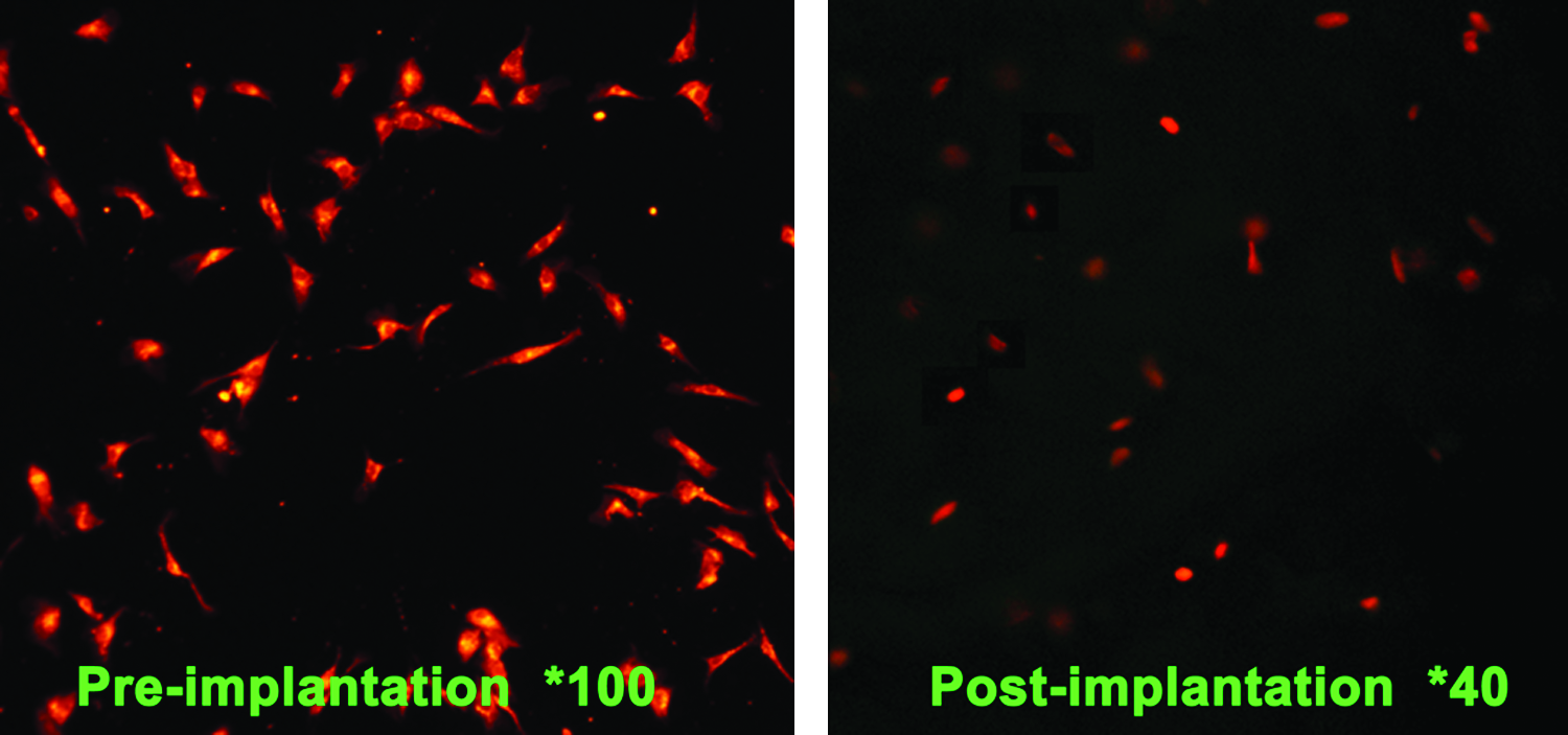
Fluorescent confocal microscopy observation of PKH-26 marked cells. NP cell demonstrated characteristic membrane-associated distribution and intense fluorescence of PKH 26. Twelve weeks after implantation, a large number of PKH-26-positive cells were found within the nucleus pulposus area providing survival evidence of the seeding cells. Color images available online at www.liebertpub.com/tea
Animal surgery
All beagle dogs survived the surgical procedure and the 12-week follow-up period. The operation time varying from 1.5 to 2 h with an average blood loss of 25 mL. No obvious complications were found and the skin incisions of all the experimented animals healed well.
Radiographic and MRI analysis
X-ray showed that the bony fusion between the intervertebral disc allograft and the adjacent host intervertebral body was observed in all three groups at 12 weeks. Disc space narrowing and osteophyte formation, which indicated the degeneration of the implanted disc, could be detected in group C 4 weeks after surgery; these changes deteriorated at the 8-week and 12-week follow-up. However, the disc height preservation in group A and B was better than group C and maintained at a stable level in the following observation, and no obvious osteophyte formation and endplate sclerosis was found (Fig. 5). AT 8 and 12 weeks postimplantation, the disc height of group A and B was significant higher than group C. (Fig. 6)
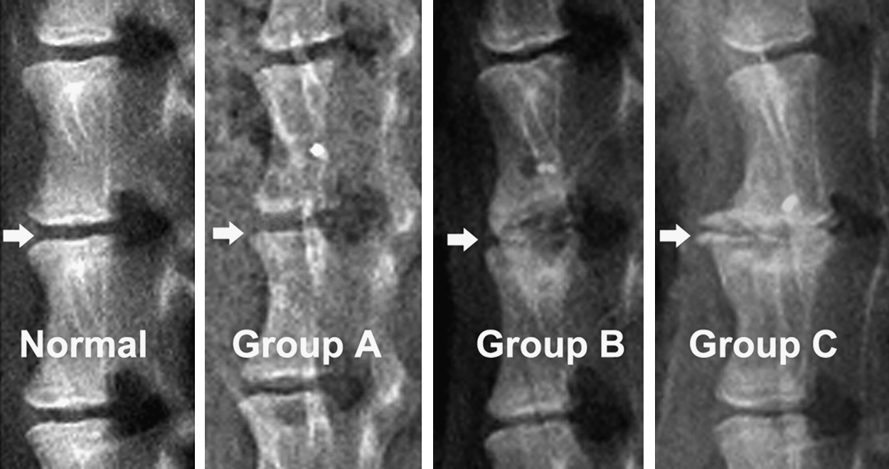
Lateral view Radiographic analysis at 12 weeks follow-up. Arrows indicate the operated segment. Fine bone fusion were observed in all three groups. Typical degenerative changes, such as space narrowing, endplate sclerosis, and osteophyte formation, could be detected in the blank control group. In the test and positive control group, however, the height and the overall morphology of the intervertebral space was preserved at a status close to a healthy intervertebral disc.
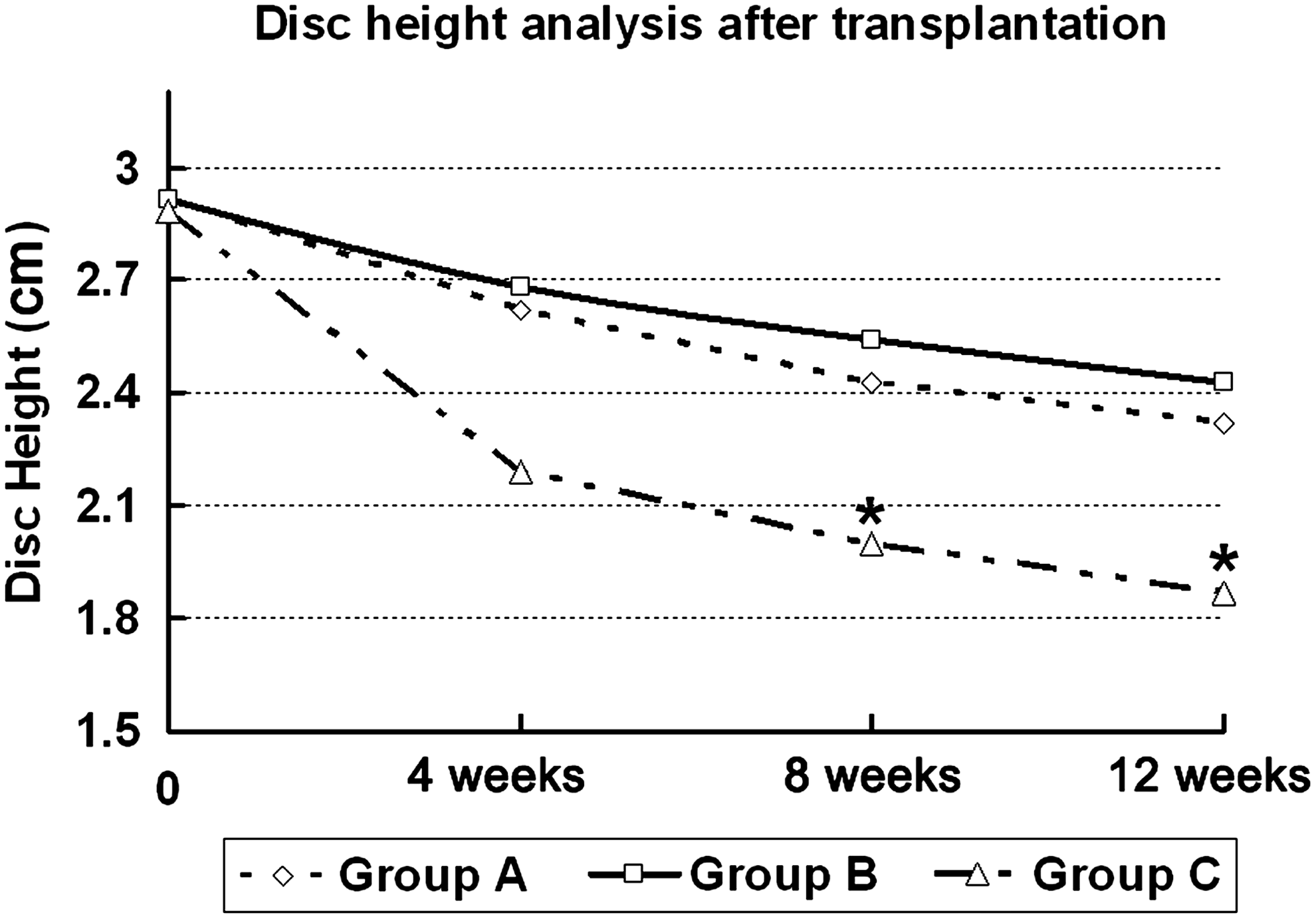
Disc height comparison of the operated segment among different groups. *indicates significant difference compare with group A and B. p<0.05. Each data point represents the mean and standard deviation of six samples.
Degenerative changes of the L4–5 disc were seen in all experimented animals by MRI examination after surgery. Group C displayed a typical black disc change, which indicated the decreasing of the water content within the NP already at a 4-week follow-up, and subsequent observation found the further descent of the NP signal intensity (Fig. 7). However, the T2 signal intensity of group A and B were higher than group C and remained at a relative stable level in the following observation. The normalized grayscale of the L4–5 disc MRI signal of group A and B was significantly higher than group C at all time points. In addition, at 12 weeks follow-up, the normalized grayscale of the L4–5 disc MRI signal of group A was significant higher than group B. (Fig. 8)
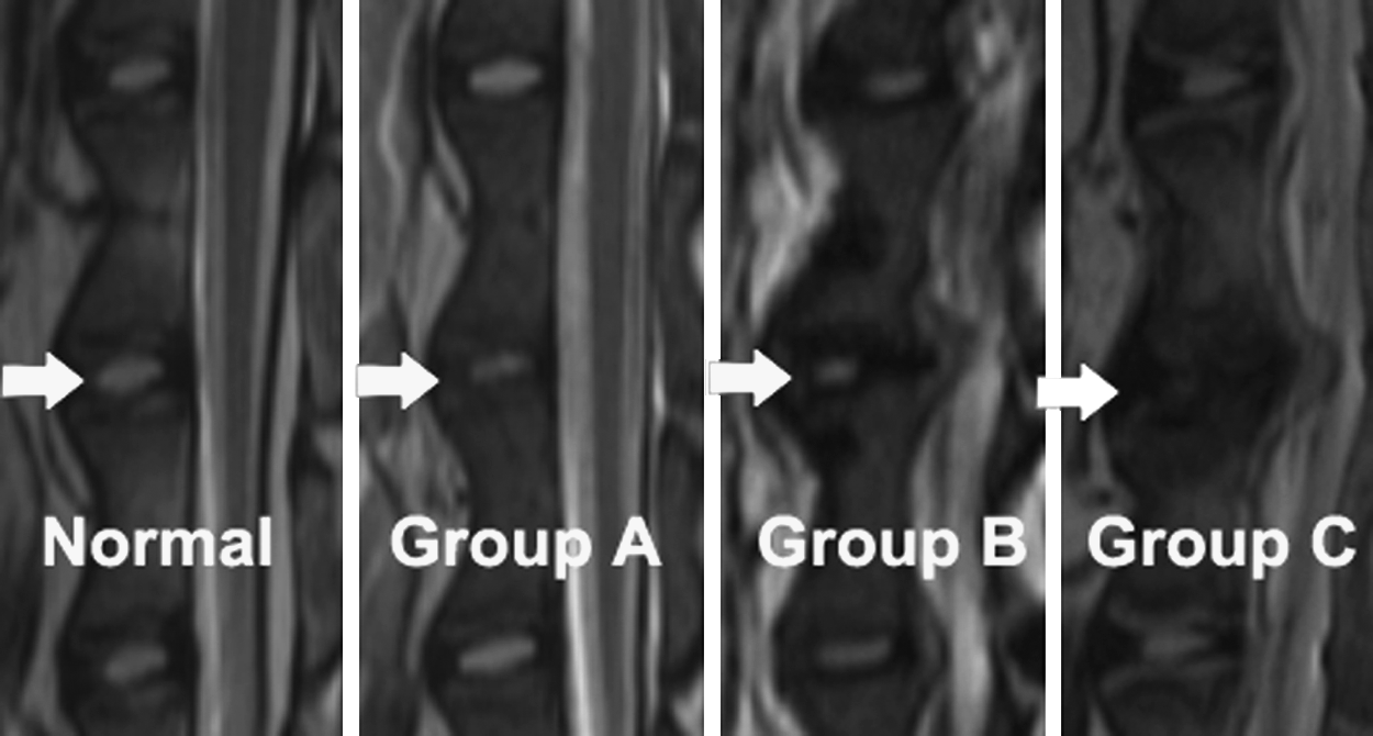
MRI examination of all groups at 12 weeks follow-up. Arrows indicate the operated segment. The T2 signal intensity and the morphology of the NP in the test group was well preserved. In the positive control group, the T2 signal intensity was also kept at a relative high level. However, typical signs of black disc changes, which indicated the decreasing of the water content in the NP were found in the blank control group. MRI, magnetic resonance images.
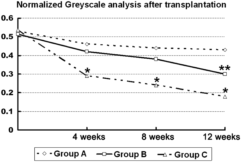
Normalized grayscale of the L4–5 disc MRI signal comparison among different groups. *indicates significant difference compare with group A and B. p<0.05. **indicates significant difference compare with group A. p<0.05. Each data point represents the mean and standard deviation of six samples.
Biomechanical analysis
Biomechanical analysis showed the range of motions (ROMs) of left–right bending were not statistically different within three groups, although there were differences displayed in the value. However, the ROM of flexion–extension and left–right rotation in group C was significantly bigger compared with group A and group B. No statistically significant difference was found between group A and group B. The detail values of the biomechanical results are displayed in Figure 9.
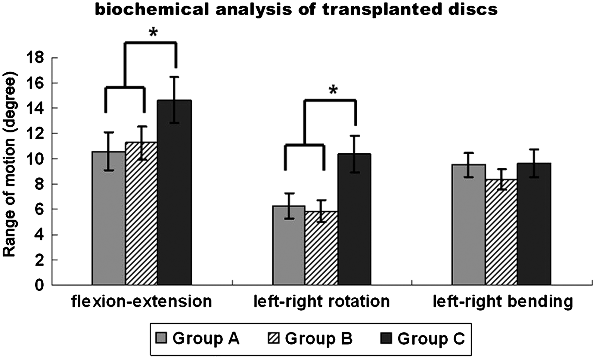
Comparison of biomechanical stability of the operated segment among different groups. *indicates significant difference between groups. p<0.05. Each data point represents the mean and standard deviation of six samples.
hTERT mRNA expressing
Positive hTERT mRNA expression was detected in the NP tissue of the transplanted allograft in group A using RT-PCR analysis at 12 weeks. In group B and C, no hTERT mRNA expression was identified. Positive GAPDH expression could be detected in all groups (Fig. 10).
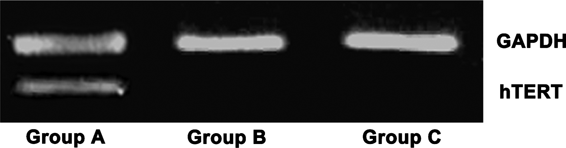
RT-PCR analysis for hTERT mRNA of nucleus pulposus tissue at 12 weeks follow-up. GAPDH was used for normalization. The positive band of hTERT was detected within the nucleus pulposus tissue of intervertebral disc allograft in group A.
Histology
The overall morphology and the nucleus pulposus structure of the intervertebral disc allograft were well preserved in group A and B 12 weeks post-transplantation. Large amount of chondrocyte-like cells was identified within the NP area, which presented in plentiful ECM substrate. In group C, however, only a small amount of ECM could be detected and the morphology of the cells in the NP area did not show a Chondrocyte-like morphology. No signs of inflammation were found in all groups (Fig. 11).

Histological analysis. NP: nucleus pulposus, AF: annulus fibrosus, CE: cartilage endplate, VB: vertebral body. The morphology of the NP and the structure of the total transplanted disc, including the NP, AF, and CE, was well preserved in the test and positive control group, large amounts of chondrocyte-like cells were observed in the NP area. In the blank control group, the normal structure of the transplanted disc was poor kept and the cells within the NP area lost their chondrocyte-like phenotype. Color images available online at www.liebertpub.com/tea
Discussion
The goal of the study was to investigate whether the intervention of NP cells or hTERT gene-transduced NP cells can prevent or delay the degeneration process after allograft total disc transplantation in a beagle dog model in vivo. X-ray showed a better preservation of disc height at 12 weeks follow-up and MRI scan showed that nucleus pulposus cells expressing hTERT could effectively preserve the water content in the NP area of the transplanted disc. Our biomechanical analysis confirmed that the cell intervention resulted in a more stable spinal functional unit than the blank control. Moreover, histology and gross morphology showed that intervertebral discs injected NP cells or hTERT-loaded NP cells have more production and secretion of an appropriate ECM than the control group. These important results strongly suggested that hTERT-loaded NP cells intervention could effectively resist the degeneration of the allogenic transplanted intervertebral discs.
Tissue engineering intervertebral disc regeneration approaches have been widely studied and showed to have the potential to effectively delay the process of intervertebral disc degeneration.21–23 All these approaches seeked to fabricate scaffolds similar to the natural intervertebral disc and construct tissue engineering composites to replace the degenerated intervertebral disc, which lost its function partly or totally. However, the implantation procedure of a solid-state tissue-engineered total disc replacement will inevitably injury the normal structure of the disc, and the union between the allograft and the host vertebral body is another troubled problem. As far as injectable tissue engineering products is concerned, the gelatinous composites lack of mechanical strengh to afford an initial stability to the disc so that their potential effect on disc regeneration might be limited. The main characteristic of our strategy is the natural structure and function of the allograft scaffold. The bony endplate can ensure a solid bony union between the implant and the host vertebral body and the natural structure might ensure the mechanical strengh and stability of the functional spinal unit. This property brought a foundation for the survival and functional preservation of our tissue-engineered allograft intervertebral disc.
Currently, strategies to regenerate the disc, focus on restoring the ability to regulate matrix production to restore the disc function. In these strategies, autologous NP cell transplantation has become one of the major techniques in attempts to prevent IVD degeneration in animal models. However, the dedifferentiation and senescence of the NP cells cultured in vitro led to the decreasing ability of secreting ECM, 24 which made the NP cells been considered difficult, for broad application clinically. To overcome this problem, hTERT was transduced into the NP cells in the present study. The purpose of these cells is not only to maintain the cell phenotypes themselves, producing ECM intended to re-establish healthy disc function, but also to provide signaling cues that ameliorate the effects of disc degeneration.25,26 Ectopic expression of the catalytic component of human telomerase, hTERT, has been shown to extend the life span of human somatic cells beyond senescence without causing neoplastic transformation, resulting in unlimited cellular growth.27,28 In our previous study, we successfully transduced NP cells using rAAV-hTERT with a 70% transfection efficiency. The population doubling time of the gene-tranduced NP cells was siginificant lower than the control. Moreover, gene transduction resulted in a better cell phenotype and morphology preservation and extended lifespan without any signs of tumorgenicity. These findings demonstrated that the transgene of the hTERT by rAAV is an effective and safety method to upregulate the activity and function of the NP cells. 18 The mechanisms may lie in the upregulation of genes involved in cellular growth, DNA repair, and chromatin remodeling as well as downregulation of apoptotic genes following telomerase overexpression.29,30 Our histology results demonstrated the typical chondrocyte-like cell type within the NP area in group A and B. The cell tracer technique by using PKH-26 provides existence evidence of exogenous cells, thus, the chondrocyte-like cell population we found in histology may involve in both the exogenous cells and the endogenous NP cells of the allograft intervertebral disc. The detailed mechanism of the interaction between the exogenous and endogenous NP cells was not identified, but the well preservation of NP structure, cell morphology, cell number, and distribution provided a solid foundation of the potential antidegeneration effect by the cell intervention pathway. On the contrary, the poor cell appearance and cell number within the NP area in group C will naturally lead to the decreasing of the physiological function, including phenotype preservation, expression of ECM, and metabolic activity. These findings confirmed our clinical research conclusion that the allograft disc transplantation without any activity stimulation may result in a progressive degeneration spinal functional unit. 17
In our surgical preparation, the bony endplate surfaces of disc were lavaged with saline solution using the Pulsavac™ wound debridement irrigation system to remove cutting debris and blood clots. We think this procedure would facilitate the nutrient diffusion into the intervertebral disc allograft by keeping micropores open and ensure the cell metabolic activity. The potential nutrition channels may lie in the cartilage endplate. In vitro disc tissue culturing has shown that cells injected into the disc could survive at least 2 months and secret the ECM. 31 Our confocal microscopy observation 12 weeks after transplantion confirmed the PKH-26-positive cells within the NP area, which strongly suggested the survival of the injected cells. Furthermore, hTERT mRNA could still be detected in the NP tissue of intervertebral disc allograft in group A by RT-PCR at 12 weeks follow-up. The persistent expression of hTERT mRNA could naturally prevent the cells from senescence and ensure the long-term outcome after allograft disc transplantation.
Disc height, segmental stability, and T2-weighted signal intensity on MRI are three major parameters for evaluating disc degeneration. Based on these parameters, our purpose to prevent the degeneration process after allograft disc transplantation using the cell intervention technique was successfully achieved in a beagle dog model. According to radiographic and biomechanical analysis, group A and B had apparently better results than group C. We did not find any significant difference in histology, disc height analysis, and biomechanical analysis between group A and B. Questions may rise on whether transduction of hTERT into NP cells make any difference. The normalized grayscale of the disc MRI T2 signal did show a significant difference between the two groups, which imply the different water content in the NP area. Decreasing of T2-weighted signal intensity is an early change and indication of degeneration clinically. 32 While disc space narrowing, osteophytes formation, and segmental instability were the succeeding middle to late stage changes. With the extension of follow-up period, changes and differences may occur between group A and B. However, this warrants further investigation.
Conclusion
The present study demonstrated that NP cells or hTERT-loaded NP cells intervention could effectively resist the degeneration of the allogenic transplanted intervertebral discs in a beagle model. The hTERT-loaded NP cells had a better antidegeneration effect on the transplanted disc than NP cells. Long-term in vivo experiments are needed to further validate our preliminary conclusion.
Footnotes
Disclosure Statement
No competing financial interests exist.