
Abstract
Hepatocytes in high density are a requisite for the functional performance of complex devices such as bioartificial liver (BAL). In addition to high cell number, efficient mass transfer is also a key parameter in such devices. High-density culture of cells and efficient mass transfer can be achieved in BAL with hollow-fiber-based bioreactors. Even though different types of hollow fibers have been tried in a BAL, prospects of using polypropylene hollow fibers are not well evaluated. In this study, a prototype of bioreactor with polypropylene hollow fibers was fabricated and evaluated for cytotoxicity and hepatocyte function. High density of HepG2/adult hepatocyte cultures was used to evaluate polypropylene hollow fiber to support the biochemical activities (albumin and urea production), ammonia detoxification, and gene expression and to provide effective oxygenation. The results confirmed that a polypropylene hollow-fiber prototype bioreactor is able to provide efficient oxygenation and supported hepatocyte functions in a high-density culture.
Introduction
Alternatives such as liver-support devices have been developed as a bridge to liver transplantation. Earlier attempts in this direction were to use charcoal or other sorbents for adsorbing and filtering toxins from blood. 5 As it is difficult to remove the complex toxic compounds using sorption therapy, these devices were not highly successful in clinical trials. 6 Development of the bioartificial liver (BAL) was initiated in 1980s, which uses hepatocytes to replace all failing liver functions. In a BAL, hepatocytes will be cultured under a well-nourished and oxygenated extracorporeal device, and patient's blood or plasma will be perfused through it. The incorporation of hepatocytes in BAL will ensure the synthetic, metabolic, and detoxification functions of the liver. Hollow-fiber membrane-, flat-plate membrane-, encapsulation-, and microcarrier-based bioreactors are currently under investigation for BAL. Among them, hollow-fiber-based bioreactors are the most commonly used.7–9 Hollow-fiber-based bioreactors are available in different configurations, which include culturing of hepatocytes entrapped inside the hollow fiber, hepatocytes adhered outside the hollow fiber, or hepatocytes attached to microcarriers with a medium perfusion either through the hollow fiber or outside it. In the proposed bioreactor, hepatocytes were cultured outside the hollow fiber with medium perfusion through the fibers.
In BAL, hollow fibers are used because of their simplicity to separate cells (hepatic cell lines or xenogenic hepatocytes) in the bioreactor from human blood or plasma. This helps to prevent possible complications due to the transfer of the xenogenic cells/hepatic cell lines to human body. By adjusting the pore size of hollow fibers, transfer of immunoglobulin's from patient's body to the cell compartment in bioreactor can also be avoided, thereby protecting cell death due to immune reactions, enhancing the efficiency of bioreactor. Hollow fibers are also used to provide adequate nutrients and oxygen to the cells in high-density cell culture.
Polysulfone, cellulose acetate, and polyester hollow fibers are being used in BAL.10–12 Polypropylene is a well-known highly hydrophobic polymer, which is being used for a variety of applications.13,14 Prospects of using polypropylene hollow fiber for BAL have not been well established. In this study, the polypropylene hollow-fiber prototype bioreactor was extensively evaluated to support the high-density culture of HepG2 cells and adult rat hepatocytes.
Materials and Methods
BAL prototype bioreactor apparatus
A BAL prototype bioreactor was fabricated using microporous polypropylene hollow fibers with 240-μm inner diameter and 30-μm wall thickness (Celgard X30 240; Hoechst Cellanese). The housing was fabricated from polymethyl methacrylate rods (acrylic), and the end caps were fabricated from polypropylene rods (Mayura Plastics). The hollow fibers were potted using a room-temperature-vulcanizing silicone elastomer, polydimethyl siloxane (adhesive) (Nusil 4905; Nusil Technologies). The interfiber distance between the hollow fibers was maintained at 100 μm (nominal), which provided a priming volume of 3 mL for a module with 60-mm length and 9-mm fiber bundle diameter. The packing density of the fiber bundle was about 50% of the chamber volume.
The unit was provided with two cell-seeding ports with standard Luer tapers conforming to the international standard international organization of standardization (ISO) 594: conical fittings with a 6% (Luer) taper for syringes, needles, and certain other medical equipment. Two perfusion ports conforming to the same Luer standards were also provided on the end caps. All fluid was sealed using silicone elastomer O'rings. Slow-infusion pump (Masterflex; Cole Parmer) with silicon rubber tubing was used for the perfusion of the medium through the bioreactor and was connected as in the Figure 1a and b. Cells were seeded in the interfiber space through the cell-seeding port (Fig. 1c), and the medium was perfused through the hollow fiber very slowly at a flow rate of 0.3 mL/min. At specific time intervals, the sampling port was used for sampling and to evaluate the performance of cultured cells.

Culture and maintenance of cell lines
The human liver hepatocellular carcinoma cell line (HepG2) was obtained from the National Center for Cell Science (NCCS, India). The Strain-L mouse connective tissue fibroblast cell line (L-929) was obtained from the American Type Culture Collection (ATCC, USA). Both cells were cultured and maintained in the Dulbecco's Modified Eagle Medium (DMEM; Sigma) supplemented with 10% fetal bovine serum (FBS; Gibco).
Direct contact assay for cytotoxicity
Materials used for the fabrication (hollow fiber, acrylic, and adhesive) were screened for cytotoxicity by direct contact as per ISO part 10993-5. For direct contact, both L929 and HepG2 cells were used. Cells were cultured to subconfluency, and the material was placed on the top of the cells. After culturing for another 24 h, cells were incubated with a neutral red solution (Sigma; 1 mg/mL) for 20 min. Wells were washed with PBS, and observed under a microscope to assess cellular morphology and neutral red uptake. Each sample was evaluated in triplicate and compared with positive (organo-tin-stabilized polyvinyl chloride) and negative control (high-density polyethylene).
Cell viability analysis
Viability of HepG2 cells cultured on hollow fiber was evaluated using a fiber module. The fiber module was set up by fixing a hollow fiber (5-cm long, 25 numbers) on a cleaned glass slide using an adhesive and was sterilized by ETO. HepG2 cells cultured in the fiber module were evaluated for their viability by double staining with fluorescein diacetate (FDA) and propidium iodide (PI). Polypropylene hollow fibers (containing cultured cells) were treated with FDA (Sigma; 10 μg/mL) for 10 min and rinsed by dipping fibers in PBS. Cells were counterstained with PI (Sigma; 1 μg/mL) for 2 min. Fibers were then rinsed by dipping in PBS and observed under a fluorescence microscope (Leica; DMI 6000).
MTT assay
The activity of HepG2 cells cultured on hollow fiber was evaluated using the fiber module. The fiber module was transferred to a sterile Petri dish, and 1×10⁵ cells (104 cells/fiber) were seeded on top of the fibers. The culture was maintained in the DMEM supplemented with 10% FBS for 5 days. Triplicate modules were used for assessing metabolic activity on 1st, 3rd, and 5th day by 3-(4,5-dimethylthiazl-2-yl)-2,5-diphenyltetrazolium bromide (MTT) assay. Cells were incubated in a culture medium containing MTT (Sigma; 1 mg/mL) for 4 h to allow the formation of formazan crystals. The formazan product was solubilized in isopropanol, and the absorbance was recorded at 570 nm using a spectrophotometer (Biotek).
Cell cycle analysis
For cell cycle analysis, HepG2 cells cultured in the fiber module were trypsinized and pipetted several times to make single-cell suspension. After passing through a cell strainer, cells were fixed in 50% methanol for 30 min at 4°C. After rehydrating, cells were treated with RNase (Sigma; 1 mg/mL in 0.2 M phosphate buffer, pH 7) for 30 min at 37°C. Cells were then centrifuged, washed, and treated with the PI solution (0.05 mg/mL; Sigma) for another 30 min at 4°C. After washing, cells were analyzed at a low flow rate (<1000 cells/s) using a flow cytometer (BD FACS ARIA). Doublets were excluded from singlets based on the width-versus-area analysis of the pulse-processed data. Relative cellular DNA content was analyzed by the height of the signal pulse of the total fluorescence emission, and relative cellular distribution in various phases of cell cycle was determined. HepG2 cells cultured in a tissue culture polystyrene plate (TCPS) were used as a control. The data were analyzed using BD FACS Diva software.
Adult hepatocyte culture
Hepatocytes were isolated from adult male Wistar rats with the approval of the institute animal ethics committee. Rats were anesthetized by injecting ketamine (60 mg/kg) and xylazine (8 mg/kg). After laparotomy, the portal vein was cannulated with a blunt needle. The liver was then perfused with an oxygenated, calcium-free perfusion buffer I (NaCl 0.69%, KCl 0.035%, KH2PO4 0.163%, and NaHCO3 0.21%) for 15 min, followed by perfusion buffer II (NaCl 0.3%, KCl 0.05%, CaCl2 0.07%, HEPES 0.24%, and collagenase type I [0.45 mg/mL]) for 10 min. The liver was dissected out; hepatocytes were dislodged into a serum-free Iscove's modified Dulbecco's medium (IMDM; Gibco) (ice-cold) by gentle shaking and passed through a nylon mesh (100 μm) to remove tissue pieces. The cell suspension was then centrifuged at 50 g for 1 min, followed by two centrifugations at 20 g for 2 min to separate hepatocytes. Cell viability was determined by trypan blue dye exclusion, and cell suspensions with >85% viability were used for the experiment. The isolated cells were cultured in the IMDM containing 2% FBS.
Adult hepatocyte/HepG2 culture in prototype bioreactor
A uniform cell suspension (adult hepatocyte/HepG2) of 5×10⁶ cells (104 cells/fiber) was seeded in to the prototype bioreactor through the cell-seeding port. The prototype bioreactor was then rotated very gently to make uniform cell distribution throughout and was incubated without disturbance at 37°C and 5% CO2. After 1 h of incubation, perfusion was started at a flow rate of 0.3 mL/min, and the culture was continued for a maximum of 1 week without any medium change. The medium was collected on every alternate days (day 1, day 3, day 5, and day 7) through the sampling port using a syringe and was stored at −80°C for evaluation of albumin and urea production.
To evaluate the effect of perfusion, albumin and urea production by HepG2 cells cultured in a prototype bioreactor (perfusion culture) was compared with cells cultured without perfusion (static culture). To assess functional performance of the prototype bioreactor, adult rat hepatocytes were compared with cells cultured in a collagen- (Type 1; Purecol) coated tissue culture plate.
Analysis of dissolved oxygen and pH
The dissolved oxygen and pH of medium in the bioreactor housed with adult hepatocyte were analyzed by sampling the medium every day. The samples were analyzed immediately after collection for dissolved oxygen and pH using a blood gas analyzer (ABL 555; Radiometer).
Albumin estimation
Albumin was estimated using a sandwich ELISA kit as per the manufacturer's instructions (Bethyl Laboratories). About 100 μL of standards and diluted samples (1:40) were added in duplicate to the antibody-coated wells. After incubating for 1 h, wells were washed four times, and 100 μL of anti-albumin detection antibody was added and incubated for another 1 h. Wells were then washed and incubated with 100 μL of horse radish peroxidase solution A for 30 min. Wells were further washed and incubated with 100 μL of 3,3′,5,5′-tetramethyl benzidine (TMB) substrate solution in dark for 30 min. The color-developing reaction was then stopped by adding 100 μL of stop solution (2N H2SO4), and the absorbance was measured at 450 nm using a spectrophotometer (Hidex). With the help of GraphPad prism software, a standard curve was drawn by a four-parameter logistic model fit, and albumin concentration in each sample was determined.
Urea estimation
Urea production by cells in a prototype bioreactor was estimated using an urea assay kit (Biochain) as per the manufacturer's instructions. The medium collected on 1, 3, 5, and 7 days was used for analysis. About 50 μL of the collected medium sample, urea standard, and control medium (blank) was added in triplicate to a 96-well plate. Two hundred microliters of the working reagent was then added to each well and incubated for 50 min at room temperature. Using a spectrophotometer (Biotek), absorbance was recorded at 530 nm, and the concentration of urea was determined.
Ammonia detoxification analysis
Hepatocytes convert ammonia to less-toxic urea that can be easily excreted from the body through urine. For analyzing ammonia detoxification capacity of adult hepatocytes in a prototype bioreactor, hepatocytes cultured for 7 days in a prototype bioreactor were treated with 10 mM ammonium chloride. The concentration of urea in the medium at 0 and 2 h after ammonium chloride treatment was estimated as described before. Ammonia detoxification was then compared with hepatocytes cultured in TCPSs.
Gene expression analysis
To assess the expression of hepatocyte key markers in a prototype bioreactor, gene expression of HepG2/adult hepatocytes was analyzed. HepG2 cells cultured on a tissue culture plate and hepatocytes cultured on a collagen-coated culture plate were used as control. Total ribonucleic acid (RNA) of cells was isolated using TRIzol reagent (Invitrogen). The cell lysate obtained was mixed with half the volume of chloroform (Merck) and centrifuged at 12,000 rpm at 4°C. The aqueous phase was collected, and RNA was precipitated using isopropanol (Merck). RNA was purified (by washing with ethanol) and dissolved in distilled water. Complementary DNA (cDNA) was then synthesized using an M-MuLV reverse transcriptase kit (Bangalore GeNei) as per the manufacturer's instructions. Gene-specific primers were used to perform polymerase chain reaction (PCR) with Red Dye PCR master mix (Bangalore GeNei) in a programmable Mastercycler (Eppendorf ). The primer sequence and PCR conditions followed are given in Tables 1 and 2. PCR products were analyzed on a 2% agarose gel, and the product size was estimated using a 100-base-pair DNA ladder. The intensity of the band corresponding to each gene was measured and compared to the reference control β-actin using Image J software. The relative intensity of the band was used to compare the expression of genes in the prototype bioreactor to control.
Hepatocyte adherence
Adherence of hepatocytes to the polypropylene hollow fibers was analyzed using an environmental scanning electron microscope (ESEM). On 7th day of culture, fibers were carefully removed from the acrylic casing of the prototype bioreactor. Fibers were rinsed by immersing in PBS and observed by an ESEM (FEI QUANTA 200) under an ESEM mode.
Statistical analysis
Data obtained from independent experiments were compared using Student's t-test. Data were expressed as mean±SD and were considered statistically significant if p-values were 0.05 or less.
Results
All the bioreactor components were first evaluated for their cytotoxicity using L929 and HepG2 cells. For further analysis of hollow fibers, fiber modules were used to evaluate the viability and proliferation of HepG2 cells. At the final stage, using HepG2 cells and adult hepatocytes, environmental conditions inside the assembled prototype bioreactor were investigated.
Cytotoxicity evaluation of prototype bioreactor
All components used for the fabrication of a prototype bioreactor such as acrylic, adhesive, and polypropylene hollow fibers were evaluated for cytotoxicity by a direct-contact method using L929 and HepG2 cells. Cells maintained morphology and showed neutral red uptake for acrylic, polypropylene fiber, and adhesive (Fig. 2).

Cytotoxic evaluation of bioreactor components: Direct-contact analysis for cytotoxicity of bioreactor components (acrylic, polypropylene hollow fiber, and nucil silicon adhesive) using L929 and HepG2 cells. Representative microscopic image of neutral red uptake by live cells is shown in figure. Label denotes the bioreactor component used for cytotoxicity evaluation (direct-contact assay by placing the material on the top of cell monolayer). The component location in the prototype bioreactor is pointed out using arrows. Scale bar denotes 100 μm. Color images available online at www.liebertpub.com/tea
Viability of HepG2 cells on fiber module
Cell viability in the fiber module assessed by FDA and PI showed that almost all cells were viable (Fig. 3).

Viability analysis of cells cultured on fiber module: Cells cultured in the fiber module were stained with fluorescein diacetate and propidium iodide. Representative microscopic image of stained cells is shown in figure. Scale bar denotes 100 μm. Color images available online at www.liebertpub.com/tea
Proliferation of HepG2 cells on fiber module
To asses growth and proliferation of HepG2 cells on fiber module, cellular activity and cell cycle analysis was performed.
The proliferation of HepG2 cells cultured in the fiber module was determined from MTT assay on days 1, 3, and 5. The cellular activity on day 3 and day 5 was significantly higher compared to day 1 (Fig. 4) (p<0.05).
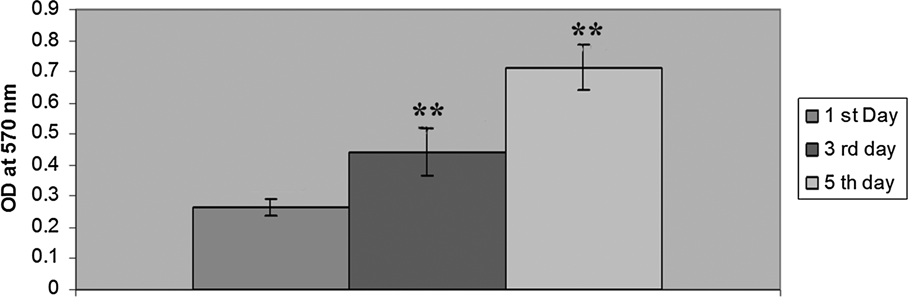
HepG2 proliferation on fiber module: Cellular activity of HepG2 cells cultured in the fiber module on days 1, 3, and 5 was assessed using 3-(4,5-dimethylthiazl-2-yl)-2,5-diphenyltetrazolium bromide (MTT) assay. Data expressed as mean±SD of three different experiments. Cellular activity was compared with that on 1st day, and significant differences (p<0.05) are denoted by **.
Cell cycle of HepG2 cells in fiber module examined by flow cytometry showed that the percentage of cells in the G0/G1, S, and G2/M phase was 55%, 23%, and 22%, respectively. In control (HepG2 cells cultured in tissue culture plate), the percentage of cells present in the G0/G1, S, and G2/M phase was 60%, 21%, and 19%, respectively (Fig. 5).

Cell cycle analysis of HepG2 cells cultured on the fiber module.
Evaluation of efficiency of perfusion in a BAL prototype bioreactor
In perfusion culture, an increase in albumin throughout the period was observed with a maximum on the 7th day (Fig. 6a). Comparison of albumin on 7th day perfusion showed significantly higher production compared to static culture (Fig. 6b). Urea estimation also showed an increase in the concentration in perfusion culture (Fig. 6c). Urea production on 7th-day perfusion was also significantly higher than that of static culture (Fig. 6d).

Evaluation of efficiency of perfusion in a BAL prototype bioreactor:
Efficiency of prototype bioreactor to support adult hepatocytes
For evaluating the efficiency of the prototype bioreactor, adult rat hepatocytes were cultured with continuous perfusion (perfusion culture) for a maximum period of 1 week. To evaluate the efficiency of polypropylene hollow fiber for high-density primary adult hepatocyte culture, the medium was collected, and dissolved oxygen was monitored every day. Dissolved oxygen remained stable throughout the culture period and was in the range of 98.7 to 99.25 (Fig. 7a). The pH of the culture medium also remained stable around neutral ranging from 7.3 to 7.5 (Fig. 7b).

Dissolved oxygen and pH inside the bioreactor.
Urea concentration increased throughout the culture period and was significantly higher on 3rd and 5th day compared to that on day 1 (Fig. 8a). Albumin concentration also increased during the culture period, and showed maximum concentration on day 7 (Fig. 8b).
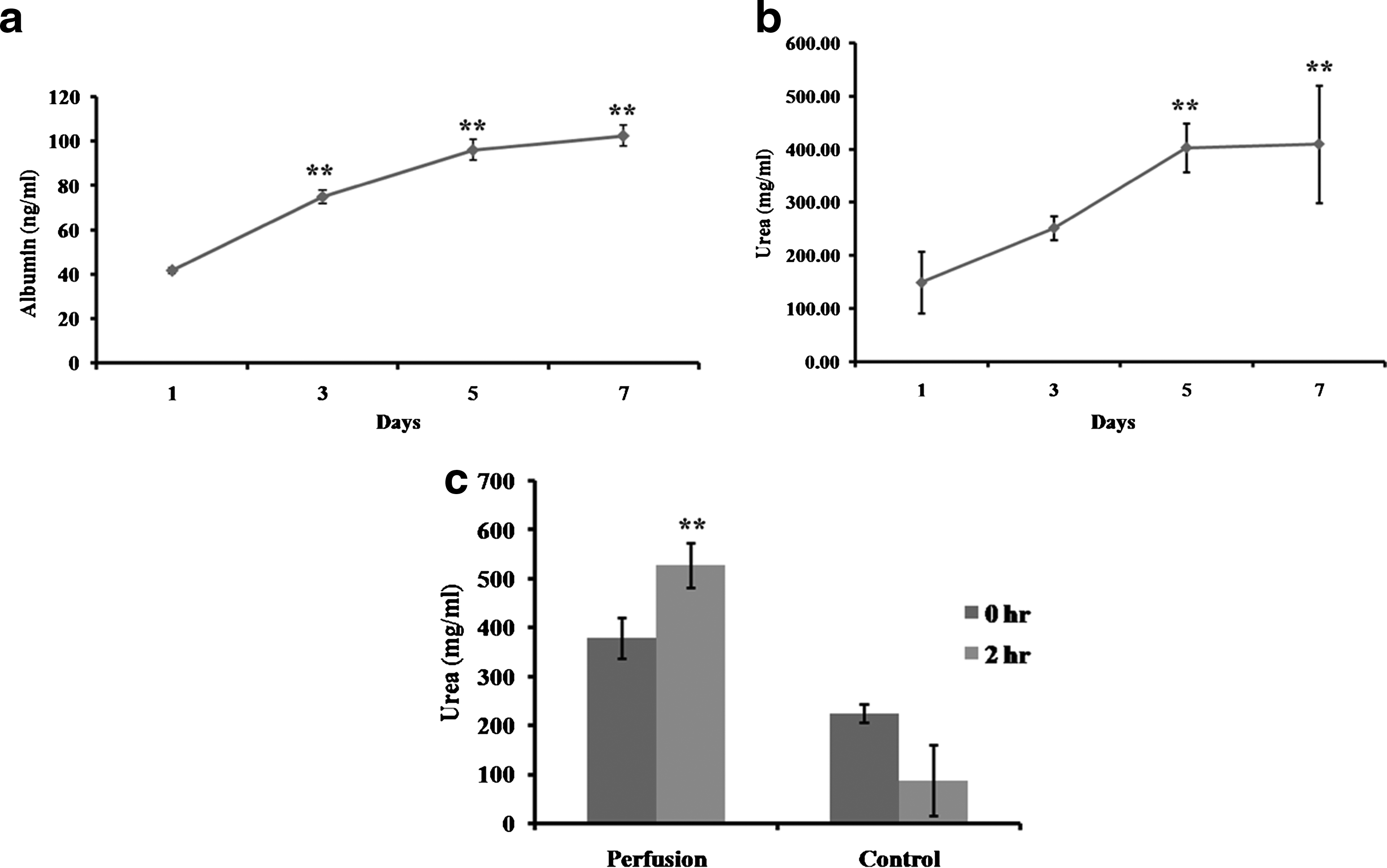
Evaluation of a prototype bioreactor to support adult hepatocytes:
To evaluate the efficiency of hepatocytes in the prototype bioreactor (perfusion culture) to detoxify ammonia, hepatocytes were treated with ammonium chloride. Urea concentration at 0 and 2 h after treatment was measured. Urea production in cells cultured on a tissue culture plate (control) was also quantified. Comparison of urea concentration at 0 and 2 h showed significantly higher concentration at 2 h in perfusion culture compared to control (Fig. 8c).
Gene expression in the BAL prototype bioreactor
HepG2 cells in the prototype bioreactor showed elevated expression of the genes albumin (albumin synthesis), α-fetoprotein (α-fetoprotein synthesis), Cytochrome P4501A1 (detoxification enzyme), carbamoyl phosphate synthetase (urea synthesis), hepatocyte nuclear factor-4α (hepatocyte transcription factor), and multidrug resistance protein 2 (bile canalicular transport protein/hepatocyte polarity) compared to control (Fig. 9a).

Gene expression of HepG2/adult hepatocyte cells in a prototype bioreactor:
Adult hepatocytes in the prototype bioreactor also showed elevated expression of albumin (albumin synthesis), Cytochrome P4501A1 (detoxification enzyme), arginase (urea synthesis), and multidrug resistance protein 2 (bile canalicular transport protein/hepatocyte polarity) compared to control (Fig. 9b).
Hepatocyte adherence
The phase-contrast image confirmed the high density of hepatocytes inside the prototype bioreactor (Fig. 10a). ESEM was used to analyze the adherence of hepatocytes to polypropylene hollow fibers in the prototype bioreactor during high-density culture. ESEM showed less hepatocyte adhesion on polypropylene hollow fibers inside the prototype bioreactor. Instead, they were located in the interfiber spaces (Fig. 10b, c).
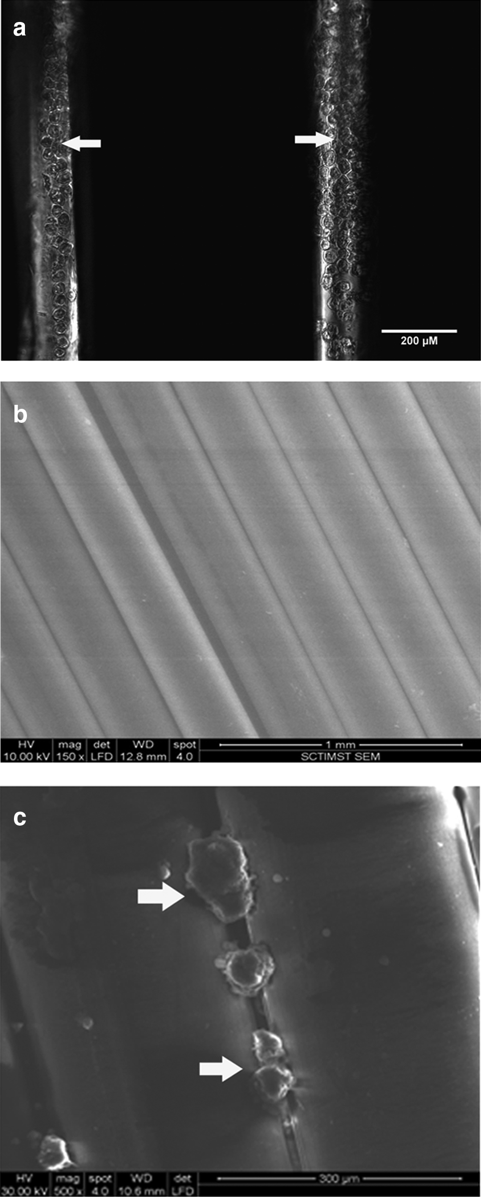
Hepatocyte adherence on polypropylene hollow fiber.
Discussion
It is considered that for an effective therapy by a BAL, at least 10%–20% of the total liver cell mass (∼109 to 1010 hepatocytes) is needed. Moreover, the total volume of a BAL has to be restricted below 1 liter due to the difficulty in draining more blood from a patient's body. Because of these issues, the main challenge of developing a successful BAL is the efficient culture of large number of cells in this limited volume. 15
The efficiency of a BAL to support a high-density culture is greatly dependent on the properties of the hollow fiber. 16 Even though polysulfone, polyethersulfone, and cellulose acetate hollow fibers are used in a BAL, prospects of using polypropylene hollow fibers are not well evaluated. 17 In a BAL, hollow fibers will be in direct contact with blood components because of the perfusion of blood or plasma. It is necessary that the hollow fiber should be hemocompatible to prevent hemolysis and platelet aggregation. 18 Polypropylene hollow fibers are well established for their hemocompatibility and so are extensively used in oxygenators.19,20 In this study, polypropylene hollow fibers were extensively evaluated for their applicability in the BAL.
In the present study, a small BAL prototype bioreactor having an internal volume of 3 mL was fabricated using acrylic, polypropylene hollow fibers, and adhesive. The bioreactor was designed to provide a suitable microenvironment for hepatocytes by mimicking the arrangement of cells and fluid flow as in the normal liver. The main component, polypropylene hollow fibers, was wound to bundles. Paraffin sections showed that fibers were spaced at 107.08±55.5 μm spirally and 136.86±67.69 μm radially in the hollow-fiber bundle. The fiber bundle was inserted into acrylic housing, and a fluid-tight seal was created by potting the ends of the bundle. The final unit with end caps allows the fluid to pass through the insides of the fibers and access to the extra capillary space within the acrylic housing. In the present design, cells were cultured in the interfiber space of hollow fibers, and the cells received nutrients, and oxygen diffused from the perfused medium inside the fibers.
For the optimum performance of a BAL, it is important that components used for fabrication should not be toxic and not affect cellular functions. To ensure cytocompatibility, all components used in the BAL prototype were screened by direct contact using L929 cells. Direct-contact cytotoxicity test results clearly confirmed the nontoxic nature of these materials. BAL is primarily intended to culture hepatocytes: cells that are highly vulnerable to toxicity due to xenobiotic metabolism.21,22 To ensure cytocompatibility to hepatocytes, components were also analyzed using HepG2 cells. The results confirmed their nontoxic nature, thus ensuring the secure growth of cells in the BAL.
Hollow fibers help to separate cells from human blood or plasma, preventing transfer of the xenogenic cells/hepatic cell lines to human body. However, cells in a BAL are prone to immune reaction due to the transfer of immunoglobulins from blood to cell compartment resulting in cell death.23,24 In the developed prototype bioreactor, polypropylene hollow fibers having a typical pore shape of 0.03×0.20 μm with an effective pore size of 0.03 μm, which provides a molecular cutoff in the range of 7500 to 800 Daltons, were used (information provided by the manufacturer). This prevents the transport of high-molecular-weight immunoglobulins to the cell compartment, thus enhancing the efficiency of the bioreactor.
To mimic the internal condition (high density of cells) of a BAL in the developed prototype bioreactor, 5×10⁶ cells were seeded within an internal volume of 3 mL. The medium was perfused slowly at a flow rate of 0.3 mL/min through the polypropylene hollow fibers. To estimate the growth of HepG2 cells inside the fiber module, cell viability, cell proliferation, and cell cycle were analyzed. Cellular viability confirmed the robust nature of cultured cells inside the fiber module. MTT assay showed a high cellular activity on day 3 and day 5, confirming the proliferation of cells in the fiber module. Cell cycle analysis further confirmed the proliferation of cells in the fiber module as indicated by increase in the S- and G2/M-phase cells. These results clearly confirm that the polypropylene hollow fiber is able to provide adequate conditions needed for the growth and proliferation of the cells.
To assess the biochemical performance of cultured HepG2 cells in a prototype bioreactor, albumin synthesis and urea production were evaluated and compared with static cultured cells. Perfusion cultured cells showed enhanced production of albumin and urea, confirming that cells were able to get enough nutrients and oxygen for their biochemical activities. Gene expression analysis further confirmed the ability of cells in a prototype bioreactor to perform protein synthesis (albumin and α-fetoprotein), detoxification (Cytochrome P450 1A1), urea production (carbamoyl phosphate synthase), polarization (MRP2), and expression of hepatocyte transcription factors (HNF4α). These results clearly confirm that a polypropylene hollow fiber has good mass transfer for oxygen and other nutrients, and is able to support growth and biochemical activities of the high-density HepG2 culture.
Physiological requirements of adult hepatocytes are different from those of hepatic cell lines.25,26 To evaluate the efficiency of the polypropylene hollow fiber to provide physiological requirements of adult hepatocytes, adult rat hepatocytes were cultured in a prototype bioreactor for 1 week. Dissolved oxygen was monitored daily and showed a steady concentration of 98% to 99% during the culture. pH of the medium also remained stable around 7.4. Hepatocytes in the prototype bioreactor were able to produce albumin and urea. Moreover hepatocytes in the prototype bioreactor showed enhanced ammonium detoxification compared to control. These results clearly show the effective oxygenation of polypropylene hollow fibers in the prototype bioreactor. These results confirm that polypropylene hollow fibers are able to even support a high-density hepatocyte culture without affecting their biochemical activity (albumin and urea production) and detoxification capability (ammonium chloride detoxification).
Membrane fouling usually occurs due to the adsorption of proteins to hollow fibers. 27 Favorable protein adsorption to the membrane surface will promote cellular adherence resulting in blockage of membrane pores, reducing the effective mass transfer of hollow fiber. Low cell adhesion on fibers under ESEM analysis indicates that the membrane pores are not blocked due to cell adhesion; instead, most of the cells were located in the interfiber space. This clearly confirms less membrane blockage of polypropylene hollow fiber due to cell adhesion even in a high-density culture, substantiating their suitability for a BAL.
As hepatocytes are anchorage-dependent cells, an approach to enhance the viability and functional performance of hepatocytes in BAL is the incorporation of an appropriate cell adhesion substrate such as membranes, microcarriers, or a biological matrix. Polyester is such a hydrophilic matrix commonly used for the enhancement of hepatocyte adhesion. 28 A major limitation for using hydrophilic matrix/fibers for perfusion of medium or oxygenation is their superior hepatocyte adherence that can hinder the mass transfer and long-term functionality of BAL. Polypropylene hollow fibers are highly hydrophobic and stable compared to other fibers. 29 It is easy to attain an uniform morphology and pore size in polypropylene hollow fibers. Moreover, polypropylene has less surface free energy and surface tension and is basic in nature compared to other fibers, thus allowing long-term functional performance of the BAL. 30
Polypropylene hollow fibers show improved bubble-point pressure and better plasma leakage characteristics, compared to hydrophilic hollow fibers. This is expected to provide enhanced longevity to the BAL system in clinic, through retaining the integrity of the fluid pathways for longer periods of time, especially when blood plasma or whole blood is perfused. Use of polypropylene hollow fibers will be an advantage for the mass transfer of nutrients and oxygen because of the less cellular adherence, reducing the possible chance of membrane blockage and mass-transfer hindrance. In the polypropylene hollow-fiber prototype bioreactor, instead of adhering, hepatocytes organized to become three-dimensional (3D) structures, a way to maintain hepatocyte functionality. Moreover, the gene expression analysis clearly confirms the enhanced functional performance of hepatocytes due to their 3D organization. The formation of large cellular aggregates is not appreciated in the BAL due to the resistance in the mass transfer of oxygen and nutrients to the central regions of the aggregates, resulting in cellular death. 31 In the present BAL prototype bioreactor, the interfiber space was around 100 μm, which limited the possible formation of large spheroids. The results clearly show that the polypropylene hollow-fiber bioreactor can accommodate a high-density hepatocyte culture, without affecting their functions, and can provide efficient mass transfer of nutrients, a major advantage in the BAL
Conclusion
A prototype bioreactor using a polypropylene hollow fiber was designed and fabricated to use as a BAL. The efficacy of polypropylene hollow fibers to support high-density culture of cells was evaluated. The results clearly showed the ability of polypropylene hollow fibers to support the proliferation and biochemical activities of HepG2 cells. Polypropylene hollow fibers were able to provide efficient oxygenation and to support hepatocyte functions in a high-density culture. This prototype has to be further scaled-up for its efficacy in preclinical experiments and its suitability for potential human therapy.
Footnotes
Acknowledgments
Authors acknowledge the Director of SCTIMST and the Head of BMT Wing for the facilities. We thank the Thrombosis Research Unit, SCTIMST, for the technical help in flow cytometry. This study was funded by the Department of Biotechnology, Government of India. The Indian Council of Medical Research, the Government of India, supported Anwar Azad with a Student Research Fellowship.
Disclosure Statement
The authors declare that there are no conflicts of interest.