
Abstract
The survival of tissue-engineered mucosa (TEM) after implantation is mostly dependent on the presence of blood vessels for continuous oxygen supply. Therefore the stimulation of vascularization of TEM is essential to improve survival in vivo. Hyperbaric oxygen (HBO) treatment, used to improve wound healing, stimulates the secretion of angiogenic factors. In this study we evaluated the effect of daily HBO treatments on TEM for 1, 3, or 5 consecutive days. Overall histology with hematoxylin-eosin staining showed no apparent changes after one treatment. After three and five HBO treatments, the basal layer became irregular and pyknotic cells were observed. Measurements of the viable epithelium showed significant thinning after one and five treatments, however, proliferation was not affected. The angiogenic factors keratinocyte growth factor (KGF), hepatocyte growth factor (HGF), basic fibroblast growth factor (FGFbasic), and placental growth factor (PlGF) were significantly increased after one HBO treatment, whereas after three treatments a significant decrease of FGFbasic and PlGF was seen. After five treatments KGF, PlGF, and vascular endothelial growth factor (VEGF) were significantly increased. One HBO treatment of TEM enhances the secretion of important angiogenic factors, hereby potentially improving the survival rate after in vivo implantation.
Introduction
T
Oxygen is a critical component in the wound healing process as it is involved in re-epithelization, collagen synthesis, angiogenesis, and oxidative killing of bacteria.4,5 Cells need a constant supply of oxygen, as they cannot store oxygen for long periods of time. The lack of oxygen results in hypoxia, which causes cell death and impairs wound healing.4,6 Hyperbaric oxygen (HBO) can be used to improve healing of different types of wounds.5,7 HBO treatment is twofold; oxygen levels increase to 100% and atmospheric pressure is increased to 2.4ATA. By increasing these two components the oxygen levels in the capillaries rises resulting in the enhanced diffusion of oxygen into surrounding tissues.4,8 Although the exact working mechanism of HBO is not clearly understood, HBO has shown to increase the release of transforming growth factors β1 and β2 (TGF β1 and β2) and (vascular endothelial growth factor [VEGF]), an important angiogenic factor.9,10 Further, TGF β1 and β2 have been reported to stimulate fibroblast proliferation in the tissue surrounding wounds and also to have angiogenic activity. These observations prompted us to use this novel and relatively simple approach to treat TEM with HBO to increase important angiogenic factors in TEM before engraftment. The aim of this study was to evaluate whether HBO treatment of TEM induces the secretion of angiogenic factors.
Materials and Methods
Cell culture
Biopsies of ∼1 cm2 were taken from the cheeks of four healthy individuals upon informed consent. Single-cell suspensions of keratinocytes and fibroblasts were obtained as described before.1,11 Briefly, keratinocytes were isolated from the epithelial sheet by overnight incubation in 0.25% trypsin-EDTA (Invitrogen) and the single-cell suspension was seeded onto lethally irradiated 3T3 fibroblast feeder layers, according to the Rheinwald & Green protocol. 12 Fibroblasts were isolated by mincing the dermis with scalpels, followed by incubation in collagenase/dispase (1.5/2.5 mg/mL, respectively; Invitrogen) solution. The cells used in this study were within passage 3 to 6.
Tissue-engineered mucosa
TEM was created as described previously.1,13 Briefly, per construct 1×10⁵ fibroblasts were spun into the lamina propria of the de-epidermized dermis (DED) 11 and 1×10⁶ keratinocytes were seeded into a steel ring (diameter 10 mm) placed onto the papillary side of the DED. After culturing under submerged conditions for 24 h, the constructs were raised to the air/liquid (A/L) interface and cultured with A/L culture medium consisting of 3:1 Dulbecco's Modified Eagle Medium (DMEM) 4.5 g/L glucose: Ham's F12 supplemented with 24 μM bovine serum albumin, 1 μM hydrocortisone, 1 μM isoproterenol, 0.1 μM insulin, 10 μM L-carnitine, 10 mM L-serine, 1 μM D L-α-tocopherolacetate, fatty acid cocktail (30 μM linoleic acid, 7 μM arachidonic acid, and 25 μM palmitic acid), 50 μg/mL ascorbic acid, 100 IU/mL penicillin, and 100 μg/mL streptomycin, for an additional 14 days. The medium was changed three times a week.
HBO treatment
TEM constructs were treated with HBO using the HBO chamber as illustrated in Figure 1A and B. 14 TEM was treated on a daily basis up to 5 consecutive days. The chamber was flushed with pure oxygen for 10 min while pressure was increased to 2.4ATA. This condition was maintained for 90 min. Next, pressure was decreased again to 1.0ATA in 5 min. Immediately after HBO treatment TEM constructs were harvested. The following groups were included: one group received a single HBO treatment; one group received three HBO treatments; and one group received five treatments. The control group did not receive HBO treatment and was harvested at the same three time points as the HBO-treated groups. Each group consisted of four TEM constructs and three independent experiments were performed.

Image of the HBO chamber used in this study
Collection preconditioned media and histology
Culture media was collected prior to harvesting of TEM, centrifuged at 400 g for 5 min at 4°C and stored at −80°C until further analysis. Next, TEM was harvested by snap freezing with liquid nitrogen. Cryosections (6 μm) were stained with hematoxylin-eosin (HE; Klinipath and Sigma, respectively) and overall morphology was assessed using a light microscope (Olympus). The thickness of the viable epithelium was determined from two consecutive images and the average thickness (μm) was measured using Hamamatsu software (Hamamatsu Photonics) by averaging 12 measurements per image, the results were displayed as mean±SEM.
Immunohistochemistry
Staining for keratin 10, 13, and 19 was done as described before.1,13 For staining of Ki67 (1:200; DAKO), vimentin (1:200; Euro Diagnostica), collagen type III (1:200; Abcam), or collagen type IV (1:200; Euro Diagnostica), cryosections were fixed for 10 min with acetone, washed thrice with PBS, and blocked for 30 min with 10% goat serum in PBS/1%BSA. After incubation with primary antibodies, slides were washed thrice with PBS and incubated with goat anti-mouse or goat anti-rabbit biotin-labeled (both antibodies 1:200; DAKO) in PBS/1%BSA for 30 min at RT, followed by incubation with Streptavidin-HRP (1:200; Southern Biotech) for 30 min at RT. About 5% 3,3′-Diaminobenzidine tetra hydrochloride hydrate (Sigma-Aldrich) in PBS supplemented with 30% H2O2 was used for visualization of positive cells. After thoroughly rinsing with tap water, slides were stained with hematoxylin for background observation. Next, sections were air-dried and coverslipped using Vectamount (Vector) and sections were assessed using a light microscope. Negative control slides were incubated with an irrelevant mouse IgG.
Quantification of proliferation
To determine the proliferation index (PI), the basal layer of the epithelium was analyzed. Images were taken from 12 randomly chosen microscopic views using a 100× magnification. The PI was established as the ratio of the Ki-67 positive cells to all cells of the basal layer (×100%), and results were displayed as mean±SEM.
ELISA assay on conditioned medium
Concentration of TEM-secreted angiogenic factors in the conditioned medium was measured using sandwich ELISA kits according to manufacturer's instructions. Factors that were studied include VEGF, PIGF, hepatocyte growth factor (HGF), keratinocyte growth factor (KGF), and basic fibroblast growth factor (FGFbasic; R&D Systems). Results are expressed as ng or pg/cm2 tissue with each sample consisting of 4 mL supernatant derived from 1 cm2 tissue.
Proliferation of human umbilical vein endothelial cells
Proliferation rate of human umbilical vein endothelial cells (HUVECs) when exposed to culture media was assessed as described previously. 13 Briefly, HUVECs were seeded in 48-well plates in endothelial growth medium (EGM; consisting of human endothelial serum-free medium supplemented with 10% FBS, 20 ng/mL FGF2, and 100 ng/mL EGF). Next, cells were cultured in starving medium (DMEM with 0.5% FCS). After 24 h, cells were cultured with air/liquid culture medium supplemented with KGF (940 pg/cm2), FGFbasic (75 pg/cm2), and HGF (25 ng/cm2) as measured with ELISA in conditioned media obtained from TEM exposed to one HBO treatment. After 48 h, cell numbers were analyzed using the CYQUANT proliferation assay (Molecular Probes) following manufacturer's instructions. The positive control consisted of EGM without growth factor supplements. The negative control consisted of A/L medium without growth factor supplements. Each assay was done in triplicate.
Statistical analysis
Data are presented as mean±SEM. Tests of normality were performed using the Shapiro–Wilk test. Statistical analyses were performed using Student t-test or Mann–Whitney U test. Statistical differences were defined as *p<0.05, **p<0.01, p<0.001, and not significant (ns).
Results
Overall morphology after HBO treatment
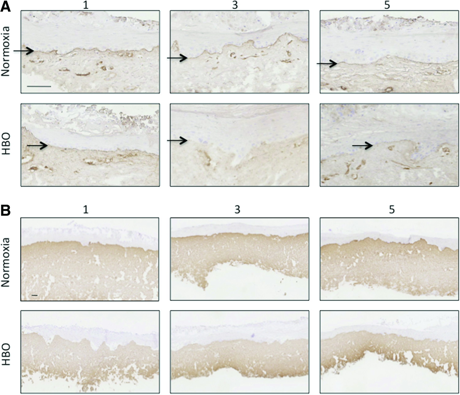
The effect of HBO treatment on TEM overall morphology was assessed using HE staining. No apparent changes in overall morphology were observed after one HBO treatment (Fig. 1C, D). After three and five consecutive HBO treatments, the basal layer became irregular and pyknotic cells were observed in the basal and intermediate layer (Fig. 1E–H). Additionally, the attachment of the epithelial layer to the underlying connective tissue appeared to be affected as gaps between these layers became apparent. Measurements of the viable epithelium showed significant thinning after one and five treatments with HBO (Fig. 1-I). Epithelial differentiation was determined using antibodies against keratins; the predominant component of the epithelial layer. Expression of K10 (Fig. 2A), K13 (Fig. 2B), and K19 (Fig. 2C) was not significantly changed by HBO treatment.
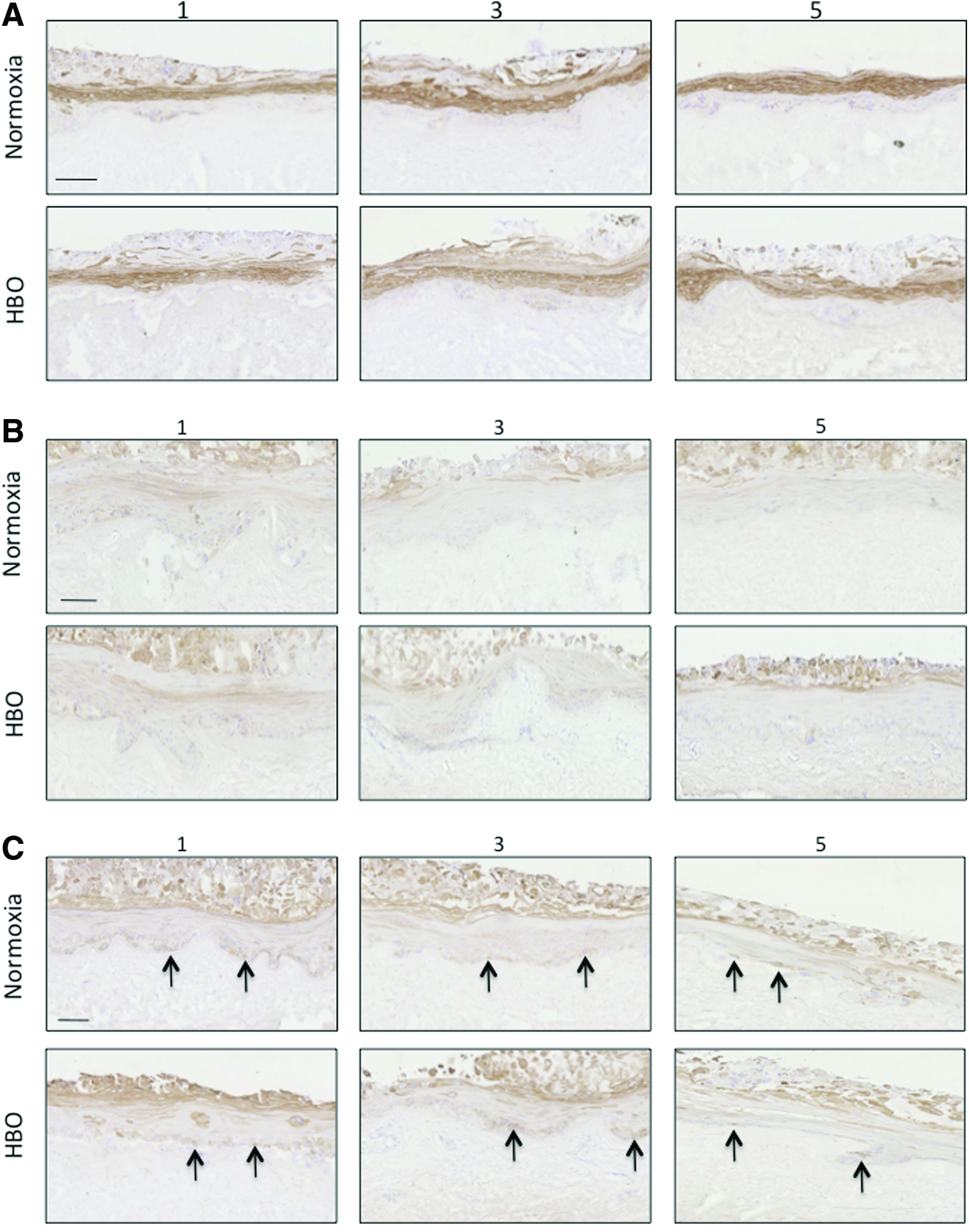
Histological appearance of TEM exposed to normoxia or HBO. Epithelial differentiation was assessed using antibodies against K10
Collagen type IV, expressed along the whole basement membrane in all control groups and after one HBO treatment, proved to be no longer expressed after three treatments. After five treatments, collagen type IV was again observed in irregular intervals in the basement membrane (Fig. 3A). Staining of collagen type III was more intense near the basement membrane in untreated TEM and after five HBO treatments, more intense staining was observed at the lower half of the lamina propria (Fig. 3B). The distribution of fibroblasts and fibrocytes studied using vimentin staining was not altered after HBO treatment. Distinction between fibroblasts and fibrocytes was established morphologically. The fibroblast/fibrocyte ratio was affected by HBO treatment as more fibrocytes relative to fibroblasts were observed after each treatment (data not shown).
Collagen type IV
Proliferating cells were observed in the basal layer of the epithelium only (Fig. 4A). After one, three, and five consecutive HBO treatments, the number of proliferating cells increased when compared with untreated TEM (Fig. 4B), albeit this increase was not statistically significant.

Cell proliferation was observed in the basal layer of the epithelium
Assessment of angiogenic factors after HBO treatment
To determine the effect of HBO on the secretion of angiogenic factors by TEM, a number of angiogenic factors were measured in conditioned media (Fig. 5A–E). A single treatment with HBO resulted in a significant increase of KGF (2.6-fold), HGF (1.4-fold), FGFbasic (2.0-fold), and placental growth factor (PlGF) (3.2-fold) when compared with untreated TEM (Fig. 3). The levels of VEGF in the conditioned media did not change after a single HBO treatment. After three HBO treatments, no changes in the levels of KGF, HGF, and VEGF were observed and levels of FGF basic (1.4-fold) and PlGF (1.8-fold) were decreased when compared with untreated TEM. After five consecutive HBO treatments, a significant increase in the secretion of KGF (4.2-fold), PlGF (1.7-fold), and VEGF (1.2-fold) was observed, whereas HGF secretion was similar to that in untreated TEM. Secretion of FGFbasic significantly decreased 1.7-fold when compared with untreated TEM.
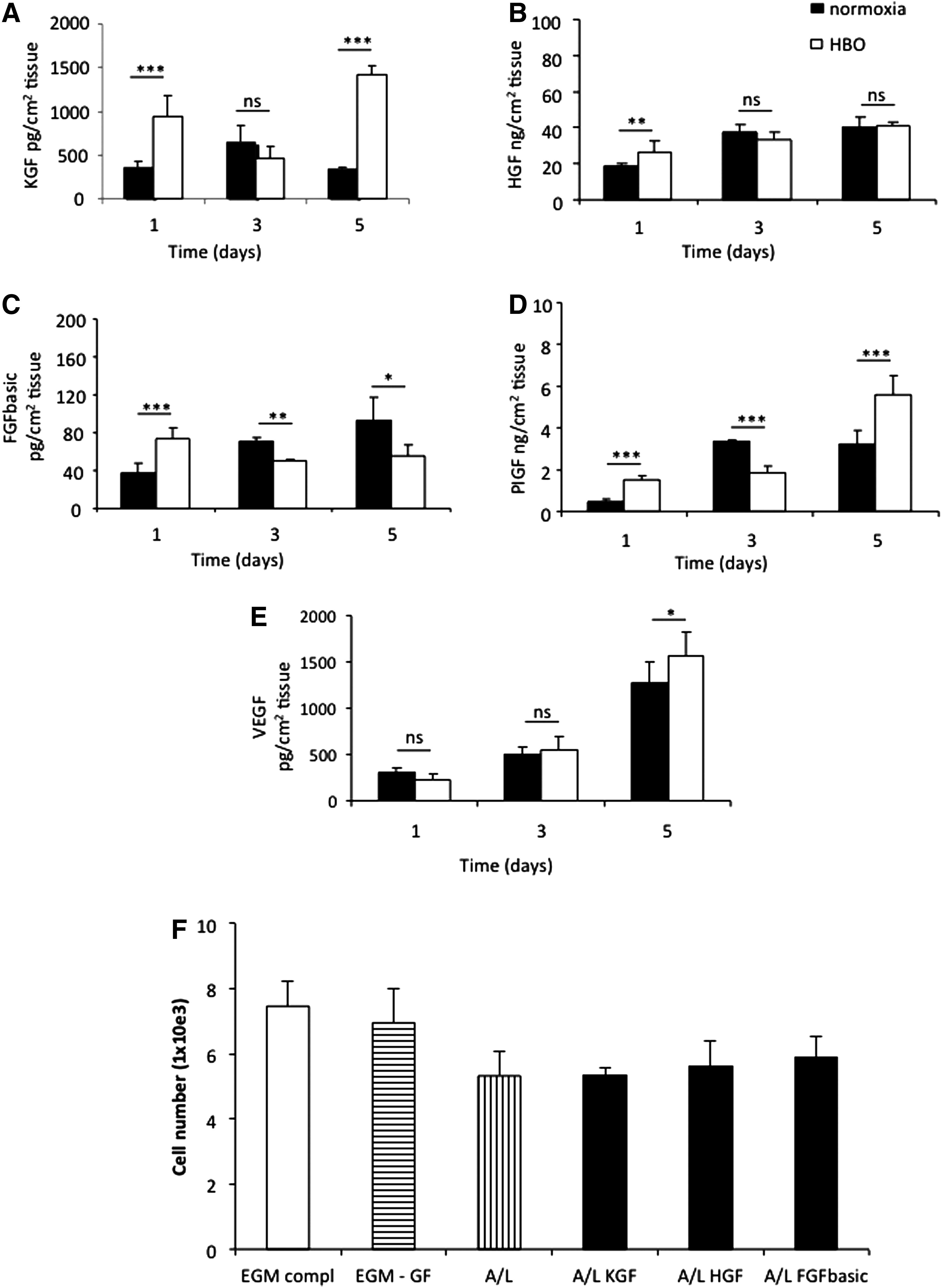
Release of angiogenic factors by TEM constructs. The secretion of KGF
Effect of growth factors secreted by TEM on endothelial cell proliferation
To analyze whether the increased concentration of angiogenic factors found in conditioned media of HBO-treated TEM was capable to stimulate endothelial cell proliferation, HUVECs were cultured with air/liquid media supplemented with KGF, HGF, or FGFbasic. Endothelial cell proliferation appeared to increase after incubation with A/L medium supplemented with FGF and HGF, although no statistical difference was found. A/L medium supplemented with KGF did not stimulate cell proliferation when compared with standard A/L medium (Fig. 5F).
Discussion
In this study we demonstrated that treatment of TEM with HBO stimulates the secretion of multiple angiogenic factors. Overall morphology was maintained and the basal cell layer remained well organized after one HBO treatment. Minor structural changes were observed in TEM after three and five HBO treatments as the first morphological changes became apparent suggesting a potential harmful effect of HBO, for example oxygen toxicity.
Oxygen tension is known to influence cell proliferation.15,16 Previous studies have reported that the balance between oxygen tension and cell proliferation is delicate. 9 In vitro studies with keratinocyte monocultures reported inhibition of keratinocyte proliferation by HBO, whereas in the same study they reported that cells seeded into 3D skin equivalents did increase proliferation due to HBO demonstrated by increased thickness of the epidermis. 17 Even though our results showed a slight stimulating effect of HBO on cell proliferation, the thickness of the viable epithelium decreased. This could indicate accelerated maturation of the epithelium and earlier onset of epithelial stratification. 4 Previous studies by Hollander et al. 6 and Dimitrijevich et al. 17 reported on the observation that HBO treatment resulted in increased keratinocyte differentiation. Our finding that the number of cells expressing K19 in HBO-treated TEM was diminutive to that in the normoxia TEM might be due to the accelerated maturation, which results in a depletion of cell numbers in the basal layer. 17
HBO is known to influence collagen synthesis, 18 although studies report contradicting findings. Studies reported an increase in collagen deposition 4 whereas others report a decrease in collagen deposition. 17 Our results showed the expression of collagen type IV after one HBO treatment and in all normoxia-treated TEM. After three consecutive HBO treatments, collagen type IV was no longer expressed, whereas after five consecutive treatments, collagen type IV was again observed. Previous work in our laboratory showed, prior to cell seeding, donor collagen type IV deposition along the whole basement membrane in the DED scaffold. 1 The observation that collagen type IV deposition was not seen after three treatments but to appear again after five treatments suggests active remodeling of the basement membrane. However, as it is not possible to differentiate between the donor collagen and the collagen deposited by the seeded cells, we cannot rule out the possibility of retained collagen from the donor.
The overall effect of HBO treatment on tissue-engineered skin equivalents has been studied before4,17 and the secretion of angiogenic factors by oral mucosal equivalents has been shown. 19 However, to our knowledge, this is the first study in which the secretion of multiple important angiogenic factors by TEM after HBO treatment is studied. Treatment with HBO is known to increase VEGF secretion in wound fluid 20 and HUVECs. 21 Aside from VEGF, HBO has been shown to induce other angiogenic factors in hind limbs of mice 22 and HUVECs. 23 We therefore studied the secretion of angiogenic factors in TEM and found significantly higher levels of PlGF, KGF, HGF, and FGFbasic in conditioned culture media of TEM after one HBO treatment when compared with conditioned culture media of TEM exposed to normoxia. Endothelial cell proliferation is considered to be an important component of angiogenesis. 24 Our finding that multiple angiogenic factors were increased after one HBO treatment, prompted us to study whether these increased concentrations were capable of inducing endothelial cell proliferation. KGF is known to be an important angiogenic factor for neovascularization. Our finding that KGF did not induce proliferation of HUVECs was also reported by Rubin et al. who showed that KGF lacked mitogenic activity on endothelial cells from large vessels 25 and by Gillis et al. who showed that KGF induced proliferation of cultured capillary endothelial cells but not HUVECs. 26 Additionally, the lack of induced proliferation could be due to the absence of the FGFR2b receptor on HUVECs, being exclusively expressed on epithelial cells, as KGF specifically acts on this receptor. Both basic FGF and HGF are known to affect proliferation and migration of endothelial cells. Our results showed that HGF alone could not significantly increase endothelial cell proliferation. This is in line with the finding of Ding et al. who reported that proliferation of HUVECs is increased by HGF in a dose-dependent manner, albeit that cell density is critical in the responsiveness of HUVEC to HGF. 27 Although HGF and FGFbasic individually did not improve endothelial cell proliferation, it could be that a combination of these factors may increase angiogenesis postimplantation. 28 Additionally, the need for oxygen is critical for survival of the graft and HBO preconditioning might shorten the hypoxic period after implantation by inducing endothelial cell proliferation and neovascular growth.
As continuous oxygen supply is essential for survival after implantation, it is necessary that blood vessels are quickly formed after implantation. HBO treatment has shown to stimulate the secretion of important angiogenic factors in TEM without effecting epithelial morphology. All together, our results suggest that preconditioning of TEM constructs with one HBO treatment prior to implantation might increase the survival rate of TEM grafts therefore making TEM an alternative tool in the reconstruction of large oral defects.
Footnotes
Acknowledgments
The authors would like to thank Dr. A. Seynhaeve for her kind gift of HUVECs and EGM, and the Erasmus MC Tissue Bank, Department of Pathology for facilitating the virtual microscope.
Disclosure Statement
No competing financial interest exists.