
Abstract
The use of growth factors in osteogenic constructs to promote recruitment of bone forming endogenous cells is not clear, while the advantage of circumventing cell seeding techniques before implantation is highly recognized. Therefore, the additive effect of the chemokine stromal cell-derived factor-1α (SDF-1α) on endogenous cell recruitment and vascularization was investigated in a hybrid construct, consisting of a ceramic biomaterial, hydrogel, and SDF-1α, in an ectopic mouse model. We demonstrated in vivo that local presence of low concentrations of SDF-1α resulted in a significant increase in recruited endogenous cells, which remained present for several weeks. SDF-1α stimulated vascularization in these hybrid constructs, as shown by the enhanced formation of erythrocyte-filled vessels. The presence of CD31-positive capillaries/small vessels after 6 weeks in vivo substantiated this finding. The SDF-1α treatment showed increased number of cells that could differentiate to the osteogenic lineage after 6 weeks of implantation, demonstrated by expression of collagen I and osteocalcin. Altogether, we show here the beneficial effects of the local application of a single growth factor in a hybrid construct on angiogenesis and osteogenic differentiation, which might contribute to the development of cell-free bone substitutes.
Introduction
B
Only a few studies21,22 show recruitment of endogenous cell populations to SDF-1α loaded scaffolds. However, the functionality in terms of vessel formation and osteogenic differentiation of recruited endogenous cell populations in bone tissue-engineered constructs in vivo is not clear. Therefore, we investigated the effect of SDF-1α loading on the recruitment of endogenous cells in ectopic hybrid constructs using a single local application. We studied the cells' potential to induce angiogenesis and to differentiate toward the osteogenic lineage in the presence of biphasic calcium phosphate (BCP) particles, which is a strong binder of bone-promoting factors and acts as a starting point for mineralization by osteoblasts in this ectopic animal model.
Materials and Methods
Cell culture
MSCs were isolated from bone marrow of female nude mice (Hsd-cpb:NMRI-nu; Harlan), according to the established protocol. 23 In short, both hind legs of each mouse were dissected and muscle and connective tissue were removed. Bone marrow was harvested by flushing of the tibias and femurs with α-MEM (Gibco, Life Technologies), supplemented with 15% (v/v) fetal calf serum (Cambrex), 100 U/mL penicillin, and 100 μg/mL streptomycin (Invitrogen, Life Technologies). The obtained cell suspension was filtered through a 70-μm filter mesh and cultured in the α-MEM, supplemented with 15% (v/v) fetal calf serum, 100 U/mL penicillin, 100 μg/mL streptomycin, 0.2 mM L-ascorbic acid-2-phosphate (Sigma-Aldrich), and 1 ng/mL FGF-2 (R&D Systems). MSCs were obtained by their adhesion to the tissue culture plastic. The medium was refreshed twice a week and cell cultures were maintained in a humidified incubator at 5% CO2 and 37°C. Passage 2 cells were used for in vivo implantation.
In vitro transwell migration assays
Migration assays were performed using transwell systems with 8 μm pore membranes (Corning Costar). To address the effect of a SDF-1α loaded plug on total cell migration, 200 μL Growth Factor Reduced Matrigel (BD Biosciences) plugs supplemented with 100 ng/mL recombinant murine SDF-1α (R&D Systems) and 20% (w/v) of BCP particles of 1–2 mm diameter (BCP-1150; Xpand) were cut in pieces and placed in the lower chambers of the 24-well plates with addition of a 500 μL expansion medium. Negative control plugs did not contain SDF-1α. About 105 isolated mouse MSCs were seeded onto transwell inserts in 100 μL of expansion medium. Plates were incubated in a humidified incubator at 5% CO2 and 37°C for 48 h. The number of cells that migrated from the top chamber to the bottom chamber as a result of SDF-1α release was counted in four randomly chosen fields. To this end, the upper sides of the membranes were carefully scraped with a cotton swab to remove adherent cells. Detached membranes were stained with hematoxylin and migrated cells were counted. Experiments were repeated twice in triplicate.
Preparation of in vivo implants
To evaluate the effect of SDF-1α on endogenous cell recruitment and vessel formation, constructs consisting of 200 μL Matrigel (BD Biosciences) plugs supplemented with 200 ng/mL recombinant murine SDF-1α (R&D Systems), were prepared for subcutaneous implantation in nude mice (n=9). As controls, empty Matrigel plugs or Matrigel plugs seeded with isolated MSCs (106 cells/mL) were used. All constructs contained 20% (w/v) of BCP particles of 1–2 mm diameter. Briefly, Matrigel was used according to the manufacturer's instructions and supplemented with each of the components for each group, transferred to 1-mL syringes, and solidified by incubation overnight in a humidified incubator at 5% CO2 and 37°C on a roller. Solidified plugs were implanted the next day.
Animals and implantation
Nine female nude mice (Hsd-cpb:NMRI-nu; Harlan) were anesthetized with 1.5% isoflurane, after which the implants were placed in two separate subcutaneous pockets in the shoulder area and one on the back. Constructs were implanted according to a randomized design. Each mouse received all constructs. The animals were postoperatively treated with the analgesic buprenorphine (0.05 mg/kg, sc; Temgesic; Schering-Plough/Merck) and housed together at the Central Laboratory Animal Institute, Utrecht University. Experiments were conducted with the permission of the local Ethics Committee for Animal Experimentation and in compliance with the Institutional Guidelines on the use of laboratory animals.
Explantation and embedding
At 1 week (n=3) and 6 weeks (n=6) after implantation the constructs were retrieved to analyze the number of recruited cells, vessel formation, osteogenic differentiation, and collagen I deposition. Samples were fixed overnight in 4% (v/v) formalin. All samples were decalcified in the Luthra's solution (3.26 M HCl and 2.65 M formic acid in distilled water) for 24 h and processed for 5-μm-thick paraffin sections through alcohol dehydration series.
Quantification of recruited cells and vascularization
To analyze the number of recruited cells into the constructs, hematoxylin/eosin (HE) staining was performed on rehydrated sections. The numbers of cells present in the SDF-1α laden constructs and empty constructs were counted in three randomly chosen fields throughout the samples; one in the lower part, one in the middle, and one in the upper part. To analyze the formation of blood vessels after 6 weeks of implantation, all three separate constructs from all animals were scored by morphology on HE stained sections. In the sections, part of the blood perfused vessels lost the erythrocytes during the embedding procedure. Therefore, all vessel structures observed in HE stained sections, blood perfused or not, were scored. No discrimination was made based on the vessel sizes.
Immunohistochemistry of CD31/PECAM-1
To show the endothelial phenotype of part of the recruited cells and blood vessel networks in the constructs, sections were stained for the endothelial marker CD31 (PECAM-1). For this, antigen retrieval on rehydrated paraffin sections was performed by incubation in a 0.1 M sodium citrate solution (pH=6.0) for 20 min at 95°C. The sections were subsequently blocked in 3% (v/v) H2O2 in phosphate-buffered saline (PBS) for 15 min and 5% (w/v) bovine serum albumin (BSA) for 1 h at room temperature. The anti-CD31 primary antibody (rabbit-anti-CD31; LifeSpan Biosciences) was incubated at 2 μg/mL overnight at 4°C, followed by the goat anti-rabbit biotinylated secondary antibody (DakoCytomation; 0.6 μg/mL) and streptavidine peroxidase (DakoCytomation; 1.4 μg/mL), each for 30 min at room temperature. The staining was developed with diaminobenzidine (DAB) and Mayer's hematoxylin was used for counterstaining. Negative controls were treated similarly, except for exclusion of the primary antibody.
Immunohistochemistry of osteocalcin and collagen I
For the evaluation of osteocalcin expression, rehydrated sections were blocked in 3% (v/v) H2O2 in PBS for 10 min and 5% (w/v) BSA for 30 min. The primary antibody against osteocalcin (rabbit anti-osteocalcin; Alexis ALX-210-333; now Thermo Fisher Scientific) was used at 2 μg/mL in 5% (w/v) BSA in PBS. As a secondary antibody, a goat anti-rabbit HRP (DakoCytomation) was used at 1.7 μg/mL in 5% (w/v) BSA in PBS.
For collagen staining, rehydrated sections were subjected to antigen retrieval steps using 1 mg/mL pronase and 10 mg/mL hyaluronidase type II for 30 min each at 37°C after which the sections were blocked with 3% (v/v) H2O2 in PBS for 10 min and 5% (w/v) BSA for 30 min. Subsequently, sections were incubated with the rabbit anti-collagen I primary antibody (Abcam, Cambridge, United Kingdom; Ab34710) at 3.3 μg/mL in 5% (w/v) BSA in PBS and a goat anti-rabbit HRP (DakoCytomation) secondary antibody at 1.7 μg/mL in 5% (w/v) BSA in PBS. All stainings were developed with DAB and Mayer's hematoxylin was used for counterstaining. Negative controls were treated similarly, except for exclusion of the primary antibody.
Statistical analysis
Statistical analysis was performed with SPSS 20.0 software. A Kruskal–Wallis test with a Mann–Whitney U post hoc test was used to compare the number of migrated cells between empty plugs and SDF-1α laden plugs in vitro. A Friedman test with a Wilcoxon signed-rank post hoc test was used to compare cell numbers at both time points after implantation, the number of vessels, and the absolute number of osteocalcin-positive cells. p-values less than 0.05 were considered statistically significant.
Results
Cell recruitment
By using SDF-1α laden plugs, we assessed the biological function of this chemokine to recruit cells of mouse origin, in vitro as well as in vivo. SDF-1α laden plugs resulted in a significant 2.5-fold increase in migration of isolated mouse MSCs in vitro, when compared to control plugs without SDF-1α (Supplementary Fig. S1; Supplementary Data are available online at www.liebertpub.com/tea). Implantation of SDF-1α containing constructs subcutaneously in mice revealed prominent endogenous cell recruitment after 1 week of implantation (Fig. 1b), while the HE staining showed that implantation of empty plugs resulted in hardly any recruitment of endogenous cells after 1 week (Fig. 1a). After 6 weeks of implantation, the presence of some cells in empty plugs was observed (Fig. 1c), whereas SDF-1α laden plugs showed abundant presence of cells inside the constructs (Fig. 1d). Quantitatively, SDF-1α laden plugs showed significantly higher endogenous cell numbers compared to empty plugs, after both 1 and 6 weeks of implantation (Fig. 1e).

Recruitment of cells in the scaffolds after 1 and 6 weeks of implantation. Cells present in empty control constructs
Since SDF-1α has been used extensively to recruit endothelial cells and induce angiogenesis, we characterized explanted constructs for expression of the endothelial marker CD31/PECAM-1 by immunohistochemistry. No CD31-positive cells or structures were found inside any of the constructs of the week 1 samples (Fig. 2b–d), whereas host vessels stained positive in the tissue surrounding the constructs. In addition, immunohistochemistry on the sections for markers of different cell types, including SSEA-4, CD105, CD45, and CD34, appeared all negative (data not shown).

CD31/PECAM-1 immunohistochemistry in the constructs after 1 and 6 weeks of implantation. No CD31/PECAM-1 expression was found in week 1 samples of a representative negative control staining for all constructs
Formation of blood vessel networks
The formation of vessels in all implanted constructs after 6 weeks was shown in HE stained sections (Fig. 3a–c). Some of the structures were perfused with erythrocytes, indicating their connection to the host circulation (Fig. 3a–c). Based on scoring, the number of vessels in the constructs after 6 weeks was significantly different between SDF-1α laden plugs and both control groups at this time point (Fig. 3g). In addition, immunohistochemical detection of abundant CD31 expression confirmed the endothelial identity of formed tubular structures throughout the constructs containing isolated mouse MSCs or SDF-1α (Fig. 3e–f), but not in empty constructs (Fig. 3d).
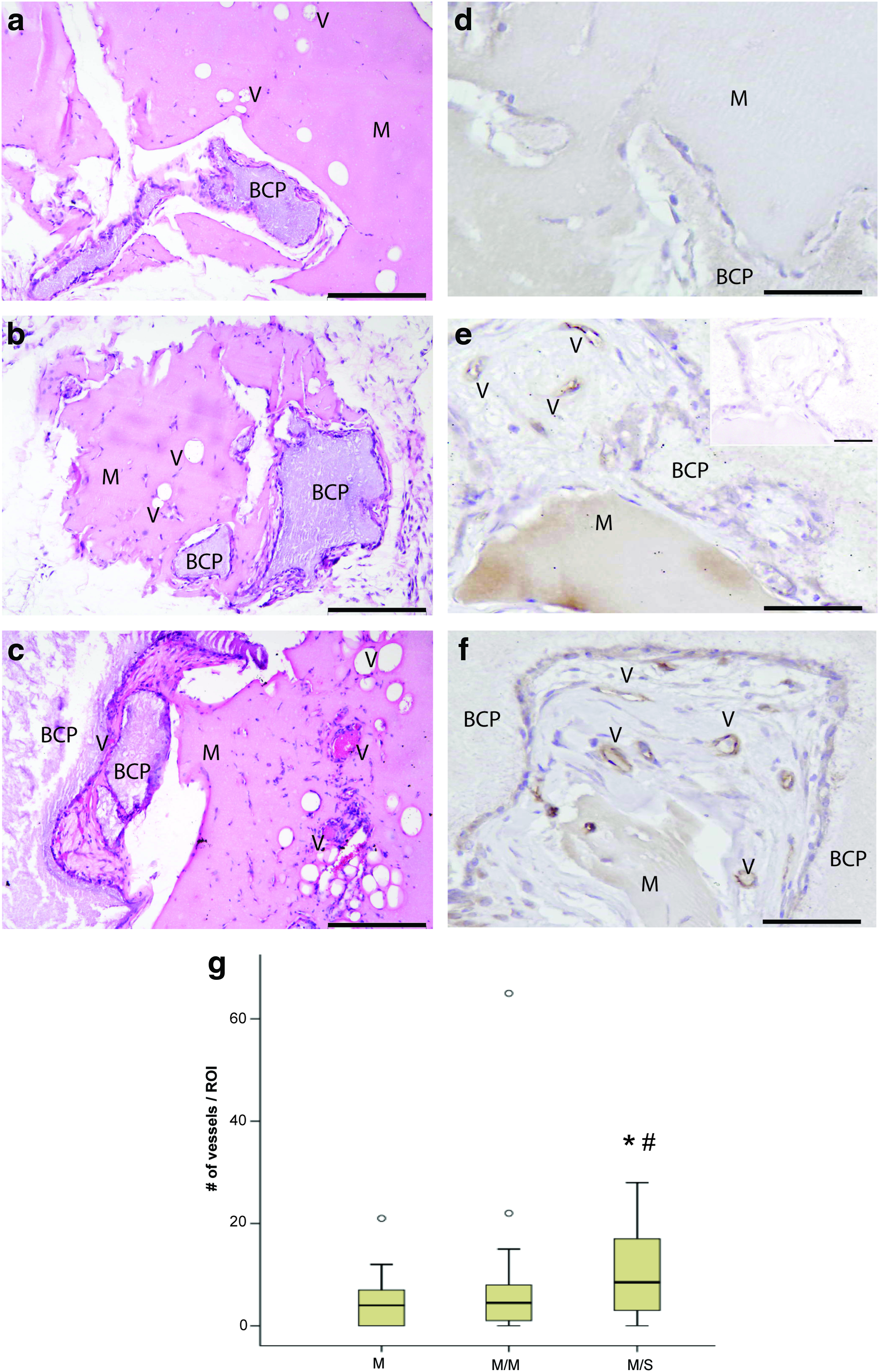
Evaluation of vessel formation in the implanted constructs after 6 weeks of implantation. HE stained sections showed vessels (V) in empty constructs
Osteogenic differentiation of recruited cells
To evaluate the osteogenic differentiation potential of the cells present in the constructs, we performed immunohistochemical analysis on paraffin-embedded sections. Analysis of osteocalcin expression revealed osteogenic differentiation of the cells in all constructs after 6 weeks of implantation, with most positive cells lining the BCP granules (Fig. 4a–c). Forty-three percent of the recruited cells in the empty plugs showed positive osteocalcin staining, whereas 48% of the recruited cells in the SDF-1α laden plugs were positive. This resulted in a significantly higher absolute number of osteocalcin-positive cells in the SDF-1α laden plugs compared to both controls (Fig. 4d). Plugs seeded with isolated mouse MSCs did not contain a higher number of osteocalcin-positive cells compared to the empty control situation (Fig. 4d). In addition, all constructs showed abundant collagen I deposition after 6 weeks in vivo (Fig. 5a–c). The collagen-rich extracellular matrix was found in high amounts in SDF-1α laden plugs (Fig. 5b), but did not show higher abundance than control constructs (Fig. 5a, c). No bone was found in the constructs containing isolated mouse MSCs or any of the other constructs after 6 weeks of implantation. However, the presence of osteocalcin and the deposition of collagen I together are indicative of osteogenic differentiation.

Evaluation of the osteogenic differentiation potential of cells present after 6 weeks of implantation. Osteogenic differentiation of cells present inside empty control constructs
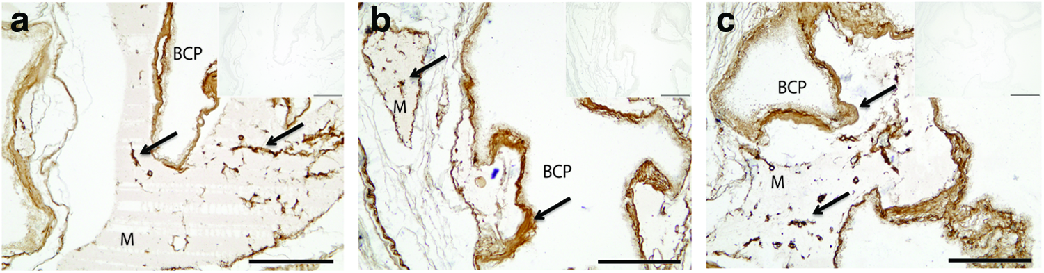
Deposition of collagen I after 6 weeks of implantation. The deposition of collagen I was observed by positive collagen I staining in brown in all three constructs (indicated by the arrows). No initial differences were observed between empty control groups
Discussion
Our results show the effectiveness of a chemokine loaded into a hybrid construct to recruit endogenous cells that have osteogenic differentiation potential. With applied SDF-1α concentrations of only 40 ng per construct, recruitment of endogenous cells already started during the first week of implantation. Significantly, higher numbers of endogenous cells were present at both one and 6 weeks in implants containing the chemokine SDF-1α when compared to empty constructs. This difference in recruited endogenous cells was attributed to both short- and long-term effects of the incorporation of SDF-1α in a hybrid construct, which may be due to protection from breakdown of the chemokine by the hydrogel. Although others show induced recruitment of endogenous cells 1 week after implantation of SDF-1α loaded constructs,21,22 we are the first to report that this effect persisted over time and that vascularization and osteogenic differentiation in vivo could be enhanced using these hybrid constructs. An earlier study from our group showed that prolonged presence of proliferating MSCs in ectopic bone-forming constructs is needed, ultimately, to form bone at these sites. 24 In addition, a continuous presence of low concentrations of SDF-1α may be favorable for bone formation in vivo, 25 although the effect of SDF-1α on cell proliferation has not been established.
Although it is known that SDF-1α is able to recruit endothelial cells as well as MSCs at early time points in vitro14,21 and in vivo,21,26 cells expressing the endothelial marker CD31/PECAM-1 could not be detected at 1 week. However, after 6 weeks, positive CD31 staining in constructs loaded with SDF-1α was found, which implicates recruitment of endothelial cells that are able to form capillary networks, delayed angiogenesis, 27 or possibly mesenchymal to endothelial transition (MET). The perfusion with erythrocytes confirms their connection to the host circulation. The presence of vessels at this later time point may indicate a delay in the differentiation of MSCs toward the endothelial lineage. 28 In addition, angiogenesis was expected to be relatively slow as a result of the degradation rate of Matrigel, which was still present after 6 weeks of implantation. However, we did find significantly induced vessel formation after 6 weeks of implantation in SDF-1α loaded constructs compared to both control constructs based on morphology in HE stained sections and therefore attribute the induced network formation in these constructs to this chemokine. These results are in agreement with previous reports on the stimulation of vessel formation in vivo by SDF-1α.18–21,29
Immunohistochemical staining for the MSC marker SSEA4 on endogenously recruited cells 21 in paraffin-embedded sections could not be detected. Nevertheless, the newly invaded cells showed expression of the osteoblast marker osteocalcin in the empty and SDF-1α laden constructs, strongly suggesting recruitment of osteoblast progenitors in these plugs.
After 6 weeks of implantation, the recruited cells showed osteogenic differentiation, as seen by osteocalcin expression and collagen I deposition. No bone was found in SDF-1α loaded constructs at this time point. It is known that the single addition of SDF-1α is not osteogenic. 26 In addition, we used more than 100-fold lower concentrations of the chemokine, compared to other reports. The SDF-1α concentrations used in this study were based on earlier reported recruitment studies.14,21 As a result, the number of recruited endogenous cells was considerable and accompanied by robust extracellular matrix deposition. We expect this to be the result of long-lasting release of the chemokine from the hydrogel, but an indirect effect by the recruited cells is not excluded. Together with the osteocalcin expression, these results suggest that osteogenic differentiation was ongoing in the constructs and that bone formation could be a matter of time. The absolute number of osteogenic cells present in SDF-1α loaded constructs was increased compared to empty constructs. Possibly, these results could be ameliorated by addition of a strong bone-forming stimulus, such as BMPs. Reports by others12,26,30 show that the additive effect of SDF-1α on BMP-2 induced bone formation. Surprisingly, the positive control constructs that contained seeded syngeneic mouse MSCs also did not show any bone formation after 6 weeks, which indicates that an animal-based effect has possibly prevented bone formation. We carefully determined the cell density of MSCs needed to induce bone formation in ectopic mouse models, 31 although these were from a different origin. Several reports show variable bone formation in vivo, depending on the origin of the MSCs.24,32,33 In addition, interindividual variation has been reported10,34 as well as different amounts of cells needed to stimulate bone formation.31,33,35
Conclusions
In summary, we demonstrated for the first time that the local administration of low concentrations of SDF-1α in the context of a hybrid hydrogel-based construct is effective in recruiting endogenous cells and that recruited cells show osteogenic differentiation potential as evidenced by extracellular matrix formation and osteocalcin expression. In addition, local application of SDF-1α also induced vessel formation. These results suggest that bone formation using cell-free applications could be made possible in the future.
Footnotes
Acknowledgments
We thank H. Yuan from Xpand for kindly providing BCP particles. This research forms part of the Project P2.04 BONE-IP of the research program of the BioMedical Materials Institute, cofunded by the Dutch Ministry of Economic Affairs, Agriculture and Innovation. We acknowledge financial support by the Anna Foundation for Scientific Research to R.M.E. (Anna Fonds; grant number: O.2011/23).
Disclosure Statement
No competing financial interests exist.
References
Supplementary Material
Please find the following supplemental material available below.
For Open Access articles published under a Creative Commons License, all supplemental material carries the same license as the article it is associated with.
For non-Open Access articles published, all supplemental material carries a non-exclusive license, and permission requests for re-use of supplemental material or any part of supplemental material shall be sent directly to the copyright owner as specified in the copyright notice associated with the article.