
Abstract
We have previously shown that nestin-expressing hair follicle stem cells from the mouse and human are multipotent and can differentiate into many cell types, including neurons and glial cells. The nestin-expressing hair follicle stem cells can effect nerve and spinal cord repair upon transplantation in mouse models. In the present study, nestin-expressing hair follicle stem cells expressing red fluorescent protein (RFP) were induced by retinoic acid and fetal bovine serum to differentiate and then transplanted together with Matrigel into the transected distal sciatic or tibial nerve stump of transgenic nude mice ubiquitously expressing green fluorescent protein (GFP). Control mice were transplanted with Matrigel only. The transplanted cells appeared neuron like, with large round nuclei and long extensions. Immunofluorescence staining showed that some of the transplanted cells in the distal nerve stump expressed the neuron marker Tuj1 as well as motor neuron markers Isl 1/2 and EN1. These transplanted cells contacted each other as well as host nerve fibers. Two weeks post-transplantation, nerve fibers in the distal sciatic nerve stump of the transplanted mice had greater expression of motor neuron markers and neurotrophic factor-3 than those in the Matrigel-only transplanted mice. Muscle fiber areas in the nestin-expressing stem cell plus Matrigel-transplanted animals were much bigger than that in the Matrigel-only transplanted animals after 4 weeks. The present results suggest that transplanted nestin-expressing hair follicle stem cells can differentiate into motor neurons and reduce muscle atrophy after sciatic nerve transection. This study demonstrates a new and accessible neuron source to reduce muscle atrophy after nerve injury.
Introduction
D
In our previous studies, we have shown that nestin-expressing hair follicle stem cells from mice and humans are multipotent and can differentiate into many cell types, including neurons and glial cells. The nestin-expressing hair follicle stem cells can effect nerve and spinal cord repair in mouse models.10–24
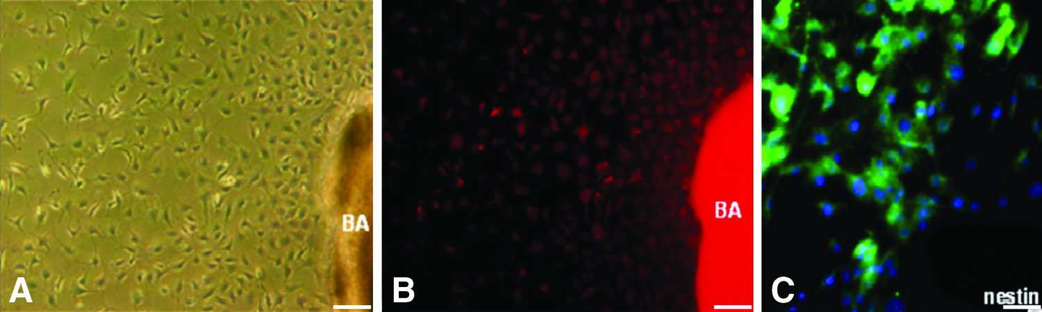
Nestin has been shown to be expressed in the hair follicle, both in the bulge area (BA) as well as the dermal papilla (DP). Nestin-expressing stem cells of both the BA and DP have previously been shown to be multipotent. Transgenic mice, in which the nestin promoter drives green fluorescent protein (ND-GFP), were used to observe nestin expression in the BA and DP. The BA had nestin expression throughout the hair cycle and to a greater extent than the DP. The cells from both regions had very long processes extending from them as shown by confocal microscopy. Both nestin-expressing DP and BA cells differentiated into neuronal and glial cells after transplantation to the injured spinal cord and enhanced injury repair and locomotor recovery. Nestin-expressing stem cells from both the BA and DP have potential for spinal cord regeneration, with the BA being the greater and more constant source. 25
It was observed in Gelfoam® histoculture over a 2-week period that the stem cells trafficked from the BA toward the DP area and extensively grew out onto Gelfoam® forming nerve-like structures.26,27
We subsequently observed that multipotent, nestin-expressing stem cells are present throughout the hair follicle and that the upper part of the follicle can produce the stem cells in large amounts that could be used for nerve and spinal cord repair. 28
Since hair follicles can be readily obtained and their nestin-expressing cells can differentiate into neuronal cells, the nestin-expressing hair follicle stem cells were evaluated in the present study for their ability to reduce nerve injury and muscle atrophy.
Materials and Methods
Mice
Nude mice (AntiCancer, Inc., San Diego, CA) were kept in a barrier facility under HEPA filtration and fed with an autoclaved laboratory rodent diet (Tecklad LM-485; Western Research Products). All animal studies were conducted in accordance with the principals and procedures outlined in the National Institutes of Health Guide for the Care and Use of Laboratory Animals under Assurance Number A3873-1. All animal procedures were performed under anesthesia using s.c. administration of a ketamine mixture (10 μL ketamine HCL, 7.6 μL xylazine, 2.4 μL acepromazine maleate, and 10 μL phosphate-buffered saline [PBS]).
Isolation of vibrissa follicles
The whisker pad of transgenic mice ubiquitously expressing the red fluorescent protein (RFP)29,30 was cut free and its inner surface was exposed. The vibrissal follicles were dissected under a binocular microscope. A small scissors was used to separate each follicle. Anagen follicles were chosen for culture.
Culture of hair follicle BA cells
The isolated vibrissal follicles were washed in PBS three times. A fine needle was used to cut off the follicle capsule. The BA was separated with a fine needle. All surgical procedures were performed under a sterile environment. A 12-well plate coated with collagen I was preincubated with DMEM-F12, containing B-27, N2, and 1% penicillin/streptomycin (all from Gibco/BRL). The separated BA attached to the bottom of the plate after 40 min. Five to eight BAs were placed in each well. A serum-free culture medium with 20 ng/mL basic fibroblast growth factor and 20 ng/mL EGF was then added to the wells. Explant floating was carefully avoided. Half of the medium was changed every other day.
After 4 to 6 days, the attached BAs were removed from the plate bottom. The cells that grew out from the BA were incubated with 0.25% trypsin for 10 min at 37°C. Fetal bovine serum (FBS) was added to inhibit trypsin. Cells were washed with the serum-free culture medium collected by centrifugation and were seeded in collagen I-coated T-25 flasks.
After two to three passages, RFP BA cells were harvested and mixed with Matrigel™ (BD Biosciences) on ice. BA cells (1×10⁷/mL) in Matrigel were seeded on a 24-well plate and incubated at 37°C. The serum-free medium was added 30 min later. Half of the medium was changed every other day.
Sciatic nerve transection and cell transplantation
Ten male GFP transgenetic nude mice31,32 were used for sciatic nerve transection. The animals were anesthetized with a ketamine mixture (see above). The right sciatic nerve was exposed, and ligatures were placed on the tibial nerve 5 mm proximal to its entry into the medial gastrocnemius. The sciatic nerve, including the tibial nerve, was severed proximal to the ligatures. The proximal nerve stump was sutured into the hip muscle to prevent reinnervation from peripheral axons in the sciatic nerve. Mice were divided into two groups. In the experimental group, RFP BA cells (5–10 μL of 1×10⁷ cells/mL), which were previously induced to differentiate by 1 μM retinoic acid (RA)+10% FBS for 1 week, were mixed with Matrigel and injected into the distal tibial nerve stump. In the other group, mice were injected only with 5–10 μL Matrigel.
Immunofluorescence staining
Cultured BA cells or frozen sections of the nerve stump were fixed in precooled 4% paraformaldehyde at room temperature for 10 min and washed with PBS three times. The main procedures for immunofluorescence staining were as follows: (1) Incubation with 0.3% Triton X-100 at room temperature for 30 min; (2) Incubation with 5% normal goat serum at room temperature for 30 min; (3) Incubation with primary antibodies at 4°C for 48 h. The primary antibodies used were nestin (rabbit, 1:100); TuJ1 (rabbit, 1:30); Isl 1/2 (rabbit, 1:100); EN-1 (rabbit, 1:100); and neurotrophic factor-3 (NT-3) (rabbit, 1:100); (4) The secondary antibody used was goat anti-rabbit IgG FITC, 1:100, incubation at room temperature in the dark for 2.5 h; (5) DAPI, 1:1000, diluted in PBS at room temperature for 5 min; (6) Slides were mounted with Fluoromount (Sigma; F4680) and observed under fluorescence microscopy.
Muscle fiber area analyses
Frozen sections of the gastrocnemius muscles in the treated and untreated mice were made and processed and stained with hematoxylin and eosin. Ten microscopic fields containing muscle fibers were selected randomly, and the area of each muscle fiber was measured. The average muscle fiber area was compared in the treated and untreated groups at different time points. The data were analyzed with the unpaired t-test. The significance level for all tests was p<0.05.
Results and Discussion
BA nestin-expressing stem cells differentiated into neuron-like cells in vitro
Many nestin-expressing stem cells arose from the hair follicle BA after 7 days of primary culture (Fig. 1). After induction with RA and fetal bovine serum (FBS), some of the cells differentiated into neuron-like cells in the Matrigel culture (Fig. 2). The percentage of differentiated neuron-like cells was approximately 60%. The cells had a large cell body with big, round nuclei in the center. Long extensions arose from the cell body that contacted the adjacent cells.
After 7 days of culture

RFP BA cells cultured in Matrigel for 7 days with retinoic acid (RA) and fetal bovine serum. Cells grew very well in the Matrigel, and some of them extended processes and appeared neuron like. The arrow and arrowhead show a neuron-like cell with a big, round nucleus in the center. Scale bar: 20 μm. Color images available online at www.liebertpub.com/tea
BA nestin-expressing stem cells differentiated into neurons in the nerve stump
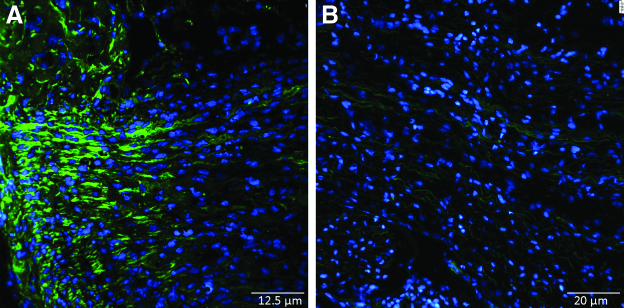
Two weeks after transplantation of the nestin-expressing RA− and FBS− pre-induced BA stem cells together with Matrigel in the transected sciatic nerve, the nestin-expressing transplanted cells grew very well in the distal sciatic or tibial nerve stump (Figs. 3 and 4). Some transplanted cells had large cell bodies and big nuclei with long extensions, which contacted each other or the host nerve fibers (Figs. 3 and 4). Two weeks after transplantation, some cells in the nerve stump were Tuj1-positive, Isl 1/2-positive, or EN1-positive (Fig. 5). Since these markers were for neurons or motor neurons and no neuron bodies exist in the peripheral nerve, the neuron marker-expressing stem cells must have been derived from the transplanted BA stem cells from the RFP mice. In the Matrigel-only transplanted group, TuJ1, Isl 1/2, and EN1 staining in the distal nerve stump was negative (Fig. 5).

Two weeks after transplantation of BA cells, mixed with Matrigel, into the sciatic nerve stump of green fluorescent protein (GFP) mice

Frozen section of the GFP nerve stump 2 weeks after RFP BA cell transplantation. Many RFP BA cells (see arrows) were found in the GFP nerve stump. Fig. 4 was obtained with the Olympus IV100 Scanning Laser Small Animal Imaging System. Scale bar: 20 μm. Color images available online at www.liebertpub.com/tea

Immunofluorescence staining of frozen sections of the sciatic nerve stump transplanted with RA− and FBS− pre-induced BA stem cells plus Matrigel or Matrigel only for 2 weeks.
In the pre-induced BA nestin-expressing stem cell plus Matrigel transplantation group, strong NT-3, immunofluorescence staining was detected in the nerve stump 2 weeks after transplantation (Fig. 6), while in the Matrigel-only transplanted group, NT-3 staining was very weak and considered negative.
Neurotrophic factor-3 (NT-3) immunofluorescence staining of frozen sections of the sciatic nerve stump transplanted with RFP BA nestin-expressing stem cells plus Matrigel or Matrigel only for 2 weeks. In the BA nestin-expressing stem cells plus Matrigel group, many NT-3-positive cells were observed as shown in panel
BA nestin-expressing stem cells reduce muscle atrophy after transplantation to the injured nerve
To evaluate whether the appearance of motor neuron-like cells in the nerve injury areas of the experimental animals had detectable therapeutic effects, we measured the muscle fiber areas in ten randomly selected microscopic fields of frozen sections of the gastrocnemius muscles in the treated and untreated mice. The muscle fiber area in the animals transplanted with RA− and FBS− pre-induced BA nestin-expressing stem cells plus Matrigel was much larger than that in Matrigel-only transplanted animals at different time points (p<0.01) (Fig. 7 and 8). In mice treated with pre-induced BA nestin-expressing stem cells, the progression and severity of muscle atrophy measured by the muscle fiber area, compared to the level in the control group, was delayed by 8 weeks (Fig. 7 and Fig. 8).

Hematoxylin and eosin (H&E) staining of the gastrocnemius muscle. Panels

Analysis of the average muscle fiber area in mice transplanted with pre-induced BA stem cells in Matrigel and animals transplanted with Matrigel only. After 2 weeks, the muscle fiber area in the animals transplanted with pre-induced BA stem cells plus Matrigel was significantly larger than that in animals transplanted with Matrigel only (**p<0.01).
The present study shows that RA− and FBS− pre-induced nestin-expressing hair follicle BA cells transplanted into the sciatic or tibial nerve stump, together with Matrigel, differentiate into motor neurons with long extensions contacting the surrounding structures. The transplanted pre-induced nestin-expressing BA stem cells reduced severe muscle atrophy, which occurred after total denervation.
The hair follicle nestin-expressing stem cells can easily be harvested from patients18,20 and are a promising cell source for clinical use. Our previous studies showed that the hair follicle BA nestin-expressing stem cells are multipotent and can differentiate into many cell types, including neurons and other cell types and effect nerve and spinal cord repair in mouse models.12,13,16,25,33
In the present study, hair follicle BA nestin-expressing stem cells were previously primed in culture by RA and FBS, mixed with Matrigel, and transplanted into the nerve stump. Compared to the Matrigel-only transplanted mice, in the mice transplanted with cultured BA nestin-expressing stem cells, primed by RA and FBS, the nerve fibers in the nerve stump were well maintained and some of the transplanted cells differentiated into motor neurons, which expressed TuJ1, Isl 1/2, or EN1. In addition, the muscle fiber area in the animals transplanted with BA nestin-expressing stem cells plus Matrigel was much larger. Thus, muscle atrophy due to nerve injury was greatly reduced by transplanting hair follicle nestin-expressing cells induced to differentiate by RA and FBS. These results demonstrated that the transplanted hair-follicle nestin-expressing cells enhanced muscle reinnervation.
Amoh et al. 13 found that the vast majority of nestin-expressing BA stem cells, which were not pre-differentiated by RA treatment, differentiated to Schwann cells after transplantation to the severed sciatic nerve. In the present study with RA pretreatment, we have found the majority of stem cells transplanted to the sciatic nerve differentiated to neurons, most of which were motor neurons. Thus, comparisons of the results of Amoh et al. and the present study indicate that pretreatment with RA drives the differentiation of the stem cells to neurons, including motor neurons, after transplantation to the sciatic nerve. These results suggest that the neurons are functional since muscle atrophy is delayed. Future experiments will include functional testing of the neurons and experiments on the mechanism of RA differentiation.
The results of the present report suggest the promising clinical potential of the use of hair follicle nestin-expressing cells to inhibit muscle atrophy after nerve injury.
Footnotes
Disclosure Statement
No competing financial interests exist.
Dedication
This paper is dedicated to the memory of A.R. Moossa, MD.