
Abstract
Electrospinning technology is an attractive process for the fabrication of a scaffold with an annulus fibrosus (AF)-like architecture for tissue engineering. Oriented and nonoriented electrospun scaffolds were prepared from poly(ester-urethane) (PU) and poly(ɛ-caprolactone) (PCL) as well as corresponding homogeneous films. Scaffolds' characteristics and mechanical properties were characterized by scanning electron microscopy, static water contact measurements, and dynamic mechanical analysis, respectively. The effect of scaffold architecture and polymer composition on bovine AF cells was investigated. PU and PCL films and scaffolds supported AF cell growth and extracellular matrix production and accumulation. Electrospun scaffolds increased the retention of collagen and glycosaminoglycan compared with films. Fiber orientation of the scaffolds promoted the AF cell phenotype with a trend toward an upregulation of matrix gene expression for oriented relative to nonoriented scaffolds. The higher yield strain of an oriented electrospun PU scaffold, compared with other scaffolds, will be advantageous for AF tissue engineering under a dynamic mechanical environment.
Introduction
T
The AF is composed of 15–25 layers, each consisting of densely aligned collagen fiber bundles with alternating angles around±30° with respect to the transverse axis of the spine.6,7 Collagen fibers (mainly type I) are embedded in a proteoglycan-rich matrix, including large aggregating (aggrecan and versican) and small interstitial (biglycan, decorin, fibromodulin, and lumican) proteoglycans. The characteristic AF structure and composition are the basis for its complex mechanical behavior, which is nonlinear, anisotropic, and viscoelastic. Modulus values for single lamella have been reported to range from 59 MPa (inner AF) up to 136 MPa (outer AF). 8
Thus, an important step toward an adapted biological IVD repair is the development of a material for AF supplementation. Different natural and synthetic scaffolds, such as atelocollagen, collagen-glycosaminoglycan (GAG), collagen-hyaluronan, and silk matrices, have been proposed for AF tissue engineering.9–12 The ideal scaffold should support cell ingrowth and should be strong enough to withstand the complex and multidirectional loading, similar to the native tissue.
Fibrous scaffolds produced by electrospinning are promising materials, since their structure can mimic the microarchitecture of the AF. Electrospun scaffolds are gaining increasing interest due to their tunable properties to represent an ideal microenvironment for a specific cell type. Indeed, fiber diameter, fiber orientation, patch porosity, and surface properties can be optimized for specific cell seeding needs.13–17 Furthermore, a wide range of polymers can be processed, and encapsulation or immobilization of different bioactive agents is possible. Recently, a number of groups have studied AF cell behavior on electrospun scaffolds using several polymers such as poly(ester-urethane) (PU),18,19 polycaprolactone (PCL),20–23 and poly(L-lactic acid). 24
Nerurkar et al. have assessed electrospun PCL scaffolds with oriented fibers and demonstrated their ability to mimic native AF tissue regarding anisotropy and nonlinearity.20,21,25 Recently, a study reported that the application of elastomeric PU may be beneficial due to the favorable mechanical properties of the electrospun scaffolds. 26
Whereas these studies have demonstrated the potential to support new AF tissue formation, they often have not comprehensively assessed biomechanical, structural, and biological parameters. Moreover, there is little information about the influence of chemical surface properties on the AF cell response, since two different polymer types have not been directly compared.
This study aimed to investigate structural and mechanical properties of PCL and PU electrospun scaffolds for AF tissue engineering and to assess their influence on AF cell behavior with regard to cell attachment, cell proliferation, cell phenotype, and extracellular matrix (ECM) production.
Materials and Methods
Scaffold and spin-coated surface production
The two polymers, PU and PCL, were processed into nonoriented and oriented electrospun scaffolds and into spin-coated films. PCL with number molecular weight (Mn)=80,000 g/mol was obtained from Sigma-Aldrich (Buchs SG, Switzerland) and used without further purification. PU was prepared as described previously by a one-step polycondenzation reaction of poly(ɛ-caprolactone diol), 1,4,3,6-dianhydro-D-sorbitol and hexamethylene diisocyanate in an N-dimethylformamide (DMF) solution.27–30 The intrinsic viscosity, the Mn, and the polydispersity of PU measured by viscosimetry and gel permeation chromatography were 1.20 dL.g–1, 238,100 g.mol–1, and 2.1, respectively. 27
For the electrospun scaffolds, the applied voltage, the gap distance, and the flow rate were kept constant, while the polymer solutions were optimized based on previous developments.16,17 Polymer solutions were prepared by dissolving 2 g of PU in 10 g of solvent mixture composed of DMF and acetone with a volume ratio of 9:1 and 3 g of PCL in 10 g of chloroform, respectively. The polymer solutions were released by an infusion pump attached to a horizontally oriented syringe fitted with a steel needle (diameter 0.8 mm), which was charged to −10 kV. The end of the needle was positioned at a distance of 0.2 m from the collecting surface and the flow of the polymer solution was set to 0.1 mL/min. For nonoriented scaffolds, the collecting surface consisted of a stationary aluminum plate covered with aluminum foil and charged to +5 kV. To produce oriented scaffolds, the aluminum plate was replaced by a mandrel (diameter 0.19 m) rotating at 1000 U/min. Scaffolds were punched out of the produced samples and were cut into a circular shape with a diameter of 10 mm. For spin-coated flat surfaces used for control groups, 1 g of PCL was dissolved in 15 mL of chloroform and 1 g of PU in 10 mL of DMF, respectively. The polymer solution (0.1 mL) was dripped on a rotating cover glass (diameter 13 mm) with the use of a syringe and the solvents evaporated at room temperature and under vacuum for 24 h. The scaffolds and the spin-coated surfaces were sterilized in a cold-cycle ethylene oxide process (37°C). The list of prepared spin-coated surfaces and electrospun scaffolds and their wettability are reported (Table 1).
Digital images were taken after 30 s directly analyzed to give an advancing contact angle (mean±SD; n=7).
Wettability was measured using a G10 angle measuring instrument (Krüss GmbH) with volume water=20 μL.
PU, poly(ester-urethane); PCL, poly(ɛ-caprolactone); SD, standard deviation.
Scanning electron microscope analysis
Surfaces and cross sections of the electrospun scaffolds were prepared for scanning electron microscope (SEM) imaging in the following way: all samples were mounted on metallic stubs with silver paint, sputter coated with gold/palladium (20 nm), and imaged using a SEM (Hitachi S-4700, Tokyo, Japan) operated at 1.5 kV, 20 μA, and a working distance of 2–3 mm. Image analysis software (Axiovision Software; Zeiss, Göttingen, Germany) was used to quantitatively analyze the scaffolds. The fiber diameter and fiber orientation were assessed interactively by evaluating 20 fibers in each surface image with a pixel size of 0.1 μm (n=6). Scaffold thickness was measured on the cross section at seven positions in each sample and then averaged (n=6).
Wettability of the spin-coated surfaces and the electrospun scaffolds were measured using a G10 contact angle measuring instrument (Krüss GmbH, Hamburg, Germany). A drop of 20 μL of water was released from the needle toward the surface. Digital images were taken after 30, 60, and 120 s by the camera and directly analyzed to give an advancing contact angle (n=7).
Mechanical testing
The mechanical behavior was characterized by uniaxial tensile testing of rectangular samples (5×35 mm). For the samples with oriented fibers, the prevailing fiber direction was in the direction of the long axis. The test was carried out at room temperature and the samples were in ambient humidity. The membrane thickness and the width were measured with a digital caliper, while the length measured as mounted between the clamps of the testing machine. Tests were performed using a dynamic mechanical analyzer (TA Instruments-Waters LLC, New Castle, DE). Samples were preconditioned with 10 sinusoidal cycles of 0–1% strain at 1 Hz. For the tensile test, a constant strain rate of 0.1%/s or 0.5%/s was applied to a strain of 100%. The Young's modulus was calculated from the slope of the linear region of the stress–strain curve. The yield stress and yield strain were determined from the intersection of the experimental data with a line parallel to the linear region and offset by +0.2% strain.
AF cell isolation and culture
Bovine caudal spines (6–9 months of age) were obtained from a local abattoir and harvested aseptically. Separate caudal spines were used for each of the five different experiments. The AF tissue was dissected out and carefully separated from the NP tissue without using the transition zone. After chopping, the tissue was sequentially digested with 0.2% pronase (Roche Diagnostics GmbH, Rotkreuz, Switzerland) for 90 min at 37°C, followed by collagenase (400 U/mL) (Worthington, Lakewood, NJ) for 13–17 h at 37°C, both in the Dulbecco's modified Eagle's medium with 4.5 g/L glucose (Invitrogen, Basel, Switzerland), 100 U/mL penicillin, and 100 μg/mL streptomycin. The resulting digest was filtered through a cell strainer (40 μm), and cells were washed and resuspended in a medium. The AF cell suspension (5×10⁴ cells/20 μL) was placed on scaffolds in a 24-well plate with a seeding density of 7.9×10⁴ cells/cm2. The scaffolds were held to the bottom of the well plate by the use of Teflon rings (inner diameter 9 mm). After allowing 30 min for cell attachment, 1 mL of growth medium (medium supplemented with 10% fetal bovine serum [Invitrogen] and 60 μg/mL ascorbic acid 2-phosphate sesquimagnesium salt hydrate) was added to the plates and constructs were incubated up to 21 days (37°C, 5% CO2, 90% humidity) with medium changes every 2–3 days. Samples were harvested after 1, 7, 14, and 21 days of culture and were processed for biochemical, gene expression, and histological analysis.
Biochemical analyses
Cell culture samples and native AF tissue for biochemical analysis were digested with 0.5 mg/mL proteinase K (PK; Roche Diagnostics GmbH) overnight at 56°C and used for DNA, GAG, and orthohydroxyproline (OHP) measurement.
The DNA content was determined from aliquots of the PK digest by the Hoechst 33258 dye assay (Polysciences, Inc., Eppelheim, Germany) using calf thymus DNA (Sigma-Aldrich) as a standard. 31 The fluorescence intensity was measured with a Victor3 Perkin Elmer (Waltham, MA) 1420 multilabel counter at an excitation wavelength of 360 nm and an emission wavelength of 465 nm.
The amount of GAG was measured with the dimethylmethylene blue dye binding assay using bovine chondroitin sulfate (both Sigma-Aldrich) as the standard. 32 Aliquots of the PK digest were used to measure the GAG content of the scaffolds. The GAG content of the culture medium, collected at each medium change, was also measured to assess the release of matrix molecules from the sample into the medium. Absorbance was measured at 535 nm.
The collagen content of scaffolds and the culture medium was determined using the OHP assay and spectrophotometry at 560 nm. 33 Aliquots of PK digests were hydrolyzed in equal volumes of hydrochloric acid overnight at 110°C. Samples were neutralized with sodium hydroxide and cleaned with a charcoal–resin mix. The standard curve was generated using L-4-hydroxyproline (Sigma-Aldrich). To each standard and sample, sodium chloride and a chloramin T solution were added, followed by incubation at room temperature for 4 min. Subsequently, the dimethylaminobenzaldehyde solution was added and samples were incubated at 65°C for 12 min. The ratio of OHP to collagen was assumed to be 1:10. 34
Gene expression
Scaffolds and polymers from spin-coated surfaces were homogenized in 1 mL of TRI reagent and 5 μL of polyacryl carrier (both Molecular Research Centre, Inc., Cincinnati, OH) using a Tissue-Lyser (Retsch GmbH & Co., Haan, Germany). RNA isolation was carried out according to the protocol of the manufacturer. Reverse transcription was performed using the Super Script® Vilo™ cDNA Synthesis Kit (Invitrogen). Real-time polymerase chain reaction (PCR) was performed on a StepOnePlus™ Real-Time PCR System (Applied Biosystems, Foster City, CA) using TaqMan™ Gene Expression Assays (Applied Biosystems) or custom-designed primer–probe sets (Microsynth, Balgach, Switzerland) with 18S rRNA as an endogenous control (Table 2). Gene expression was analyzed according to the ΔΔCt method, with expression levels normalized to the corresponding day 0 samples. Gene expression data at 14 and 21 days were not shown as they did not significantly differ between test groups.
fw, forward; rev, reverse.
Histology
Samples for histology were fixed in 70% methanol at 4°C and incubated in 5% D (+) sucrose solution in PBS for 12 h at 4°C before embedding them in the Jung tissue freezing compound and cryosectioning at 10 μm (Microm HM560 CryoStar; Thermo Scientific, Waltham, MA). To visualize cell distribution and ECM accumulation, sections were stained with Toluidine blue.
Statistics
Statistical analysis was performed using the software package SPSS (version 19; SPSS, Inc, Chigaco, IL). Structural and mechanical data and DNA, GAG, and OHP results were analyzed using a general linear model with repeated measures. p-Values were adjusted according to the Bonferroni's method. Gene expression results were analyzed by the Kruskal–Wallis test. The significance level was defined at p<0.05. For scaffold characterization and mechanical testing, two different batches for oriented scaffolds and three different batches for nonoriented scaffolds of each group were analyzed in duplicates. Five experiments each with a different donor were run in triplicates for biochemical and gene expression analysis. The six different groups were compared at each time point and each group was compared between different time points. Significant differences in cross-comparison, when polymer composition and surface structure are modified simultaneously, are not reported.
Results
Scaffold preparation and characterization
The structure and fiber orientation of the scaffolds were examined by SEM (Fig. 1). The fiber diameters were in the range of micron size, 3.10±1.49 μm and 2.17±1.53 μm for nonoriented and oriented PCL scaffolds, and submicron size, 0.92±0.36 μm and 0.84±0.32 μm for nonoriented and oriented PU scaffolds, respectively. The processing of the PCL scaffolds led to a bimodal distribution of fiber diameter and therefore to a higher variation, interquartile range 2.35 μm, than for the PU scaffolds, interquartile range 0.44 μm. The pores formed between overlying fibers appeared interconnected for all scaffolds, whereby the nonoriented PCL scaffolds showed the least dense structure (Fig. 1a). Both nonoriented PCL and PU scaffolds contained randomly distributed fibers without a particular orientation. The scaffolds that were produced on a rotating mandrel showed a high degree of fiber orientation. Alignment analysis showed that oriented PCL and PU scaffolds had 64% and 68%, respectively, of their fibers aligned within a±10° angle. The thickness of the scaffolds was 314.9±43.7 μm and 374.5±83.2 μm for nonoriented and oriented PCL, and 257.9±109.0 μm and 224.5±114.1 μm for nonoriented and oriented PU, respectively. There was a trend toward a higher thickness for PCL compared with PU scaffolds, but this was not statistically significant. The wettability evaluation of the scaffolds showed, Table 1, that spin-coated flat films for both polymer types have comparable wettability, while electrospun PCL scaffolds tended to have decreased wettability and electrospun PU scaffolds tended to have an increased wettability with respect to aqueous media.

Representative scanning electron microscope (SEM) images showing morphological structures of
Mechanical properties
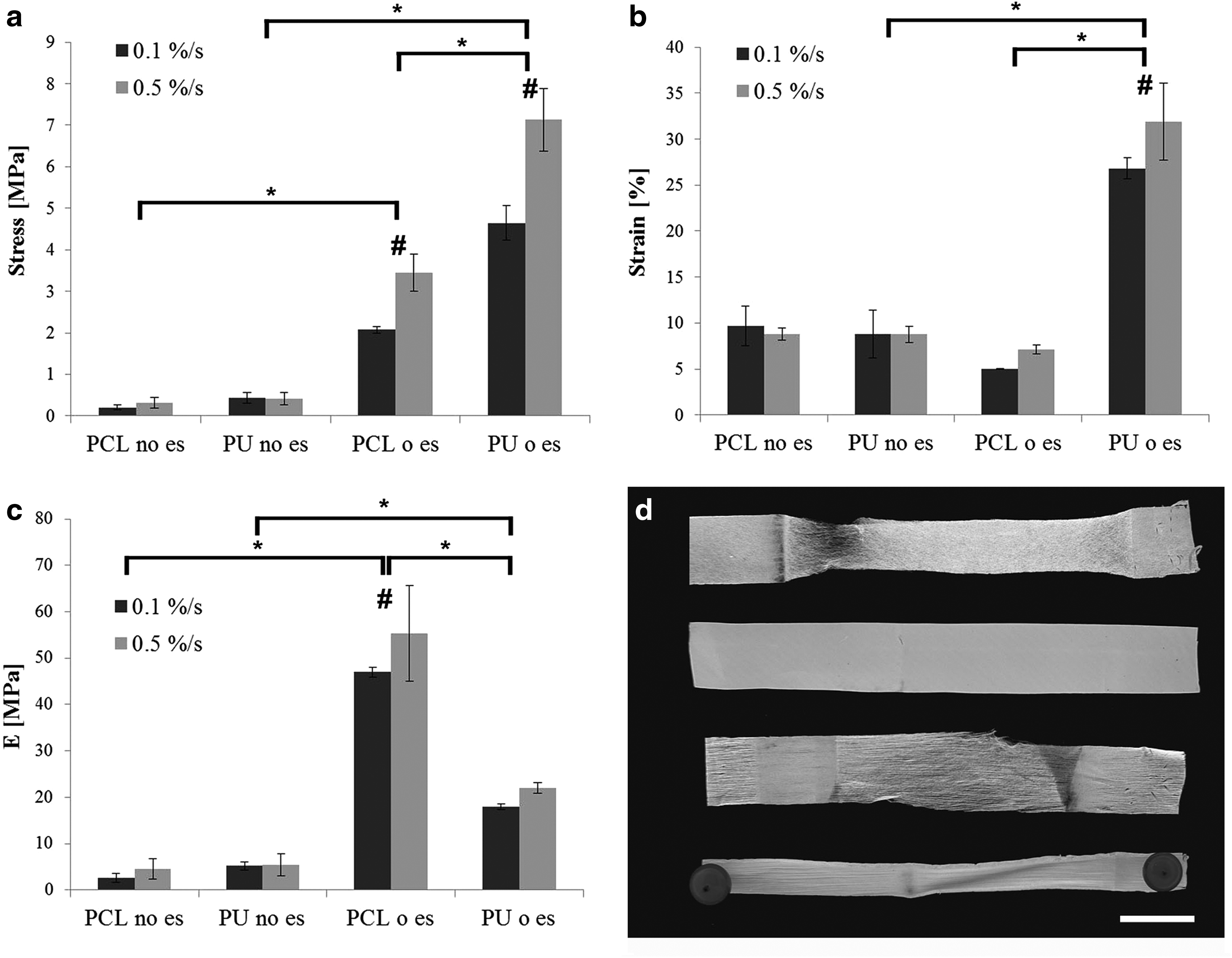
The mechanical behavior of the electrospun membranes was determined by tensile testing with applied strain rates of 0.1%/s and 0.5%/s. The experimentally acquired yield stress, yield strain, and Young's modulus are shown in Figure 2. The yield stress was significantly higher for oriented PCL, 2.07±0.08 MPa at 0.1%/s and 3.45±0.45 MPa at 0.5%/s, and oriented PU scaffolds, 4.65±0.42 MPa at 0.1%/s and 7.13±0.75 MPa at 0.5%/s, compared with their corresponding nonoriented scaffolds. The oriented PU scaffolds had a significantly higher yield strain value, 26.81%±1.16% at 0.1%/s and 31.92%±4.21% at 0.5%/s, than the other three scaffolds. In contrast, the oriented PCL scaffold was significantly stiffer than all other scaffolds, with a Young's modulus of 47.00±1.09 MPa at 0.1%/s and 55.33±10.33 MPa at 0.5%/s. Comparing the two test protocols, significant differences could only be found between the oriented scaffolds. After a strain of up to 100% was applied, PCL fibers ruptured close to the clamp, and therefore, a failure of the scaffold occurred. Moreover, a change to an elongated and narrower shape could be observed for the nonoriented PCL scaffolds (Fig. 2d).
Biochemical assays and cell–scaffold interaction
PCL versus PU film surfaces
Polymer films were prepared to assess if AF cell behavior would be different on PCL and PU made of PCL macrodiol surfaces. Comparable surfaces were produced by spin coating and drying in an analogy to the electrospinning process. AF cells proliferated with a similar rate over time in culture and produced GAG (Fig. 3) and OHP, an indicator for collagen (Table 3), to a comparable amount on both PCL and PU flat surfaces. In addition, differences in RNA expression could not be observed between the two polymer types regarding all tested genes.
The values are given as mean±SD. n=3 donors (triplicates per donor). Total OHP content containing OHP retained in scaffolds and OHP released to the medium (OHP tot), OHP retained in scaffolds (OHP scaff) and the ratio of GAG retained in scaffolds to OHP retained in scaffolds (GAG scaff/OHP scaff) in comparison to human and bovine AF tissues.
p<0.05 for PCL no es and PCL o es.
p<0.05 for PU no es and PU o es.
AF, annulus fibrosus; GAG, glycosaminoglycan; OHP, orthohydroxyproline.
Cell seeding, proliferation, and matrix production
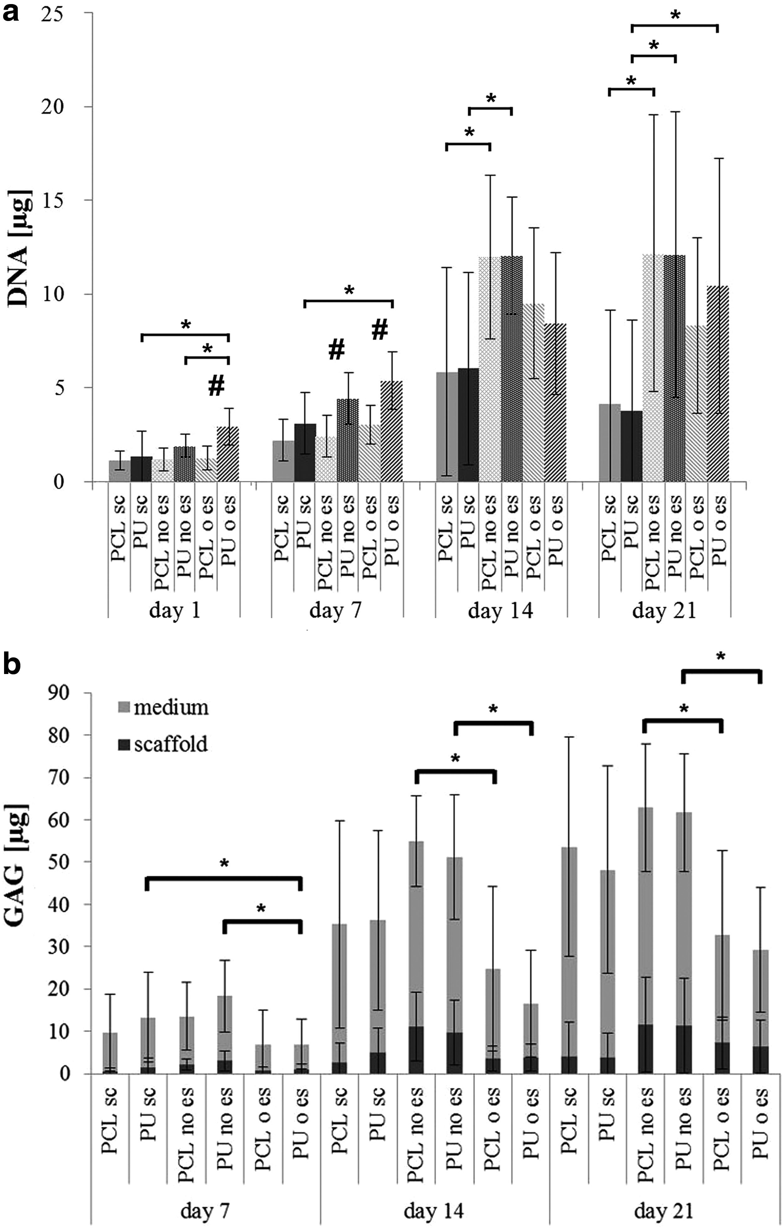
The cell seeding efficiency was evaluated by measuring the DNA amount in scaffolds after 1 day of cell culture. Oriented scaffolds showed a significantly higher DNA amount for PU than for PCL. Nonoriented scaffolds showed also a trend toward higher DNA values for PU compared with PCL. The effect of higher seeding efficiency for nonoriented and oriented PU scaffolds continued up to day 7. After 14 days of cell culture, there was no significant difference between the scaffolds. Independent of polymer type and scaffold architecture, the DNA content increased in all constructs up to day 14, after which a plateau was reached (Fig. 3a). The ECM production increased in all constructs in a time-dependent manner. In contrast to DNA, all groups showed an increase in the GAG (Fig. 3b) and collagen (data not shown) content through day 21. Regarding GAG production, PCL and PU nonoriented scaffolds showed significantly increased values compared with oriented scaffolds, for both GAG retained in the scaffolds and GAG released into the medium (Fig. 3b). The total OHP measured for all groups, including film surfaces, ranged from 32.0 to 40.7 μg/sample. However, OHP retained in the scaffolds was three times higher than OHP retained on film surfaces (Table 3).
Gene expression
Gene expression was measured after 7, 14, and 21 days of cell culture relative to freshly isolated bovine AF cells (day 0). The collagen type I gene expression remained constant throughout culture, while there was a downregulation of aggrecan and collagen type II expression and an upregulation of elastin expression over time in all scaffolds. Oriented PU scaffolds retained a higher collagen type II expression compared with the respective film surface at day 7 (Fig. 4), whereas collagen I and aggrecan expression did not differ between the scaffold types at any time point. At day 7, the oriented PU scaffolds also showed the highest expression levels of biglycan, decorin, and versican. There were no differences in the expression values of these genes at later culture time points.

Relative mRNA expression of AF cells after 7 days of cell culture with spin-coated PCL (PCL sc), spin-coated PU (PU sc), nonoriented PCL (PCL no es), nonoriented PU (PU no es), oriented PCL (PCL o es), and oriented PU (PU o es) scaffolds. Collagen type 1 (COL1), collagen type 2 (COL2), aggrecan (AGG), biglycan (BGN), decorin (DCN), elastin (ELN), and versican (VCAN) are shown. All data were normalized to freshly isolated AF cells (day 0). n=3 donors (triplicates per donor).
Histology
To visualize cell distribution, scaffold cross sections were prepared and stained using Toluidine blue. PCL scaffolds showed an increased cell ingrowth into the scaffolds from day 7 to 21. In contrast, barely any cells could migrate into the PU scaffolds and cells were most abundant on the surface of the scaffolds (Fig. 5).

Representative Toluidine blue stained cryosections after 21 days of cell culture with
Discussion
The creation of oriented micron and submicron fibrous scaffolds is an interesting approach for mimicking the size and orientation of collagen fibers in the native AF tissue and ultimately may provide a suitable microenvironment for AF tissue engineering. Numerous studies targeting AF tissue engineering with the use of electrospun scaffolds, made of several polymer types, have been reported recently.20–22,24,25 However, to our knowledge, the concomitant influence of scaffold architecture and polymer composition on AF cell behavior has not been investigated. In this study, we aimed to determine the appropriateness of electrospun PCL and PU scaffolds for AF reparative treatment, regarding their effect on AF cell behavior and their ability to mimic the native AF tissue structure and mechanical properties.
First, PCL and PU films were prepared by a spin coating process with a slow evaporation method using hydrophobic solvents. These control surfaces were prepared to investigate the influence of the surface chemistry. The film fabrication procedure was analogous to the electrospinning process and comparable polymer chemistry was obtained at the films and fiber surfaces for PCL and PU, respectively. 35 The biological assessment of AF cells seeded onto the PCL and PU film surfaces did not reveal any significant difference between the two surfaces. On both surfaces, AF cells could proliferate and produce a GAG-rich ECM. We conclude that both the PCL and PU films elicited a similar response; hence, the scaffold fiber diameter and orientation are likely to be the main parameters influencing cells after seeding onto the PU and PCL electrospun scaffolds.
From both materials, porous scaffolds could be prepared by electrospinning, resulting in fiber diameters in the range of micron size for PCL and submicron size for PU. Scaffold fiber orientation was achieved, while the fiber diameter size within one polymer group was maintained. Furthermore, mechanical properties could be tuned by changing the polymer composition or fiber orientation. Modulus values approach those reported for similar electrospun materials and native AF tissue, thereby providing compatibility with the tissue surrounding a repair region.26,36 The higher yield strain value and consequently the better resistance to stretch of the oriented PU scaffold, compared with the other prepared scaffolds, could be advantageous in a dynamic mechanical environment, preventing rupture of the scaffold during the initial tissue integration phase of the repair.
Compared with the spin-coated polymer surfaces, improved cell growth and retention of the ECM could be achieved in the electrospun scaffolds. Moreover, for collagen type 2 and biglycan, a significantly higher upregulation of the gene expression could be reached with oriented PU scaffolds compared with the PU spin-coated surface. The initial cell-seeding efficiency was higher for electrospun PU than for PCL scaffolds. This may be explained either by better cell attachment on submicron-sized fibers than on micron-sized fibers, 37 an increased surface area due to a decreased fiber diameter, 38 and/or by a decreased hydrophobicity of PU compared with PCL electrospun scaffolds, leading to difference in wettability and a wider seeding area (Table 1). Oriented electrospun scaffolds are intended to mimic the collagen fiber alignment of the native AF. Baker et al. reported values for GAG retained in nanofibrous PCL scaffolds that were slightly increased in oriented compared with nonoriented scaffolds with both mesenchymal stem cells (MSCs) and meniscal fibrochondrocytes after 14 days of cell culture using a hundred times higher seeding density. 39 Other investigators using bovine AF cells with a 30 times higher seeding density showed that the produced GAG amount relative to DNA tended to be higher with the orientation of the electrospun PCL fibers after 14 days of cell culture. The fiber diameter of the scaffolds was in the range of micron size and therefore comparable to the present study. 23 However, we observed a higher cell proliferation rate and a higher GAG production for nonoriented compared with oriented scaffolds as time in culture increased. The oriented scaffolds used in the present study showed a close packing of the fibers due to their parallel alignment during the production process, which reduced the interpore connectivity and thus the available area for the deposition of a new matrix.
To assess the preservation of the AF cell phenotype while culturing the cells on electrospun scaffolds, gene expression relative to freshly isolated cells was determined. We noted that the AF cell phenotype was maintained regarding collagen type 1, while collagen type 2 and aggrecan gene expression were downregulated. This may imply that the cells were not extensively influenced by the micron and submicron fibrous 3D structure of the prepared scaffolds. Due to a limited cell ingrowth into the scaffold, the majority of the cells may have grown in a monolayer on top of the scaffold rather than interacting with the 3D structure. Nevertheless, the decrease in type 2 collagen could be delayed in oriented PU scaffolds. Regarding the proteoglycans—biglycan, decorin, and versican, and regarding elastin, the cell phenotype was maintained throughout culture. Furthermore, the oriented PU scaffold also shows increased expression of some of these molecules at the beginning of culture, suggesting a positive initial effect on AF cells. A trend toward a higher upregulation for collagen type 1, aggrecan, elastin, and versican mRNA could be observed for oriented relative to nonoriented scaffolds after 7 days of cell culture. Thus, scaffold architecture stimulates the AF cell phenotype; by fiber orientation, a higher stimulation and therefore an upregulation of mRNA expression could be achieved.
Mwale et al. reported proteoglycan to collagen ratios measured as the GAG to hydroxyproline ratio, for native human AF tissue with values between 1.5 and 2.2 depending on the age (60–80 years, respectively, 15–25 years). 40 For the native bovine AF tissue from the same species as we used cells in the described study, we observed a lower value for the GAG to hydroxyproline ratio of 0.63±0.17. Values in the same range were reported by Mizuno et al. for native AF sheep cells. 41 In the present study, all groups showed a respective ratio of around 2.4 at day 14 that decreased to 2.2 by day 21 of cell culture. Since the collagen synthesis takes longer than the GAG synthesis it is difficult to reach ratios for tissue-engineered constructs similar to the native tissue after such a short time culture period. However, we could report decreasing ratio values over culture time which means that the ECM characteristics develop in the direction of the targeting native AF tissue.
Whereas the use of AF cells allows studying the effect of the scaffold on cell behavior, the need for an alternative to AF cells arises in clinics, due to the scarcity of healthy autologous cells and the fact that invasive surgical procedures are required for their isolation. MSCs would be an attractive alternative for this application, as they are readily available from bone marrow or adipose tissue. A previous study demonstrated the potential of culturing MSCs in oriented nanofibrous scaffolds. 42 However, research with the aim of directing stem cells toward a discogenic phenotype has been hindered by the lack of specific markers. It is not yet known which factors will be effectively influencing the differentiation of MSCs into AF cells. 43 Nevertheless, the data of the present study in terms of AF cell phenotype and matrix production can be used as a reference and as a control for further studies with MSCs to insure cell differentiation in the direction toward the fibrocartilage cell phenotype.
Whereas electrospun fibrous scaffolds present a suitable foundation for the engineering of collagen-rich tissues, one significant drawback lies in an inherently small pore size. To assess the degree of cell ingrowth into the scaffold, histological cross sections were prepared. Thereby, a clear association between an increased pore size due to a larger fiber diameter of the PCL scaffold and an increased cell migration into the scaffold could be observed. Hence, the challenging aim is to improve the porosity while keeping the fiber diameter small for a better cell attachment and for retaining adequate mechanical characteristics. Strategies to increase the porosity include the use of composite scaffolds, application of ultrasonication, and tailoring of the fiber diameter.44–46 An attractive option could be to combine electrospun scaffolds with cell-infiltrated hydrogels.
Conclusion
We comprehensively characterized electrospun scaffolds, based on fiber orientation and polymer composition, and demonstrated their potential for AF tissue engineering. Oriented PU scaffolds exhibit ECM retention, AF cell phenotype stimulation, and superior mechanical properties compared with electropsun PCL scaffolds or films. In future, these features can be further improved with increased porosity of the scaffolds. The higher yield strain of oriented electrospun PU scaffolds compared with other scaffolds will be advantageous for AF tissue engineering under dynamic mechanical loading.
Footnotes
Acknowledgments
The S.G., S.J.F., M.A., and D.E. are supported by a consortium grant from the AO Exploratory Research Board. Carole Boissard and Pierre-Etienne Bourban (EPFL, Lausanne, Switzerland) are gratefully acknowledged for their support with the mechanical testing, Markus Glarner and Christoph Sprecher for their help with the PU synthesis and scaffold characterization, respectively.
Disclosure Statement
The authors declare that they have no competing financial interests.