
Abstract
There is an unmet clinical need for a biomaterial sealant capable of repairing small annulus fibrosus (AF) defects. Causes of these defects include painful intervertebral disc herniations, microdiscectomy procedures, morbidity associated with needle puncture injury from discography, and future nucleus replacement procedures. This study describes the enhancements of a fibrin gel through genipin crosslinking (FibGen) and the addition of the cell adhesion molecules (CAMs), fibronectin and collagen. The gel's performance as a potential AF sealant is assessed using a series of in vitro tests. FibGen gels with CAMs had equivalent adhesive strength, gene expression, cytomorphology, and cell proliferation as fibrin alone. However, FibGen gels had enhanced material behaviors that were tunable to higher shear stiffness values and approximated human annulus tissue as compared with fibrin alone, were more dimensionally stable, and had a slower in vitro degradation rate. Cytomorphology of human AF cells cultured on FibGen gels exhibited increased elongation compared with fibrin alone, and the addition of CAMs to FibGen did not significantly affect elongation. This FibGen gel offers the promise of being used as a sealant material to repair small AF defects or to be used in combination with other biomaterials as an adhesive for larger defects.
Introduction
L
Healthy spinal function and regeneration are likely to require restoration of mechanical functionality of a spine unit through repair of the ruptured AF.5,6 The gold standard treatment for painful IVD degeneration is spinal fusion surgery, which reduces the spinal motion, and while total disc replacement has increased in practice,7–9 it does not restore native IVD mechanics and is not a living repair. Several hydrogels and biomaterial scaffolds have been developed, including commercially available NP replacements. 5 These strategies also require some form of puncture or incision through the AF, compromising mechanical integrity and resulting in implant expulsion or IVD degeneration. Therefore, their success would likely depend on an effective AF sealant.
AF closure repair strategies offer promise to repair damaged IVDs and slow accelerated degeneration from this defect. Sealing AF defects following the removal of herniated IVD material during microdiscectomy is a preventative approach that could slow or avert further degeneration of the IVD. Sealing the mechanically compromised AF can also help to restore pressurization and prevent IVD infiltration with proinflammatory cytokines, which could prevent painful conditions and offer a niche more amenable to repair and regeneration. Injectable biologics and cell therapies that are both currently available and in development offer the greatest potential for IVD regeneration, yet they all rely on AF repair techniques to close the needle puncture injuries or other small defects. AF competence is important for reestablishment of mechanical stasis and prevention of progression of IVD degeneration supported by studies that report a lower percentage of herniation reoccurrence resulting from smaller defects.6,10,11
There are few effective methods for AF repair, and those in current use are limited to sutures and modified sutures, which do not restore mechanical properties or compensate for loss of AF tissue. 12 Sutures that show mechanical promise 13 may not be practical to install in the tight confines of the spinal surgical space, and sutures did not provide lasting repair in vivo in animal models. 14 Polyethylene AF closure devices showed signs of expulsion and endplate damage following injection in a goat model. 15 An injectable sealant approach for AF closure is therefore a very attractive option. There are a number of adhesives that are commercially available, including cyanoacrylates (Superglue), gluteraldehyde-albumin (Bioglue), and fibrin glue (BIOSTAT BIOLOGX®, Tisseel®, Evicel™, and Crosseal™), but none have been shown to be suitable for AF repair. Fibrin sealants have shown promising results in porcine models by facilitating structural, compositional, and mechanical repair of surgically damaged IVD, and promoted the production of anti-inflammatory cytokines.16,17 However, fibrin sealants alone have relatively low stiffness and degrade quickly at high concentrations. Adhesive sealant biomaterials can also be used in combination with fibrous biomaterials to create an AF patch or other composite scaffolds capable of repairing larger AF defects. This study analyzes a fibrin gel with a genipin crosslinker for improved shear stiffness, resistance to degradation, and adhesion to AF tissue.
The objective of this work is to develop an easy to use, injectable, and tunable biomaterial with comparable shear properties to native AF tissue to be used to seal AF defects. The shear modulus of the AF is a strong determinant of mechanical function of the IVD18,19 and can play an important role in AF delamination.20,21 Therefore, the shear properties of native AF tissue were considered a relevant target design property for evaluating an AF sealant. A genipin crosslinked fibrin hydrogel (FibGen) offers promise as an adhesive AF sealant. 22 Fibrin is Food and Drug Administration (FDA) approved and genipin is a plant-based chemical crosslinker with low cytotoxicity used with a variety of materials, including fibrin for articular cartilage engineering. 23 FibGen is a tunable material with comparable mechanical properties to native AF tissue, and although cells remained viable on the gel, the relatively slow proliferation and presence of abnormal cells with rounded morphology motivated further optimization. 23 Preliminary tests were done to reduce the genipin concentration, while maintaining the mechanical properties, and concentrations of 6 and 11 mg/mL genipin were chosen for evaluation. To further improve cytomorphology, the key extracellular matrix proteins, collagen and fibronectin, were added to the formulation. It was hypothesized that the addition of these proteins would maintain the mechanical properties of this gel, while improving cell morphology and viability. Characterization of the in vitro properties of these gel formulations with stiffness, adhesion, in vitro degradation, gelation kinetics, gene expression, and contraction analyses was performed. The addition of cell adhesion molecules (CAMs) to FibGen to promote an AF cell phenotype and enhance cell proliferation on the gels was also assessed.
Materials and Methods
Gel fabrication
Five different gel formulations were characterized in this study and labeled by the final concentration of genipin and the addition of CAMs as follows: 0 mg/mL (uncrosslinked fibrin gel), 6 mg/mL, 11 mg/mL, 6 mg/mL+fibronectin, and 6 mg/mL+collagen. Depending on the experiment, the volume of the gel used varied from 50 to 400 μL. Unless otherwise stated, each gel was thoroughly hand mixed by adding each component by a pipette, then slowly dispensing and gently mixing the solution with the pipette tip within a mold or on glass slides. Fibrin was made by combining fibrinogen and thrombin, both isolated from bovine plasma (Sigma-Aldrich, St. Louis, MO), dissolved in PBS at 37°C at 200 mg/mL and 100 U/mL, respectively. Thrombin was mixed with fibrinogen at a ratio of 1 U per 5 mg fibrinogen. In the four crosslinked groups, genipin (Wako, Richmond, VA) dissolved in DMSO (Fisher Scientific, Waltham, MA) was mixed with thrombin before adding the thrombin mixture to the fibrinogen to achieve a final concentration of genipin of 6 or 11 mg/mL. In fibronectin-modified groups, fibronectin isolated from bovine plasma (Sigma-Aldrich) was homogeneously mixed with fibrinogen for a final concentration of 50 μg/mL. This concentration was chosen based on the manufacturer's recommendations for three-dimensional cell culture. In collagen-modified groups, collagen type I isolated from rat tail tendon (BD Biosciences, San Jose, CA) was combined with 10×PBS, dH2O, and NaOH in amounts indicated by the manufacturer's instructions, and then mixed by the pipette with fibrinogen, thrombin, and genipin to achieve a final concentration of 0.08 wt% or 0.8 mg/mL. The collagen concentration was chosen based on pilot studies that tested the maximum collagen concentration that could be achieved while allowing ease of mixing. Unless otherwise stated, all gels were allowed to set for 24 h before testing or cell seeding.
Shear stiffness and gelation testing
A rheometer with parallel plate geometry (TA Instruments, New Castle, DE) was used to evaluate the shear stiffness and gelation kinetics of all five gel formulations. For mechanical testing, cylindrical gel specimens from custom teflon molds (5-mm ø, 1.5-mm thick, n=8) were used to characterize the shear properties of the gels over dynamic frequency and strain sweeps following methods previously described by Schek et al. 23 For gelation kinetics testing, the Winter–Chambon Criteria were employed as described by Jiao et al. 24 Fifty microliters of freshly mixed gel was placed onto the rheometer. Using a 1-mm gap between plates, multifrequency tests were performed from 0.5 to 5 Hz at 3% strain. These testing parameters were chosen based on preliminary strain sweep tests, to establish the minimum phase angle of 3% strain, and frequency sweep tests to establish the range of frequencies (0.5, 2, 3.5, and 5 Hz). Multiple frequency tests established the gelation time as the time point when the loss tangent [Tan (δ)] responses of all frequencies tested converged (Fig. 1). 24 Gelation kinetics testing was performed at 37°C testing for all formulations (n=4), and 6 mg/mL genipin gels were also tested at 25°C and 4°C to observe the effect of temperature on gelation time.
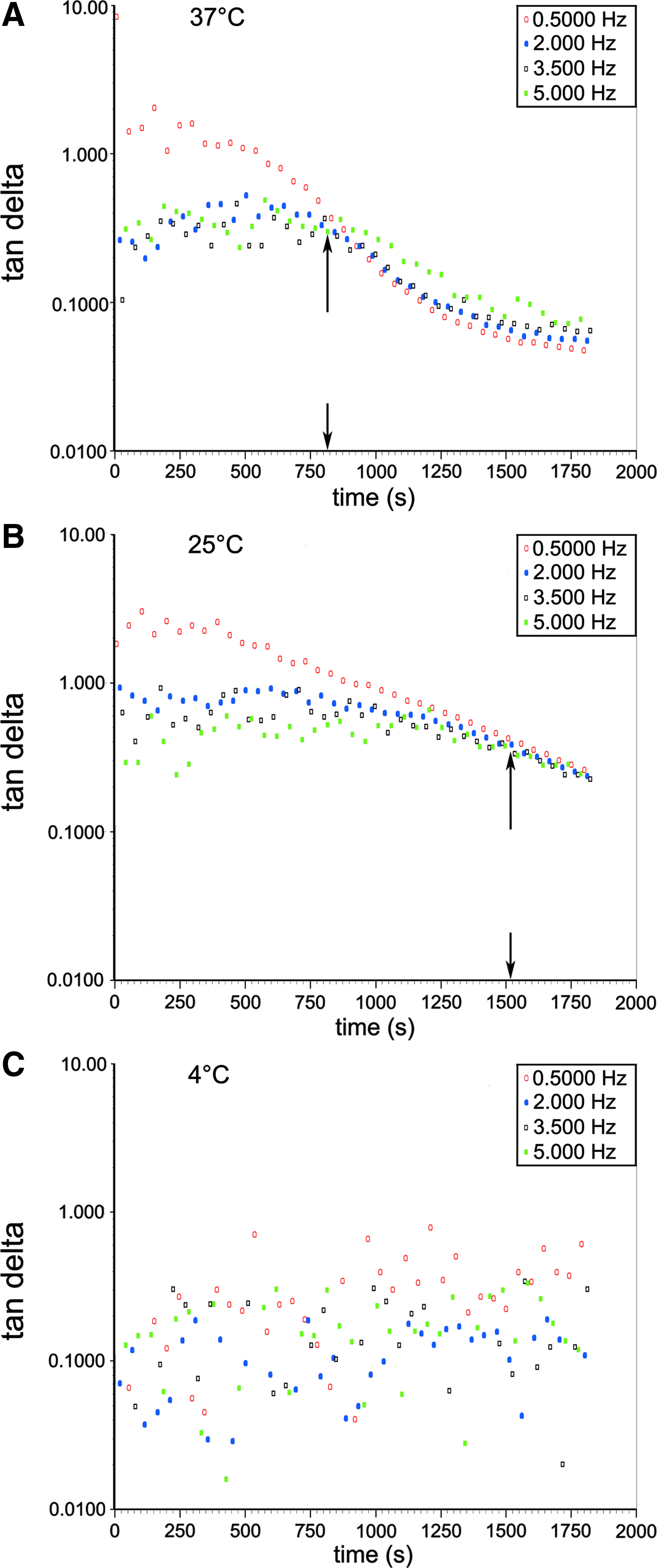
Representative gelation kinetic testing results showing temperature dependency: loss tangent [Tan (δ)] is plotted as a function of time for 6 mg/mL genipin gels at multiple frequencies tested at
Adhesion testing
Bovine caudal IVDs were dissected and 8-mm-diameter specimens were punched out radially from the IVD and then cut to a thickness of 3 mm. Samples were subpunched to create a 3-mm-diameter defect, which was then filled with freshly mixed gel, or as a control, the punched out AF tissue core was reinserted back into the defect without an adhesive. The release of any AF prestress when subpunching the samples resulted in a snug press fit in the control samples. 25 Damaged AF samples repaired with FibGen gels were left at room temperature for 1 h during the initial set period, and then at 4°C overnight. Samples were then placed in a confined chamber with centered 3.5 diameter holes at the top and bottom, and were indented with a 2.8 mm flat-ended tip at a rate of 0.01 mm/s until failure, following the methods described by Maher et al. 26 The peak load recorded was used to calculate adhesion strength based on the cylindrical surface area of gel that abutted the AF tissue.
In vitro degradation testing
In vitro degradation was measured using samples from molds (5-mm ø, 1.5-mm thick) of each gel formulation. Individual gels were weighed (Wi) and placed in sealed well plates with 1 mL of PBS at 37°C for 1, 3, 7, 10, 14, and 21 days to obtain the wet weight (Ww) and then lyophilized to obtain the dry weight (Wd) (n=4 for each time point and formulation). The equilibrium weight swelling ratio (Ww/Wd) and in vitro degradation ((Wi−Ww)/Wi) were calculated at each time point.
Contraction testing
The dimensional stability of gels was evaluated using freshly mixed 50 μL droplets (n=4 for each formulation) placed between glass slides in a humidified environment. Images were captured over a 60-min time period, where t=0 represented mixing of the gel, to record contraction of the sample during gelation.
AF cell culture
Gels were prepared aseptically using 150 μL to make a 14-mm diameter and 1 mm droplet for imaging, and 400 μL of gel for PCR in 12-well plates. Human AF cells were seeded on the gels at a density of 12,500 cells/mL in 2 mL of high glucose DMEM with 10% FBS (Gibco, Carlsbad, CA), 0.5% fungizone, and 50 μg/mL ascorbic acid (Sigma-Aldrich). AF cells were obtained from autopsy and surgical samples (n=6, P1–P2, average donor age of 52±19). Human subject samples were obtained with consent using protocols approved by the Institutional Review Board, and samples were deidentified for use on this project. The cell culture medium was exchanged every 3–4 days until the day of imaging (day 7 or 21) when gels were rinsed with PBS and incubated with a solution of 4 mM Calcein-AM and 0.2 μL/mL Hoechst in PBS (Invitrogen, Carlsbad, CA) for 15 min. Samples were imaged using a fluorescent inverted Zeiss microscope (excitation/emission; Calcein=494/517 nm and Hoechst=350/461 nm). Five representative images were obtained per gel. Six gels were made per formulation, each seeded on gels with a different human AF cell donor population, for a total of 30 images per formulation. ImageJ (NIH) was used to calculate the average cell count per mm2. For qualitative assessment of cell morphology, a Leica SP5 DM confocal microscope was used to image the F-actin structure of cells stained with Alexa Fluor 488-phalloidin (Invitrogen) at 5 U/mL and Hoechst 7 days after seeding (n=2).
Additional cell culture was performed on two individual human donor AF cell populations labeled HU7 and HU26 (n=2, P2-P3, average donor age of 64±24) to assess further changes in cell morphology and quantify cell proliferation at days 1 and 3. Gels were prepared using 400 μL to make a 2-mm-thick and 8-mm-diameter droplet. Cells were seeded on the gels as previously described here. To reduce variability that may have been caused by hand mixing, gels were mixed using a double-barreled syringe with a mixing tip 2-K MIXPAC system (Sulzer Mixpac AG, Haag, CH). The first few microliters were discarded from the syringe to avoid air bubbles and ensure homogeneous mixing of the components. The gels were then incubated in Calcein-AM and Hoechst dye and images were taken at days 1 and 3 as previously described (n=4 gels per formulation). Cell morphology analysis was performed for each image using the Zeiss AxioVision AutoMeasure analysis software to calculate cell roundness and elongation. Elongation as measured by feretmax and feretmin was calculated by the AutoMeasure analysis software by determining the length of an outlined object at 32 angle positions, similar to a sliding caliper, and assigning the maximum and minimum values as feretmax and feretmin, respectively. Elongation was represented as the ratio of feretmax to feretmin, where a larger ratio indicates a more elongated cytomorphology and a ratio close to 1 indicates a rounded cytomorphology (Fig. 6C). Additional plated gels were used to quantify concentrations of dsDNA using the QuantiFlour dsDNA system (Promega, Madison, WI) at day 1, 3, and 7 (n=4). Plated cells were lysed using 100 μL of RLT buffer (Qiagen, Limburg, NL) and then analyzed for dsDNA content.
Gene expression was measured using real-time reverse-transcription PCR (qRT-PCR). Human AF cells (n=6, P1-P2, average donor age of 58±15) were first lysed at 7 and 21 days after seeding using the RLT buffer and RNA was extracted with the Qiagen RNEasy Kit (Qiagen). Gene expression of housekeeping (18S), anabolic (COL1), catabolic (MMP13), and inflammatory (IL-1β) genes was profiled using predesigned and optimized human TaqMan gene expression assays (Applied Biosystems, Carlsbad, CA). Relative fold changes in gene expression were calculated normalized to 18S and relative to 0 mg/mL genipin (fibrin). Analysis was performed using the 2−ΔΔCT method. 27
Statistics
A two-way ANOVA with Tukey's post hoc testing and unpaired t-test was used to detect significant effects of gel formulation and time point, respectively (GraphPad Prism). A repeated-measures two-way ANOVA was performed to detect significant effects of gel formulation and time for HU7 and HU26 cell populations. One-way ANOVA with Tukey's post hoc testing established effects of gel formulation endpoint measurements. In all cases, p<0.05 represents a significant effect, and data are represented as mean±SD.
Results
Shear stiffness testing
For brevity, only the dynamic shear modulus (|G*| at 1 Hz and 10% strain) obtained from the frequency sweep is reported. Other frequencies and magnitudes showed similar trends for gel formulation. The dynamic shear modulus magnitude (|G*|) of all FibGen gels was significantly higher compared with 0 mg/mL genipin gels and there was no difference between FibGen gels and native AF tissue (Table 1). Results are compared to previously obtained values for human AF tissue. 23
FibGen formulations are given with genipin concentration, so that 0 mg/mL refers to fibrin alone.
Shear modulus of fibrin was significantly different from AF tissue, ap<0.005 with shear modulus values for human AF tissue from Schek et al. 23
Gelling time was shorter for fibrin than all other groups, bp<0.05.
The 11 mg/mL genipin formulation exhibited a significant contraction from 1, t=0 condition, cp<0.05.
Adhesion strength was significantly lower for the AF press fit control than all FibGen groups, dp<0.005.
AF, annulus fibrosus; ND, not determined.
Gelation testing
Gelation occurred in ≤15 min for all gels. It is notable that the 0 mg/mL genipin gel formulation had the fastest gelation time of 202 s (Table 1). The addition of genipin caused a nonlinear effect on the gel time, and the addition of collagen extended the gelation time. A temperature dependence was shown when gelation tests were performed at 25°C and 4°C as compared with 37°C using 6 mg/mL genipin gels (Fig. 1). Slower gel times were observed at 25°C and no observed gelation occurred at 4°C.
Adhesion testing
A representative load curve demonstrates a toe region, linear stiffness zone, and then an abrupt failure point at which adhesion strength was calculated (Fig. 2). FibGen had comparable adhesion strength to fibrin alone (0 mg/mL genipin), and all formulations were significantly greater than the press fit control (Table 1). The failure strains, calculated by the indenter depth normalized to the thickness of the sample, were close to 100% for all samples and no significance was found between groups (Table 1).
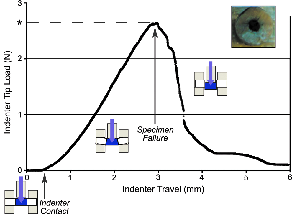
Representative loading curve for push-out tests with cross-sectioned schematic of test at different stages (unloaded, loading, and failure). Adhesion strength (Table 1) was calculated from the load at failure (*) and adhesion surface area of gel (n=6). Top view of gel-filled sample (inset). Color images available online at www.liebertpub.com/tea
Contraction testing
A minimal amount of gel contraction was observed for all samples. The largest amount of contraction was observed in the 11 mg/mL genipin gels (10%) and the smallest in the 6 mg/mL genipin and 6 mg/mL genipin+fibronectin formulations (≤3%). The darkening of color over the 60-min test period signifies the increased formation of genipin crosslinks (representative images of testing using 6 mg/mL genipin formulation, Fig. 3).
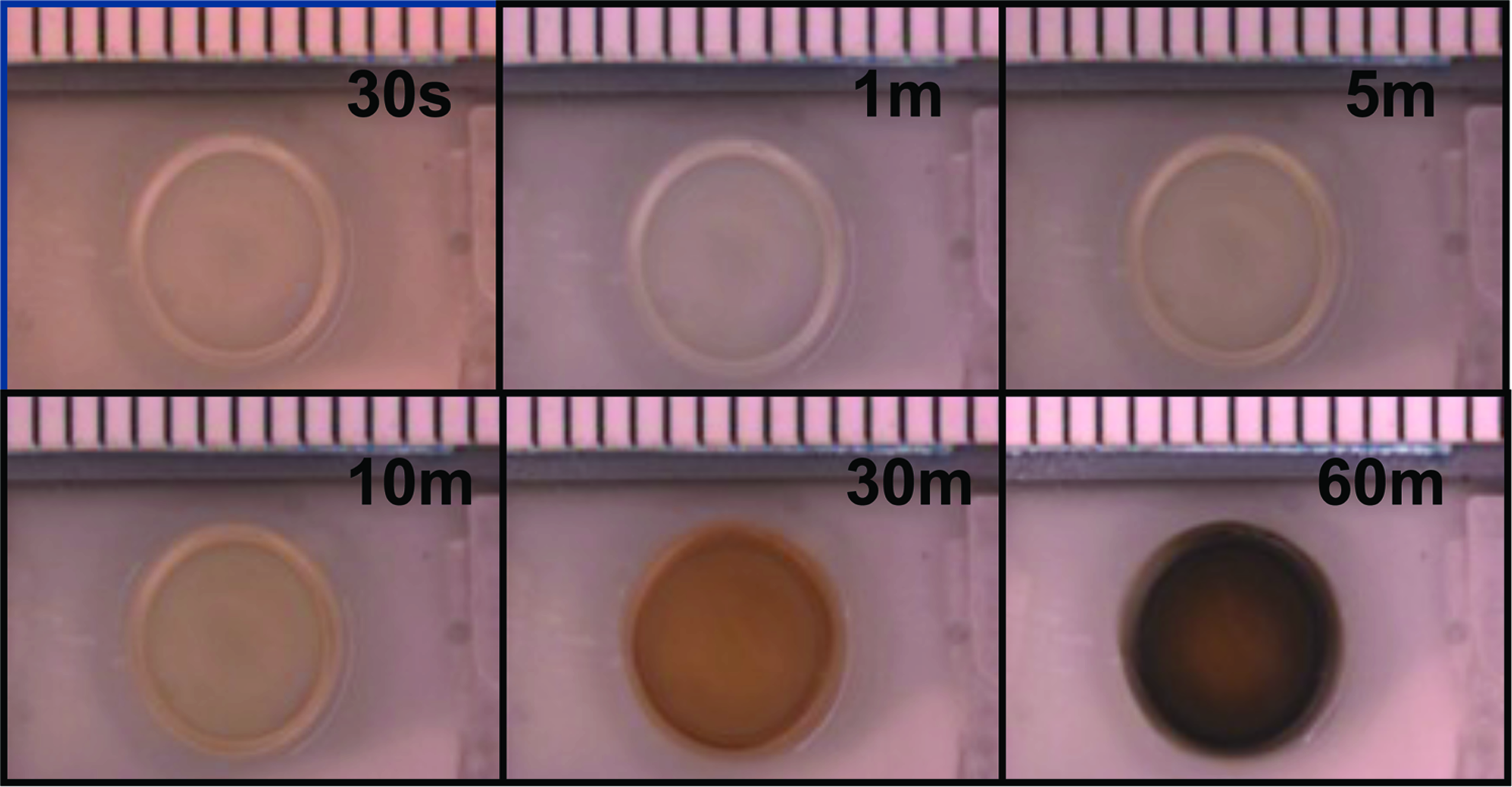
A representative FibGen gel (6 mg/mL genipin) used in contraction testing shown at acquired time points during the testing period (0.5, 1, 5, 10, 30, and 60 min). Graduation marks represent 1 mm. Genipin crosslinking can be observed as a darkening in color. FibGen gels exhibited dimensional stability for the 1-h testing period (Table 1). Color images available online at www.liebertpub.com/tea
In vitro degradation testing
Degradation and swelling ratios for all FibGen formulations remained stable for the entire test period (days 1, 3, 7, 10, 14, and 21) with day 21 values given (Table 1). Average values of degradation are normalized to day 1 and are reported as mean±SD for all specimens. In contrast to FibGen formulations, fibrin alone was completely degraded by 7 days with degradation values of 0.87±0.05 (day 1) and 0.89±0.05 (day 3) and swelling ratio values of 9.2±0.7 (day 1) and 10.1±0.8 (day 3). Therefore, 0 mg/mL genipin data were not available for day 21.
AF cell culture
Representative confocal images show decreased cell density in the FibGen groups as compared with fibrin, which was recovered with the addition of CAMs (Fig. 4). The apparent decrease in cellularity with FibGen as compared with fibrin and FibGen with CAMs was also observed in the quantitative dsDNA and cell count data and propagated at all time points, however, this was not significant (Fig. 5). There was an increase in cellularity with days in culture that varied somewhat between the patient and measurement technique, yet was significant at day 3 as compared with day 1 for most groups when measured with dsDNA. Cell count data exhibited similar trends as dsDNA measurements.
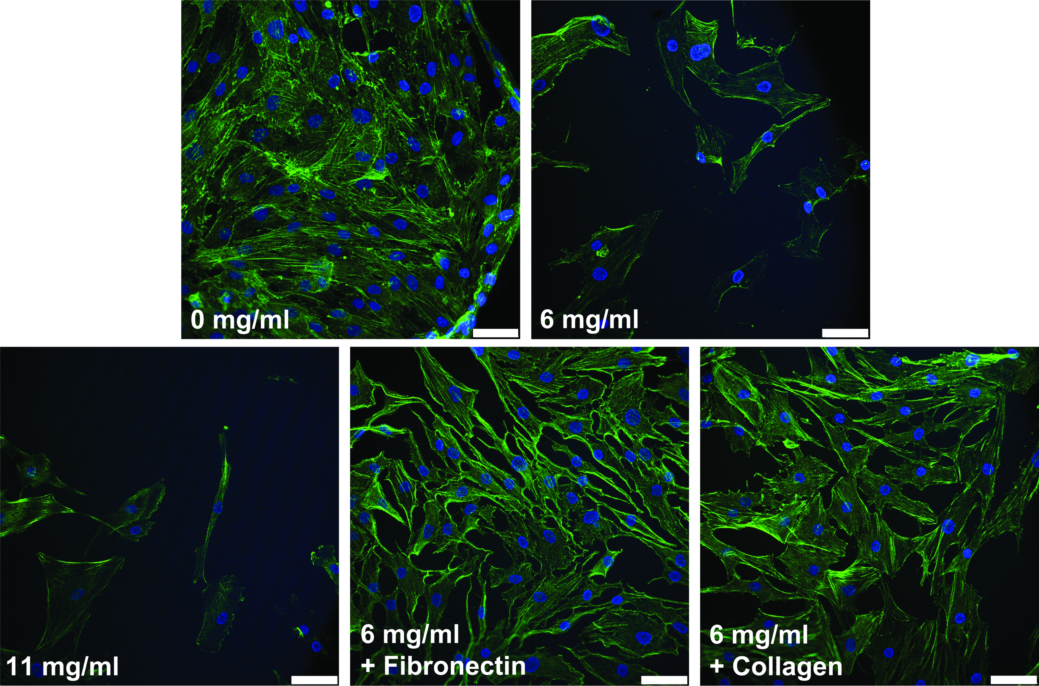
Representative confocal images of human annulus fibrosus (AF) cells seeded on gels at day 7. FibGen formulations are given with genipin concentrations. Two-dimensional image represents a stack of 25 slices (3 μm each) using a 20× dipping lens with 405 Diode (blue Hoechst-stained nuclei) and Argon 488 (green phalloidin-stained actin) laser excitation. Although a slight increase in cellularity can be observed in CAM enhanced gels as compared with 6 and 11 mg/mL genipin formulations for this donor population, this was not significant in quantitative observations (Fig. 5). Scale bar=50 μm. Color images available online at www.liebertpub.com/tea

Cell proliferation quantified by dsDNA concentration and cell count for early and late experimental times. FibGen formulations are given with genipin concentrations, so that 0 mg/mL refers to fibrin alone. dsDNA quantification
Cell morphology analysis at day 1 and 3 demonstrated interpatient variability in cell shape responses between two different patients. Calcein-AM staining showed cytomorphology of cells, and differences in proliferation and elongation were observed for cells from different patients (Fig. 6). Elongation increased for cells on groups containing the genipin crosslinker from day 1 to 3 for patient HU7, and was increased for all groups compared with gels composed of fibrin alone (Fig. 6D). Elongation was greater for patient HU26 than patient HU7, and was increased for all groups compared with gels composed of fibrin alone (Fig. 6D). Elongation was greater for patient HU26 than patient HU7 starting at day 1, and while there was a similar trend of increased elongation from day 1 to 3 for all groups, this was not significant (Fig. 6E). For HU26, cells cultured on gels containing collagen exhibited a significant decrease in elongation from day 1 to 3.
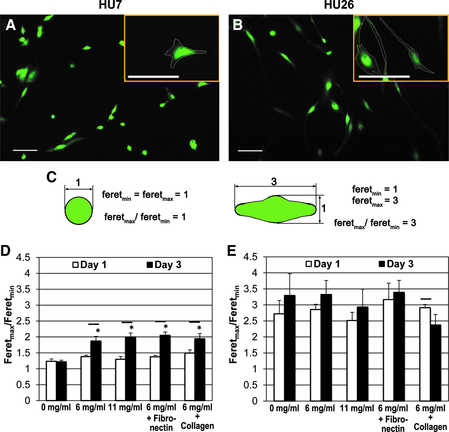
Cell morphology analysis during early culture of experimental gel compositions using human AF cells from two different patients demonstrates interpatient variability in cell shape responses. Calcein-AM staining of human autopsy AF cells from patient HU7
The qRT-PCR results showed no difference between 0 mg/mL genipin (fibrin) control and all gel formulations for COL1, IL-1β, or MMP13 at days 7 and 21(Table 2). There was a slight upregulation for all gels at day 21 for IL-1β, however, this was not significant.
SD calculations reflect±error on a log scale following the method of Livak and Schmittgen. 27 No differences were found in gene expression of cells seeded on FibGen gels as compared to fibrin gels.
Discussion
A newly formulated crosslinked fibrin-genipin gel was developed as an injectable sealant for repair of AF defects. This new formulation included addition of fibronectin and collagen as CAMs to enhance cell proliferation and phenotype. The gel was thoroughly evaluated in vitro for performance parameters associated with shear stiffness, adhesion strength, gelation rate, dimensional stability, cytocompatibility, and in vitro degradation.
Fibrin gels are a well accepted and utilized material in surgery and tissue engineering. The importance of genipin as an addition to a fibrin gel used as an AF sealant was shown by an increase in shear modulus to match that of native AF tissue and a stability of formulation. The lack of difference in contraction profiles, adhesive properties, and gene expression in inflammatory and catabolic proteins between FibGen formulations and fibrin alone (0 mg/mL genipin gels) strengthens the case for genipin as a safe and effective crosslinking agent. Good performance in shear stiffness, in vitro degradation, adhesion, and cytocompatibility of all FibGen gels with CAMs demonstrates its promise as an injectable sealant for AF repairs.
The addition of fibronectin or collagen to FibGen gels had an unbiased effect on cell proliferation and morphology compared with fibrin gels alone, however, incorporation of these CAMs did not alter the mechanical or adhesive properties. There was no significant difference between cell counts on different gel formulations, which was associated with the expected high variability of human AF cells, but quantification of dsDNA showed some significance between groups, although variable between patient samples. Images of cell morphology were used to show differences in cell behavior. Interpatient variability of cell responses to different gel compositions is clear and may be due to the variability caused by cell donor's age and condition. The differences in cell morphology observed between patients highlights this variability. At days 1 and 3, cell proliferation with time showed opposite trends between cells from different patients. The presence of long cell processes, which is marked by an elongated AF cell morphology, is important for proper AF cell function.28,29 Whereas elongation increased for most groups between day 1 and 3 toward a more AF phenotype, cells seeded on collagen enhanced gels trended toward a more rounded morphology for patient HU26. This cell response may be due to the difficult mixing of the collagen enhanced gels, which polymerize quickly. The varibility observed between patients highlights the importance of robust cell response characterization using multiple cell donors for in vitro characterization of the FibGen material. There were no significant differences between collagen or fibronectin enhanced gels for all quantitative assays, although the addition of fibronectin offers an easier mixing method compared with collagen. Whereas cell proliferation was variable between patients, observed cytomorphology, increased elongation compared to fibrin, and ease of mixing suggest that the the inclusion of fibronectin to FibGen may serve as a practical enhancment of cytocompatibility without affecting mechanical or adhesive properties.
Gelation times were recorded at ∼15 min or less for FibGen, which was considered an acceptable working time for gel injection, and within the time frame (4–15 min) specified by ISO Standard 5833/1-1999 E for injectable materials (i.e., bone cement). Fibrin exhibited the fastest gel time, whereas gelation for all FibGen formulations was longer than for fibrin alone, which may be associated with competing crosslinking mechanisms between thrombin and genipin. However, it is likely that FibGen gel times could be accelerated with the addition of more thrombin in future formulations.
The base FibGen formulation was not statistically different from formulations with CAMS. The addition of fibronectin had nonsignificant trends of improved cell proliferation without diminishing any other properties. Therefore, fibronectin can remain in consideration as a CAM to increase cell adhesion, particularly at early time points, and definitely shows feasibility of adding bioactive molecules to the base FibGen formulation. Results with collagen did not warrant the added technical challenges in mixing and associated increased variability in cell adhesion. Although it was hypothesized that the CAMs would improve the cellular response to the FibGen materials, it is possible that the genipin crosslinker modified the adhesion molecules in such a way as to reduce their bioactivity. Variability in cell proliferation data due to nonuniform mixing was tested by performing additional experiments that utilized a syringe to uniformly mix the gels across all samples. No differences were observed between hand mixing and syringe mixing, although syringe mixing was faster, eliminated human variance in mixing procedures, and more closely mimicked that which would be utilized clinically. Hand and syringe mixed gels were uniform in both visual appearance and cell adhesion across the surface of the gel. SEM analyses and high-performance liquid chromatography are required to assess the structural uniformity of adhesion and interactions of FibGen proteins and CAMs with the native AF tissue. In particular, intermolecular crosslinking of FibGen with collagen in the adjacent tissue may be an important functional mechanism, given the genipin is believed to form intra- and intermolecular crosslinks with a cyclic structure within collagen fibers in biologic tissue. 30 This study builds on the evidence that fibrin can be modified to enhance its performance as an AF sealant. It was previously shown that it is possible to modify fibrin with genipin to tune material behaviors. 23 The current study provides many additional material characterizations of FibGen and further demonstrates the ability to modify fibrin with the addition of the CAMs, fibronectin and collagen. Moreover, these findings suggest that other bioactive molecules may be similarly incorporated into the various FibGen gel compositions. All the concentrations of FibGen used in this study are approximately a factor of six lower than that previously reported by our group. 23 Interestingly, there is negligible difference in the shear modulus between these formulations. The lower concentration of genipin in this study may explain some of the differences in cytomorphology between studies, where Schek et al. found a more rounded cytomorphology of AF cells with the addition of genipin, while the current study exhibited a more elongated AF cell morphology.
FibGen formulations exhibited acceptable cell morphology and density without negatively impacting gel stiffness, adhesion, and geometric and degradation stability. It has been shown that healthy IVDs are subjected to NP pressurization well above 1 MPa in vivo. 31 The pushout test in this study has very different boundary conditions from intradiscal pressure measurements, so is not directly comparable. Nevertheless, the adhesion strength values of 59–81 kPa are an order of magnitude lower than intradiscal pressures and indicate that material enhancements to strengthen FibGen will be important. However, the high values of adhesive failure strain suggest that FibGen has adequate toughness to seal small AF defects. Future in situ biomechanical and in vivo testing are required to evaluate FibGen in more clinically relevant conditions. The qRT-PCR data indicated there were no differences in the gene expression of the selected proinflamatory, catabolic or anabolic proteins between genipin crosslinked gels and fibrin alone. The three-gene panel of qRT-PCR measurments provides an initial screen and more genes and time points may discern greater differences between groups. The data suggest that CAM additions could increase AF cell proliferation on FibGen gels, which is a key attribute for tissue regeneration, yet these trends were nonsignificant and dominated by biological variance in fibrin and human AF cells. Future investigation is warranted to assess performance with in situ biomechanical testing, in vivo degradation tests, and in vivo biocompatibility analyses to characterize the inflammatory response.
In conclusion, fibrin can be modified with the addition of crosslinkers and CAMs to tune biomechanical and biological behaviors. The current FibGen gel formulation displayed excellent performance for adhesion, shear stiffness, dimensional and compositional stability. The sub-15-min gel time and slow in vitro degradation rates further demonstrate that the material can be easily mixed and injected, motivating future in situ and in vivo performance testing. The addition of CAMs demonstrated the feasibility of incorporating bioactive molecules into FibGen. Although in our studies the base FibGen formulation was equivalent to those with CAMs, the role of CAMs in FibGen requires further validation and characterization to elucidate possible cell–matrix interactions at the molecular level. Overall, this biomaterial offers promise for sealing small AF defects and may be useful as an adhesive in combination with other biomaterials for repairing larger AF defects. Future in vivo and in situ testing is warranted to better assess performance, biocompatiblity, and degradation rates.
Footnotes
Acknowledgments
This work was supported by NIAMS/NIH R01 AR057397, NIAMS/NIH F32 AR062455, and the AO Exploratory Research Board of the AO Foundation. The authors thank Dr. Samuel Cho and Dr. Sheeraz Qureshi for providing human surgical specimens used for isolating AF cells as well as Ilana Stock.
Disclosure Statement
No competing financial interests exist.