
Abstract
Biodegradable hydrogels with three different water contents were prepared through the glutaraldehyde crosslinking of gelatin with an isoelectric point of 5.0 under varied reaction conditions. The objective of this study is to investigate the effect of time period of basic fibroblast growth factor (bFGF) release that is modified by the hydrogel water content, on the bone regeneration. A bone fracture was prepared at the femur bone of mice, while gelatin hydrogels incorporating bFGF or bFGF solution was applied at the fracture site. The gelatin hydrogels incorporating bFGF exhibited significantly stronger bone regeneration than the bFGF solution, although the extent depended on the water content of hydrogels. Bone regeneration induced by gelatin hydrogels incorporating bFGF increased with a decrease in their water content. Histological and biochemical examinations indicated that the area of bone tissue newly formed and the level of osteogenic genes were enhanced for the hydrogels incorporating bFGF of lower water content compared with those of higher one. It is possible that the hydrogels that slowly degraded released bFGF for a longer time period, resulting in an enhanced bone regeneration. It is concluded that the time profile of bFGF release is one of the factors contributing to the bFGF-induced bone regeneration.
Introduction
I
Materials and Methods
Materials
A gelatin sample with an isoelectric point (IEP) of 5.0 (Mw=99,000), prepared through an alkaline process of bovine bone, was kindly supplied by Nitta Gelatin Co. An aqueous solution of bFGF (Mw=17,000, IEP=9.6, 10 mg/mL) was kindly supplied by Kaken Pharmaceutical Co. Glutaraldehyde (GA), glycine, and other chemicals were obtained from Wako Pure Chemical Industries, Ltd. Plank-Rychlo's solution is aqueous solution of 0.3 M aluminum chloride and 5 wt% formic acid in HCl (3 vol%) for decalcification. 23
Preparation of gelatin hydrogels
A gelatin derivative was prepared as described previously. 22 Briefly, aqueous solution of 5 or 10 wt% acid gelatin containing various concentrations of GA (Table 1) was cast into a polytetrafluoroethylene mold. Crosslinking was allowed to proceed for 12 h at 4°C, and the resulting hydrogel sheets were immersed in 50 mM glycine aqueous solution at room temperature for 1 h to block the residual aldehyde groups of GA. The hydrogels were rinsed three times with double-distilled water (DDW) at room temperature, followed by freeze-drying and sterilized with ethylene oxide gas. The hydrogel weight was measured before and after complete drying under a vacuum condition, and the water content (the wt% of water in the wet hydrogel to the wet hydrogel) was calculated from the two weights.
In vivo degradation test of gelatin hydrogels
To evaluate the in vivo degradation profile of gelatin hydrogels, the implantation of 125I-labeled hydrogels was performed according to the method previously reported. 24 Briefly, 20 μL of [125I] Bolton–Hunter reagent solution in benzene was completely evaporated at room temperature. The resultant solid reagent was redissolved into 1 mL of phosphate-buffered solution (PBS, pH 7.5) and the resulting solution (20 μL) was dropped onto a freeze-dried gelatin hydrogel, followed by leaving at 4°C overnight to introduce 125I into the amino groups of gelatin. The radioiodinated hydrogels were washed with DDW thoroughly to exclude the uncoupled, free 125I molecules till to make the DDW radioactivity to the background level. Following the implantation into the back subcutis of mice, the hydrogels were taken out at different time intervals to count the radioactivity remaining by the gamma counter (Auto Well Gamma System ARC-380 CL; Aloka Co., Ltd.). Each experiment was independently performed for three samples at each sampling time.
In vivo release test of bFGF from gelatin hydrogels
bFGF solution was radioiodinated according to the conventional chloramine-T method as previously described. 25 Briefly, 5 μL of Na125 I was added into 200 μL of bFGF solution (150 μg/mL) in 0.5 M potassium PBS containing 0.5 M NaCl. Then, 100 μL of the same buffer containing 0.2 mg/mL chloramine-T was added to the solution mixture. After vortex mixing at room temperature for 2 min, 100 μL of PBS containing 0.4 mg sodium metabisulfite was added to the reacting solution to stop the radioiodination. The solution mixture was passed through a PD-10 desalting column (GE Healthcare Life Sciences) to remove the uncoupled, free 125I molecules from the 125I-labeled bFGF using PBS as an eluting solution. The solution containing 100 μg of bFGF (20 μL) was then dropped onto a freeze-dried gelatin hydrogel, followed by leaving at 4°C overnight to obtain hydrogels incorporating 125I-labeled bFGF. Following the implantation into the back subcutis of mice, the hydrogels were taken out at different time intervals to measure the radioactivity remaining by the gamma counter. Each experiment was independently performed for three samples at each sampling time.
Animal experiments
C57BL/6 mice (6-week-old, male) were purchased from Shimizu Laboratory Supply. During the experiment, all animals were maintained in cages with free access to food and water and 12 h lighting. All the animal experiments were performed according to the Institutional Guidance of Kyoto University on Animal Experimentation and under permission by animal experiment committee of Institute for frontier Medical Science, Kyoto University.
The experimental bone fracture was performed according to the procedure with a locking needle described previously. 26 The mice were anesthetized by the intraperitoneal injection of pentobarbital sodium solution (35–40 mg/kg body weight). The operative site either on the left or right leg was shaved, and the lateral parapatellar incision was performed to mobilize the patellar tendon and to dislocate the patella medially. At the intercondylar notch, an intramedullary canal was opened by the injection needle of 24 gauge. Additionally, the greater trochanter was drilled retrogradely over the intramedullary cavity by using the same injection needle. After removal of the needle, the femur was fractured using a side-cutting diamond disk and a high-speed micromotor under an abundant irrigation with sterile saline solution. A 24-gauge injection needle was used for the locking of bone fracture. The rotation stability of bone was achieved by flattening the tip and the distal end. The locking needle was 0.55 mm in diameter and 15 mm in length, whereas the spearhead and the distal flattening were about 1.0-mm wide and 0.1–0.2-mm flat. The bone fracture was fixed by locking needle inserted intramedullary. Gelatin hydrogels with different water contents incorporating 100 μg of bFGF or 100 μg bFGF solution were applied around the bone fracture while the overlying muscles were repositioned with an absorbable suture (monodiox 6-0; Alfresa Pharma Co., Ltd). Then, the skin was closed with a nonabsorbable suture (nylon 4-0; Bear Medical Systems, Inc.). Each experimental group was composed of three mice. The animals were sacrificed by the intravenous administration of sodium pentobarbital at an overdose 2 and 4 weeks after application. The femur bone was retrieved and fixed in 4 wt% paraformaldehyde solution in PBS to assess the bone regeneration by μCT and histological examination.
Histological examinations of bone tissues regenerated
The samples were fixed with 4 wt% paraformaldehyde in PBS at 4°C for overnight 2 and 4 weeks after application. For the histological examination, the samples were decalcified with the Plank–Rychlo's solution for 2 days, embedded in paraffin, and sectioned at 4-μm thick at the defect site, followed by staining with hematoxylin and eosin and Masson's trichrome. The images were taken under a microscope (AX80 Provis; Olympus Ltd.).
μCT of bone regenerated
The bone mineral density (BMD) was measured and three-dimensional (3D) images of bone regenerated in the femur bone were visualized by the CT scans (X-RAY CT System, Latheta LCT200; Hitachi Aloka Medical, Ltd.). Samples were scanned continuously with increments of 48-μm thickness for slices and the voxel size was 24×24 μm2. The two-dimensional (2D) images were reconstructed and submitted to the VG Studio MAX 2.0 software (Volume Graphics GmbH) for processing to produce the 3D images of bone regenerated. The BMD of each bone fracture was measured at the 24×24×6000 μm3 region of interest. This instrument was calibrated automatically.
Quantitative real-time polymerase chain reaction
Total RNA was purified from the femur bone using an RNeasy Plus Mini Kit (Qiagen) according to the manufacturer's protocol. cDNA was generated from 1 ng of whole RNA using a SuperScript VILO cDNA Synthesis Kit (Invitrogen). cDNA was analyzed for content using an SYBR Green-based, quantitative fluorescent PCR method (Applied Biosystems). Fluorescence was detected with an Applied Biosystems 7500 Real-Time PCR System (Applied Biosystems). The primers summarized in Table 2 were used. The following polymerase chain reaction (PCR) conditions were used: 95°C for 15 min followed by 40 cycles of 95°C for 15 s, 60°C for 30 s, 72°C for 30 s, followed by 72°C for 10 min. 18s was used as a housekeeping gene. Relative quantification of genes involved with osteogenesis by using the (ddCt) comparative threshold cycle method. The data for the treatment groups are expressed as fold change relative to the group of bFGF-free solution.
Statistical analysis
All the data were analyzed by Student's t-test to assess statistical significance between experimental groups and p<0.05 was accepted as significant. Experimental results were expressed as the mean±standard deviation of the mean.
Results
Controlled release of bFGF from gelatin hydrogels and hydrogel degradation
Figure 1A shows the time profiles of bFGF released from gelatin hydrogels. An initial burst in bFGF release was seen for the first day, but thereafter 65–75% of radioactivity was released out from the hydrogels. The time period of bFGF released prolonged with a decrease in the water content of hydrogels.

Figure 1B shows the relationship of radioactivity remaining between bFGF incorporated in gelatin hydrogels and the hydrogels of release carrier. Irrespective of the type of gelatin hydrogels, a linear relationship of the radioactivity remaining was observed. The time profiles of bFGF release were well correlated with that of hydrogel degradation.
Bone regeneration at the femur fracture
Figure 2 shows the 3D images of bone tissue regenerated in the femur fracture 2 and 4 weeks after application. At 2 weeks, cartilage tissues were regenerated, but little bone tissue was observed around the fracture. At 4 weeks, new bone tissues were regenerated for all the groups, although the extent of gel 3 group was greater than that of other groups. In relation to the union rate, at 2 weeks after application, the period of the bone regeneration is in the reparative phase. It was difficult to recognize the bone regeneration by μCT. At 4 weeks of application, for all groups, the union of fracture site could be recognized by μCT. Based on this, the union rate was 0% after 2 weeks of application and 100% after 4 weeks of application in all groups.
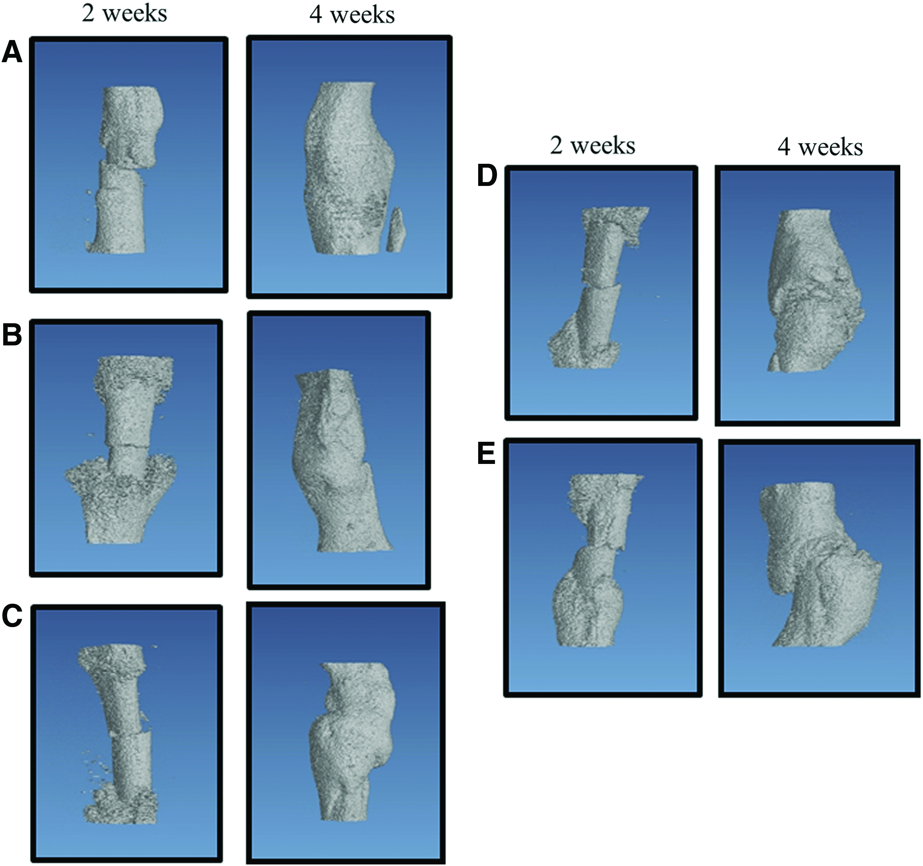
Three-dimensional images of femur fracture of mice 2 and 4 weeks after application of bFGF-free solution
Figure 3 shows the BMD values of femur fracture after treatment. Higher BMD was seen at the bone tissue regenerated in the group of gel 1, 2, and 3 than the group of bFGF-free and bFGF solution 2 weeks later. At 4 weeks, significantly enhanced BMD was found for the group of gel 3 compared with other groups.
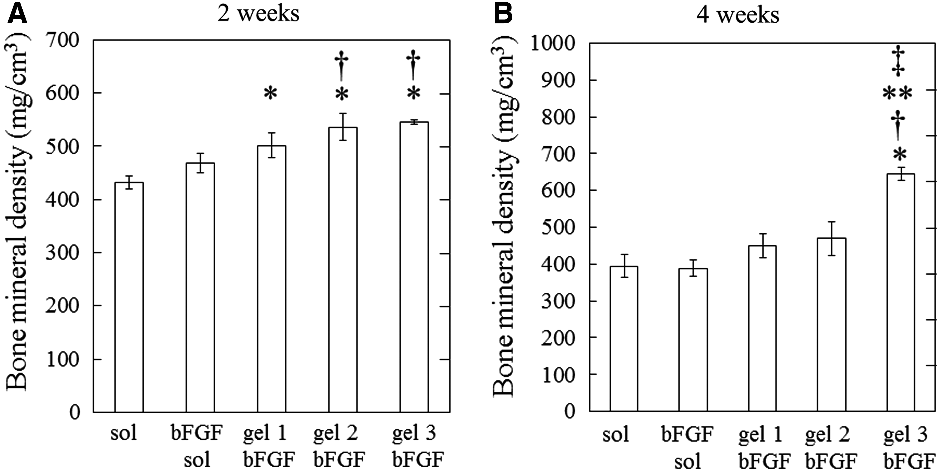
The bone mineral density of bone regenerated of the femur fracture of mice 2 and 4 weeks after application of bFGF-free solution, 100 μg of bFGF solution, gel 1 incorporating 100 μg of bFGF, gel 2 incorporating 100 μg of bFGF, and gel 3 incorporating 100 μg of bFGF. *p<0.05, significantly against the value of bFGF-free solution; †p<0.05, significantly against the value of bFGF solution; **p<0.05, significantly against the value of gel 1 incorporating 100 μg of bFGF; and ‡p<0.05, significantly against the value of gel 2 incorporating 100 μg of bFGF.
Figure 4 shows the histological section of femur fracture 2 and 4 weeks after application. At 2 weeks, cartilage tissues were observed around the fracture site, especially for the groups of gel 2 and gel 3. At 4 weeks, new bone tissues were regenerated around the fracture site for all the groups. The group of gel 3 exhibited significantly strong bone formation compared with other groups.
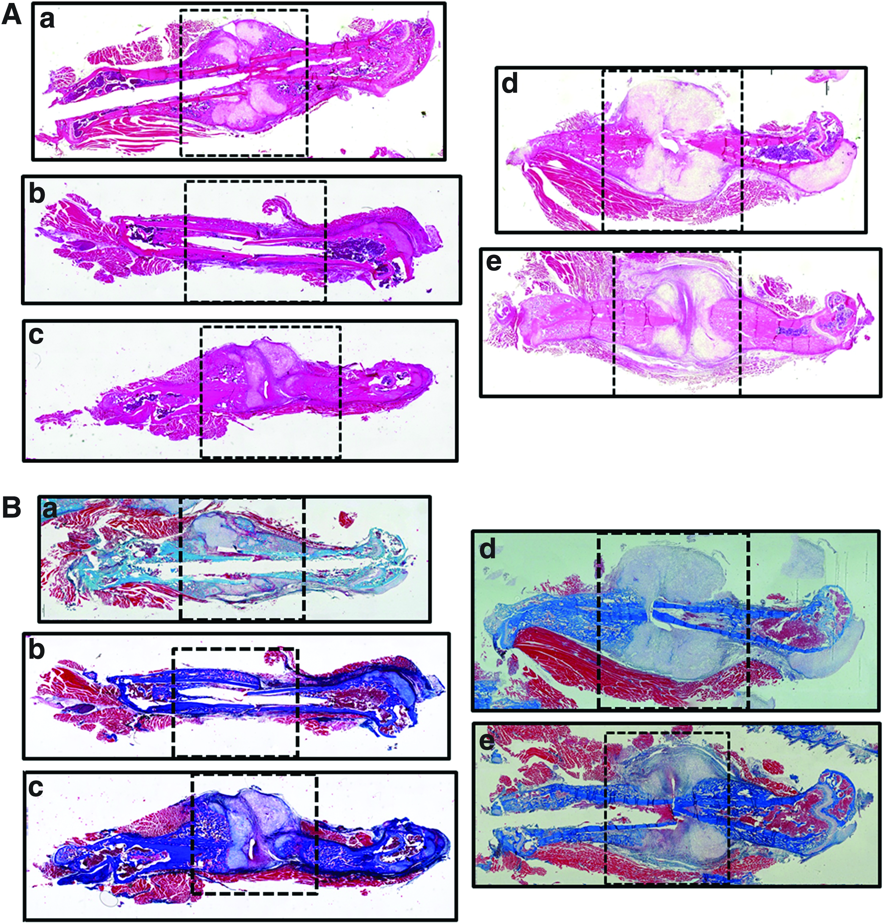
Histological sections of the femur fracture of mice 2

Expression of osteocalcin and Runx2 mRNA in the femur bone fracture
To investigate whether bFGF locally applied could affect mineralization, the mRNA expression levels of osteocalcin and Runx2 were estimated 1, 2, and 4 weeks after application. Both of expression levels for the groups of gel 1, gel 2, and gel 3 were significantly higher than those of bFGF-free or bFGF solution 2 weeks after application. The levels for gel 2 and 3 groups were significantly higher than those of bFGF-free, bFGF solution, and gel 1 groups 2 weeks after application. However, 1 week after application, there was no significant difference in the mRNA expression levels of osteocalcin and Runx2 for all groups.
Discussion
The present study investigates whether or not the time period of bFGF released from gelatin hydrogels affects the healing of a femur fracture bone, in particular on endochrondral ossification. The results demonstrate that the controlled release of bFGF for a longer time period significantly increased the area of bone tissue newly formed, the BMD at the bone fracture, and the mRNA expression of osteoblast-related osteocalcin and Runx2.
bFGF is a growth factor to trigger the proliferation of capillary endothelial cells in addition to osteoblasts. 27 Weiss et al. reported that bFGF stimulated cells involved in osteogenesis and angiogenesis in a vascularized bone graft. 28 Indeed, angiogenesis was observed around the skull defect of bFGF-treated rabbits during the initial period. It is reported that bFGF may be proliferative both for preosteoblasts 29 and marrow cells.30,31 A positive effect of bFGF on the osteogenesis in the bone marrow has also been found in vivo. 32 The higher number of osteocytes incorporated into the bone site newly formed by bFGF may be one reason to explain the phenomenon. It has been suggested that bFGF, in fact, acts mainly on endosteal cells, because the increased activity was observed in the endosteum, but not in the periosteum after the intravenous administration of bFGF to rats.10,33 In conclusion, bFGF is able to promote orthopic osteogenesis in the mice fracture model, although to a limited degree. This is because bFGF itself has a proliferative effect on cells committed to the osteogenic lineage.
Our previous researches have demonstrated that the hydrogels prepared from gelatin with an IEP of 5.0 could release bFGF in a controlled fashion.14,22 The gelatin hydrogels release bFGF either in the diffusion or hydrogel degradation manner. bFGF without interacting with gelatin molecules of hydrogel would be initially burst-released from the hydrogels by the simple diffusion mechanism. The remaining bFGF is released as a result of gelatin hydrogel degradation. The water content of hydrogels that affects their degradability could be easily changed only by altering the concentration of gelatin and GA used for the crosslinking reaction (Table 1).
In addition to the time profile of bFGF release, that of hydrogel degradation should be considered. The hydrogel remaining in the bone defect will impair physically the process of bone regeneration induced in the defects. It is demonstrated that bone tissue formation is also influenced by the degradation profile of carrier itself. 34 To break through the issue, in this study, the hydrogels were not applied into the bone defect, but the site around the defect.
Locally administered bFGF in solution is rapidly eliminated from the applied site either passively by simple diffusion, or actively by proteolysis or antibody neutralization in the tissue.9,35–37 It is well recognized that endogenous FGF is stored in the extracellular matrix (ECM) in vivo, and released in the tissue to exert its biological activity following the ECM degradation by proteases. 38 Previous studies have revealed that osteogenic differentiation is sensitive to the dose and duration of FGF exposure.39,40 It would be ideal to supply the growth factor for the time period long enough to promote the proliferation and differentiation of osteogenic cells. The in vivo release tests show that bFGF was retained at the site of hydrogels applied for a prolonged time period. Animal experiments with 125I-labeled bFGF or gelatin demonstrated that the retention period of bFGF in the body was in good accordance with that of gelatin hydrogels. This indicates that bFGF was released as a result of in vivo degradation of the hydrogel matrix. 36 The hydrogel of release carrier with an optimal degradation profile will contribute to an optimal release profile of bFGF incorporated to achieve bFGF-induced bone regeneration. The merits to use the controlled release of osteogenic growth factors are not only the efficient induction of bone regeneration, but also the decrease in the dose.
Many research groups have used the same femur fracture model to evaluate bone regeneration. The researches have demonstrated the high reproducibility and reliability of the fracture model.41–43
The present study clearly demonstrates that combination with a good carrier enables bFGF to enhance the osteogenic functions, leading to an enhanced healing of femur bone fracture. It is likely that the controlled release of bFGF from the gelatin hydrogel takes place in the tissue around the bone fracture site. We can say with certainty that the bFGF released induced the bone regeneration at the femur fracture. The bFGF-incorporated gelatin hydrogels with lower water content showed higher BMD than those of higher one (Fig. 3). The prolonged acting of bFGF must be necessary to allow osteogenic cells to proliferate and activate for bone regeneration.
To evaluate the effect of bFGF on the osteogenic differentiation of cells, the mRNA expression of osteocalcin and Runx2 was examined. The bFGF released enhanced the mRNA levels although they were higher for the hydrogels for longer time periods of bFGF release. Osteocalcin of a non-collagenous matrix protein is synthesized by mature osteoblasts.44–47 Runx2 is known to be the critical transcription factor for the development and differentiation of osteoblasts.48–51 Runx2 upregulates bone sialoprotein and osteocalcin, two major components of bone ECM synthesized exclusively by osteoblastic cells. 52 Previous researches indicated that the Runx2 expression and phosphorylation are activated by the FGF signaling via the mitogen activated protein kinase pathway or the protein kinase C pathway.53,54 Formation of a Runx2-smad osteogenic complex and subnuclear targeting are structurally and functionally inseparable for the execution and completion of BMP2 signaling for osteoblastogenesis. 55 The osteogenic differentiation would be promoted by the bFGF release, resulting in an enhanced bone regeneration.
As FGF signaling exerts a direct effect on Runx2 activation, these functional relationships may enable researchers to unravel the overall differentiation control mechanism by which a single FGF2/FGFR signaling event can reciprocally induce promotion and inhibition of osteoblast differentiation in a stage-dependent manner. Canalis et al. demonstrated that the stimulatory effect of bFGF on the osteoblastic collagen synthesis was indirect and secondary to the mitogenic effect. In addition, bFGF caused a direct inhibitory effect on the osteoblastic function manifested by a decrease in type I collagen synthesis, when calvariae were continuously treated with bFGF. This inhibitory effect of bFGF was selective for bone collagen synthesis, indicating that it was not due to a generalized toxic effect. 8 Although in vitro experiment, Debiais et al. reported that bFGF induced opposite effects on osteocalcin synthesis and matrix mineralization in early and late cultures. bFGF inhibited osteoblast differentiation markers in less-mature cells and increased late-osteoblast markers, such as osteocalcin, and matrix mineralization in more mature cells indicates that the response of cells to bFGF varies with the stage of cell maturation. 56
It is apparent from Figure 5 that the time period of bFGF release may be essential to promote bone induction activity. The bone regeneration at the fracture for the group of bFGF solution was similar to that of bFGF-free solution (Fig. 5). The ability of gelatin hydrogels incorporating bFGF to induce bone regeneration greatly depended on their water content (Figs. 3, 4C, 4D, and 5). It is likely that the fast-degraded hydrogel could neither prolong the in vivo retention of bFGF nor protect the fracture from the ingrowth of fibrous tissue. When the rate of hydrogel degradation is too slow compared with that of bone regeneration at the femur fracture, it is likely that the hydrogel remaining at the fracture physically impairs bone regeneration, even though bFGF is released over the long time period. Our previous studies experimentally confirmed that gelatin hydrogels with a low degradability physically impaired bone regeneration induced by gelatin hydrogels incorporating osteogenic growth factor both at the ulna and skull defects of rabbits.57,58 The osteoinduction activity of hydrogels with a water content of 85.1 wt% was significantly higher than that of hydrogels with lower water content. The threshold period of hydrogel degradation to start interfering bone regeneration is unclear at present and optimal time periods that enhance bone formation in vivo should be investigated.

mRNA expression of osteocalcin
In this study, we successfully examined the time profile of the degradation of gelatin hydrogel using the radioactivity agents in vivo. Considering the release mechanism, it is likely that bFGF molecules are released out being associated with water-soluble gelatin fragment. The bFGF is released as the result of hydrogel degradation. We previously examined the profile of the degradation of gelatin hydrogel and of the release of bFGF using the radioactivity in vivo.22,25
We demonstrated that vascularization was remarkable around the implantation site of gelatin hydrogels incorporating bFGF by histological examination. The amount of tissue hemoglobin, which is a measure of bFGF-induced neovascularization, notably increased within 1 day of implantation of gelatin hydrogels incorporating bFGF. 59
We also demonstrated that a rabbit model of a skull bone defect was employed to evaluate the in vivo efficacy of gelatin hydrogels incorporating bFGF in bone formation. When implanted into a skull defect, the gelatin hydrogel incorporating bFGF accelerated bone regeneration at the skull defect and almost closed the defect after 12 weeks of implantation and enhanced the BMD of the skull defect. 13
Moreover, we previously demonstrated that gelatin hydrogel incorporating bFGF enhanced tissue survival of fingertip amputations and restoration of blood flow of Buerger's disease in clinical study.60,61
Although in vitro studies and clinical trials provide adequate techniques and instruments to improve our knowledge on bone repair, animal studies still represent an essential tool to analyze the biology of fracture healing. 62 Accordingly, numerous mammalian species, ranging from the mouse to the sheep, have been introduced as models to study fracture healing.63–65
Because of a more primitive bone structure without a haversian system, small rodents, like mice and rats, are thought to be less appropriate for bone healing studies compared with larger animals. In contrast to larger rodents and other mammalians, small rodents use resorption cavities for bone remodeling during fracture healing, and this process of remodeling has been shown similar to the haversian remodeling in larger animals.
The small size of mice is challenging for the development of a fracture model. Therefore, particularly large long bones, such as the femur and the tibia, are thought to be the most appropriate for studies on fracture healing.65,66
In contrast to the tibia, the mouse femur is a tubular bone with a relatively consistent inner and outer diameters. Accordingly, different fracture sites within the diaphyseal part of the bone show comparable callus responses. Because of the straight longitudinal axis, standardized fracture stabilization is easier in the femur. It is commonly argued that murine fracture models do not require biomechanical standardization because the predominance of research using mouse models has focused on the molecular aspects of fracture healing.
Nevertheless, the most established imaging techniques to study murine fracture healing are high-resolution radiography and 2D and 3D μCT. Using conventional X-ray techniques, the size and radiologic density of the fracture callus can be analyzed in living animals and resected bones.67–69 Common parameters to be evaluated by the μCT are the tissue mineral density, total callus volume, and bone volume fraction of the callus.70,71 In addition, the assessment of in situ messenger RNA expression can be additionally supported by semiquantitative techniques, such as Northern blot analysis and reverse-transcription PCR.72,73
In our study, the controlled release system for bFGF is achieved to enhance in vivo bone regeneration with mice femur fracture model. Moreover, the quantification measurement of BMD demonstrated a significant increase in the bone regeneration for mice femur fracture. Taken together, these findings indicate that the gelatin hydrogel incorporating bFGF provides a benefit that will enable to achieve bone regeneration for mice femur fracture.
Footnotes
Disclosure Statement
No competing financial interests exist.