
Abstract
Fish scales, which consist of type I collagen and hydroxyapatite (HA), were used to fabricate a bioabsorbable bone pin in this study. Fresh fish scales were decellularized and characterized to provide higher biocompatibility. The mechanical properties of fish scales were tested, and the microstructure of an acellular fish scale was examined. The growth curve of a myoblastic cell line (C2C12), which was cultured on the acellular fish scales, implied biocompatibility in vitro, and the morphology of the cells cultured on the scales was observed using scanning electron microscopy (SEM). A bone pin made of decellularized fish scales was used for the internal fixation of femur fractures in New Zealand rabbits. Periodic X-ray evaluations were obtained, and histologic examinations were performed postoperatively. The present results show good cell growth on decellularized fish scales, implying great biocompatibility in vitro. Using SEM, the cell morphology revealed great adhesion on a native, layered collagen structure. The Young's modulus was 332±50.4 MPa and the tensile strength was 34.4±6.9 MPa for the decellularized fish scales. Animal studies revealed that a fish-scale-derived bone pin improved the healing of bone fractures and degraded with time. After an 8-week implantation, the bone pin integrated with the adjacent tissue, and new extracellular matrix was synthesized around the implant. Our results proved that fish-scale-derived bone pins are a promising implant material for bone healing and clinical applications.
Introduction
F
Metallic and polymeric internal fixation devices are frequently applied to bone fractures. However, the inappropriate mechanical strength and/or deleterious degradative products may limit clinical applications. Biodegradable fixators are mostly constructed of synthesized polymers (e.g., polyglycolide [PLA], polylactide [PGA], polycaprolactone, and polydioxanone). 8 To avoid secondary surgery, biodegradable fixators are alternatives for internal fixation and reduce the total costs of medical treatment compared with the use of metallic devices. Biodegradable fixation devices can keep fractured bones in place while being gradually degraded and eventually replaced by regenerated bone tissue. The stresses can be transferred to the surrounding bone tissues and not concentrated in the injury site. However, disadvantages of biodegradable devices include inappropriate degradation time, insufficient mechanical properties, a resulting acidic environment after degradation, and foreign-body reaction during bone remodeling. 9 Moreover, a recent study reported that the degradation of biodegradable devices will most likely not be accompanied by the restoration of normal trabecular bone, such as when PGA and PLA screws are used. 10 Therefore, a new biodegradable material should be developed to benefit fracture fixation.
To solve these foregoing problems, natural polymers, such as collagen, cellulose, and chitin, are used to fabricate orthopedic fixation devices with better biocompatible properties. Among these materials, collagen has shown to have more diverse biomedical applications. Collagen and hydroxyapatite (HA) are the major components of human bones; therefore, these two substances were reported to be the materials of choice for bone substitutes. 11 Collagen can be obtained from human, porcine, bovine, ovine, and fish origin. In particular, the major components of fish scales are type I collagen and HA, which resemble the composition of human bone tissues. 12 Further, the mechanical properties of collagen-based materials can be adjusted by the level of crosslinking and the choice of crosslinking agent. 13 Therefore, the stability of fish-origin collagen could be improved by physical or chemical crosslinking methods prior to further applications.14,15 However, the possible toxicity of residual crosslinking agent during the crosslinking method cannot be ignored16,17 because many crosslinking agents, such as glutaraldehyde and carbodiimide, prove to be cytotoxic in nature. The residual crosslinking agents and their specific crosslinks give rise to differences in sensitivity to hydrolysis and enzymatic breakdown and in direct release of products. 18 Glutaraldehyde toxicity of crosslinked collagen, whose effects can be quenched by glutamic acid and induce apoptosis, has been demonstrated using glutaraldehyde components on the surface of a material. 19 To solve these foregoing problems, the stratified layers of collagen fibrils within fish scale provide superior mechanical properties.12,20 The native collagen structure of fish scale may be considered to develop a new orthopedic fixation device with better mechanical properties and stability.
Another major component of fish scale is HA, and it is reported that screws have better stability when coated with HA. 21 In addition, HA-coated, tapered external-fixation pins can improve the strength of the bone–pin interface in osteoporotic bone, 22 which reveals the importance of HA in bone remodeling. Thus, fish-scale-derived materials with HA components shall have great potential for bone tissue repair and clinical applications.
In this study, fish scales were decellularized and characterized, and the biocompatible properties of the acellular scales were determined. Finally, the decellularized fish scales were used to fabricate bone pins, which were implanted into femur fractures of animals, to evaluate the potential of fish scales in bone regeneration.
Materials and Methods
Decellularization of fish scales
Grass carp fishes were obtained from a commercial dealer in Taipei. The fishes were stored at 4°C and immediately transported to the laboratory. Fresh scales were harvested and subsequently cleaned in distilled water. A decellularization solution containing 0.05 M Tris-buffer (Sigma-Aldrich) and 0.1 M Triton X-100 (Sigma-Aldrich) was used to remove the cellular components of the fish scales at 4°C for 3 days. Finally, the decellularized scales were rinsed with 70% ethanol and stored in sterilized phosphate-buffered saline (PBS) at 4°C before further application.
Cell nucleus staining of decellularized scales
The decellularization process of fish scales was confirmed using cell nucleus staining. Acellular samples were fixed in 3.7% neutral-buffered formaldehyde solution (Sigma-Aldrich) for 15 min at room temperature and then permeabilized with 1% Triton X-100 (Sigma-Aldrich) for 5 min. After washing twice with PBS, the decellularized scales were incubated with 4′,6-diamidino-2-phenyl-indole (DAPI; Sigma) at room temperature for 20 min. The acellular fish scales were then examined using a fluorescence microscope (Olympus BH Series) at an excitation wavelength of 350 nm.
Analysis of decellularized scales using a thermogravimetric analyzer
For the characterization of fish scales, a thermogravimetric analyzer (TGA; Perkin Elmer Pyris Diamond) was used to analyze the decomposition patterns of the organic and inorganic contents in the samples. The decellularized fish scales were further decalcified using a 0.5 M EDTA solution, and acellular fish scales with or without decalcification were examined by TGA.
Mechanical property test
The mechanical properties of decellularized scales were determined. The test samples were prepared according to the ASTM D638-10 standard test method for tensile properties of samples using a laser system (VLS-4.6; UNVERSAL). The specimens possessed a gage section length and width of 10 and 0.78 mm. This test method is useful to evaluate the mechanical properties of materials. All samples have not been preconditioned. The tensile strength of each sample was determined using a universal testing machine (H1KS; Tinius Olsen) with a 100-N load cell, and the maximum frame capacity is 1 kN. The test was performed at a crosshead speed of 6 mm/min at room temperature, and measurements were performed for six independent samples. The Young's modulus was calculated using the stress–strain curve by extrapolation of the initial, linear portion of the curves. The fracture stress for the ultimate tensile strength was observed using the stress–strain curve. The results were expressed as the mean±SD of six experiments.
Evaluation of cell proliferation on decellularized scales
A mouse muscle myoblastic cell line, C2C12 (CRL 1772; ATCC), was cultured and expanded in high-glucose Dulbecco's modified Eagle's medium (DMEM, 4.5 g/L glucose, 12100-061; Gibco) supplied with 4 mM L-glutamine, 1% antibiotic (15240-062; Gibco), and 10% fetal bovine serum (15622-029; Gibco) in an incubator at 5% CO2 and 37°C.
The decellularized scale was cut as a disc with a diameter of 11 mm and placed into a 24-well culture plate. C2C12 cells were seeded at a density of 8000 cells per fish scale and cultured for 1, 2, and 3 days. At predetermined intervals, the cell proliferation was determined by cell number counting. The control group of this test was the same number of C2C12 cells cultured in a 24-well culture plate for the same intervals. This test was performed in six replicates for each group.
Scanning electron microscopy examination
Scanning electron microscopy (SEM, JSM-5610; JEOL) was used to examine the surface morphology of the scale and after cell attachment. After culture for 3 days, C2C12 cells cultured on the decellularized fish scales were washed twice with PBS to remove loosely adherent cells, and the samples were fixed in 2.5% glutaraldehyde in PBS for 10 min and then dehydrated by treatment with a series of graded ethanol solutions. The samples were further critical-point dried (model E3100; Quorum Technologies), sputtered with gold ions using an SEM coating system (Sputter Coater 11430; SPI), and finally examined using SEM.
Preparation of a pin implant from fish scale
The decellularized fish scales were prepared as a pin shape in a needle mold followed by a heat-drying process. The bone pin was fabricated by 40°C heating to dry out the water for 16 h. Subsequently, the fish scale pins were sterilized by 18-kGy gamma ray irradiation and used as internal fixation for small bone fractures.
Animal study
Experimental protocols and surgical procedures of the animal study were approved by the Faculty of Mackay Memorial Hospital Commission for Animal Experimentation. Twenty-four New Zealand White rabbits (6-months old and 3 kg in weight) were included in this study. Animals were maintained in accordance with the International Council for Laboratory Animal Science guidelines for the care and use of laboratory animals. Rabbits were marked to permit individual identification and kept in their cages for 14 days prior to the beginning of experiments to allow for acclimatization to laboratory conditions.
The operation was performed under general anesthesia. 23 In brief, animals were treated with nothing-per-oral 12 h before surgery to prevent inhalation pneumonia during operation, and Zoletils (0.8 mg/kg body weight; Virbac) was injected intramuscularly to induce anesthesia. After adequate skin preparation and sterilization, a 4-cm longitudinal incision was made over the dorsum of the forearm followed by meticulous dissection. The radius was exposed and a transverse fracture was made using an electric saw (Fig. 5c). The internal fixation was performed using standard procedures. The fracture was reduced properly and fixed with clamps, and the hole for the implant was drilled obliquely by a metallic pin to cross the transverse fracture site. The fish scale pin was inserted into the drilled, measured hole to keep the fracture stable (Fig. 5d). After the just explained procedures were performed, the wound was closed in layers, and the rabbits were returned to a padded cage until spontaneous and purposeful movement was noted. The left forelimb of each rabbit was treated, and a right forelimb radial fracture was created without internal fixation as a control.
The animals were examined individually on a daily basis throughout the study period, and all observations were systematically recorded. Additional observations were recorded if the animals displayed signs of toxicity. Animals found in a moribund condition and those showing severe pain or enduring signs of severe distress were humanely sacrificed. When animals were sacrificed for humane reasons or found dead, the time of death was recorded. At 4, 8, 12, and 24 weeks (n=6 for each time point) postoperation, the animals were euthanized, and the specimens were harvested and preserved for further examinations.
Radiologic analysis
Rabbits underwent standardized, serial radiography immediately after surgery at 4, 8, 12, and 24 weeks. All radiographs were size standardized, the harvested specimens received radiographic analysis, and the radiographic images were digitized. The radiographic pictures were used to evaluate the healing of the fractures with time.
Histological analysis
After radiographic analysis, the samples were fixed in a 10% neutral buffer formalin solution, decalcified in hydrochloride, and embedded in paraffin wax. Consecutive sections were cut from the paraffin blocks into 6-μm slides, and the sections were deparaffinized and stained with hematoxylin and eosin (H&E) to assess the cell/matrix morphology. Other sections were stained with Masson's Trichrome staining for collagen-fiber staining. 24
Results
Morphology of decellularized fish scales
DAPI staining revealed identified cell nuclei with blue fluorescence and that the cells were distributed homogenously in the untreated fish scales (Fig. 1a). After decellularization, the cells were removed and no cell nuclei were found on the acellular fish scales (Fig. 1b). Based on SEM of the cross-section of a fish scale, the stratified layers of collagen fibrils (Fig. 1c) stacked tightly with an orthogonal arrangement. A lateral view of the fish scales revealed that the morphology of the stratified collagen layers was not influenced after the decellularization process (Fig. 1d).

Morphology of fish scale surface with and without decellularization.
Cell proliferation and morphology on decellularized fish scales
C2C12 myoblasts grew well on the fish scale, and confluent cells were found (Fig. 2a). Higher magnification views of the scales showed that the cells adhered onto the surface along collagen fibrils with a specific pattern (Fig. 2b). Abundant extracellular matrixes were also observed. The cell proliferation was examined by cell number counting, and the results showed increasing growth with time, which reveals that the cells proliferated normally on the fish scales (Fig. 2c).

Growth of C2C12 myoblasts on decellularized fish scale. Morphology of fish scale examined by SEM.
Thermal analysis
In the results of the TGA analysis, the total weight was decreased as the temperature increased. For the decellularized scales, the first peak was located between 49°C and 100°C, and the initial weight decrease represented the water loss from the scales (Fig. 3a). The second peak at 320°C was attributed to the decomposition of the organic components of the fish scales. Finally, the weight of the residual inorganic components was 31.2% of the original fish scales when the temperature was raised to 1000°C. Compared with the decellularized scale, the weight of the inorganic compositions was decreased to 0% in the decalcified fish scales (Fig. 3b).
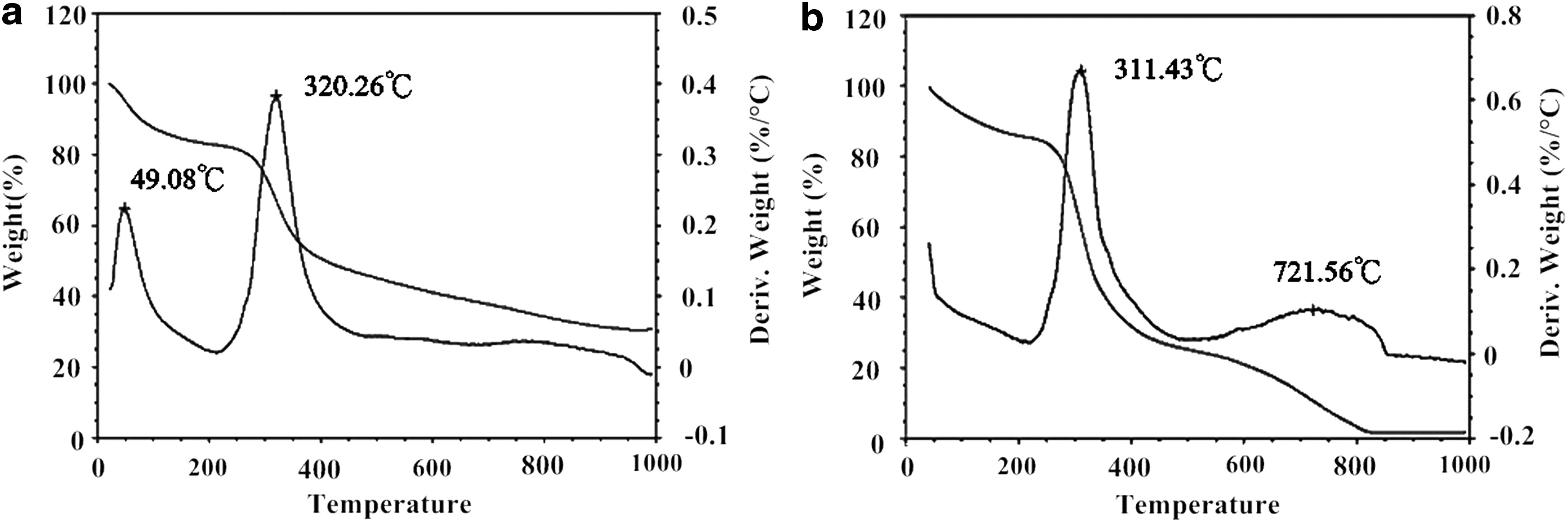
Thermal properties analyzed by TGA.
Mechanical property of the fish scales
A test sample for mechanical strength was selected from the area of the scale as shown in Figure 4a, and the sample was clamped onto the sample holder of the universal testing machine (Fig. 4b). The stress–strain curves were obtained and used for Young's modulus calculation (Fig. 4c), which was measured as 332±50.4 MPa, and the tensile strength was measured as 34.4±6.9 MPa for the decellularized scale samples.
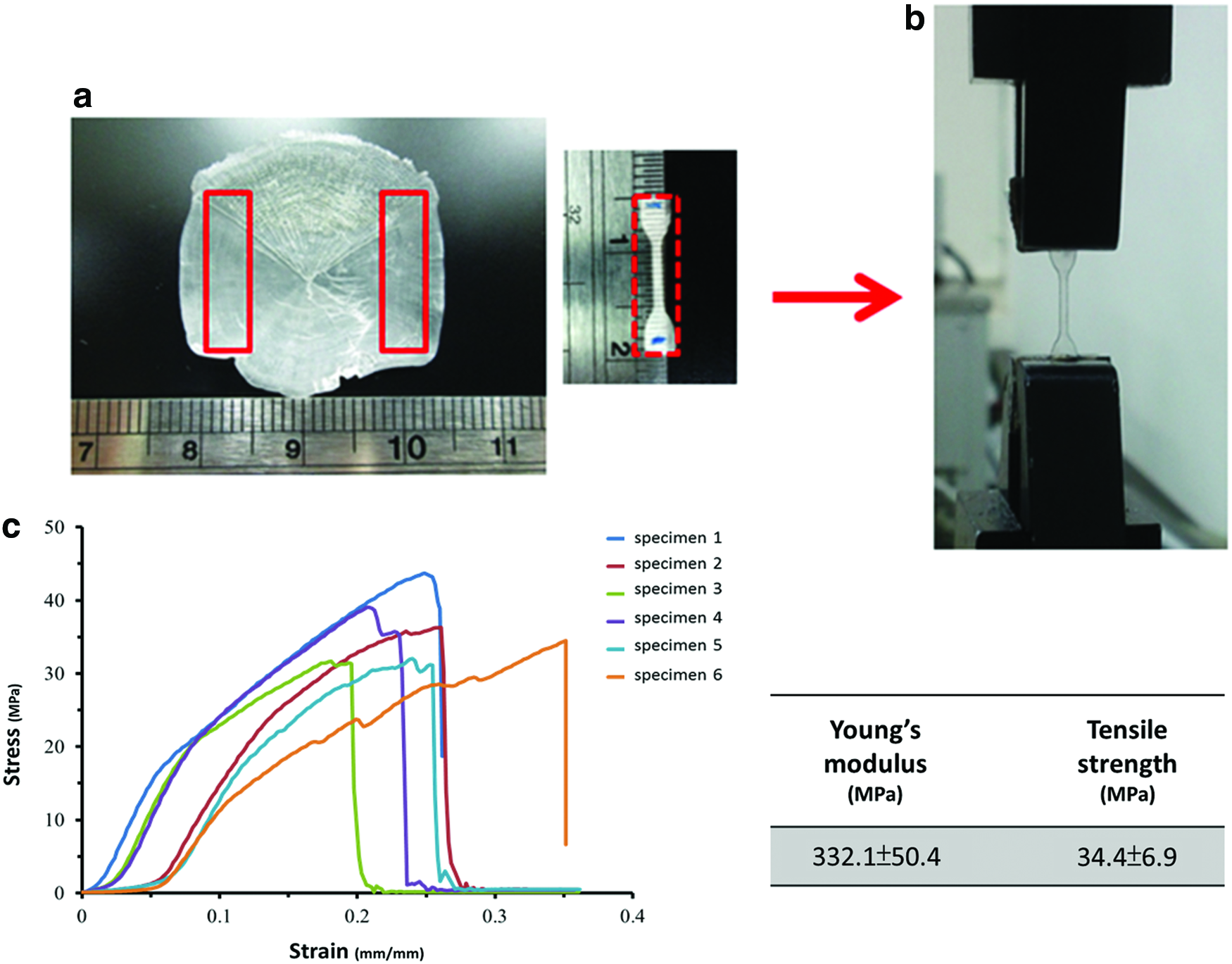
Mechanical test on fish scale.
Bone pin preparation and animal study
Figure 5a shows the needle mold for bone pin preparation, and pins with a length of 2 cm and diameter of 1 mm were obtained (Fig. 5b). Twenty-four experimental rabbits recovered well postoperatively. The bone fracture was created in the radius (right forelimb, control group, Fig. 5c), and the fish scale bone pin was implanted (left forelimb, Fig. 5d). The radial fracture of the forelimb (Fig. 5e) and bone pin implantation (Fig. 5f) were visualized using X-ray images. During the observation periods, no abnormal clinical signs were noted in any rabbit, and no animal died during the experimental period. Twenty-four experimental rabbits tolerated the presence of the fish scale pins well, and no infection was observed.
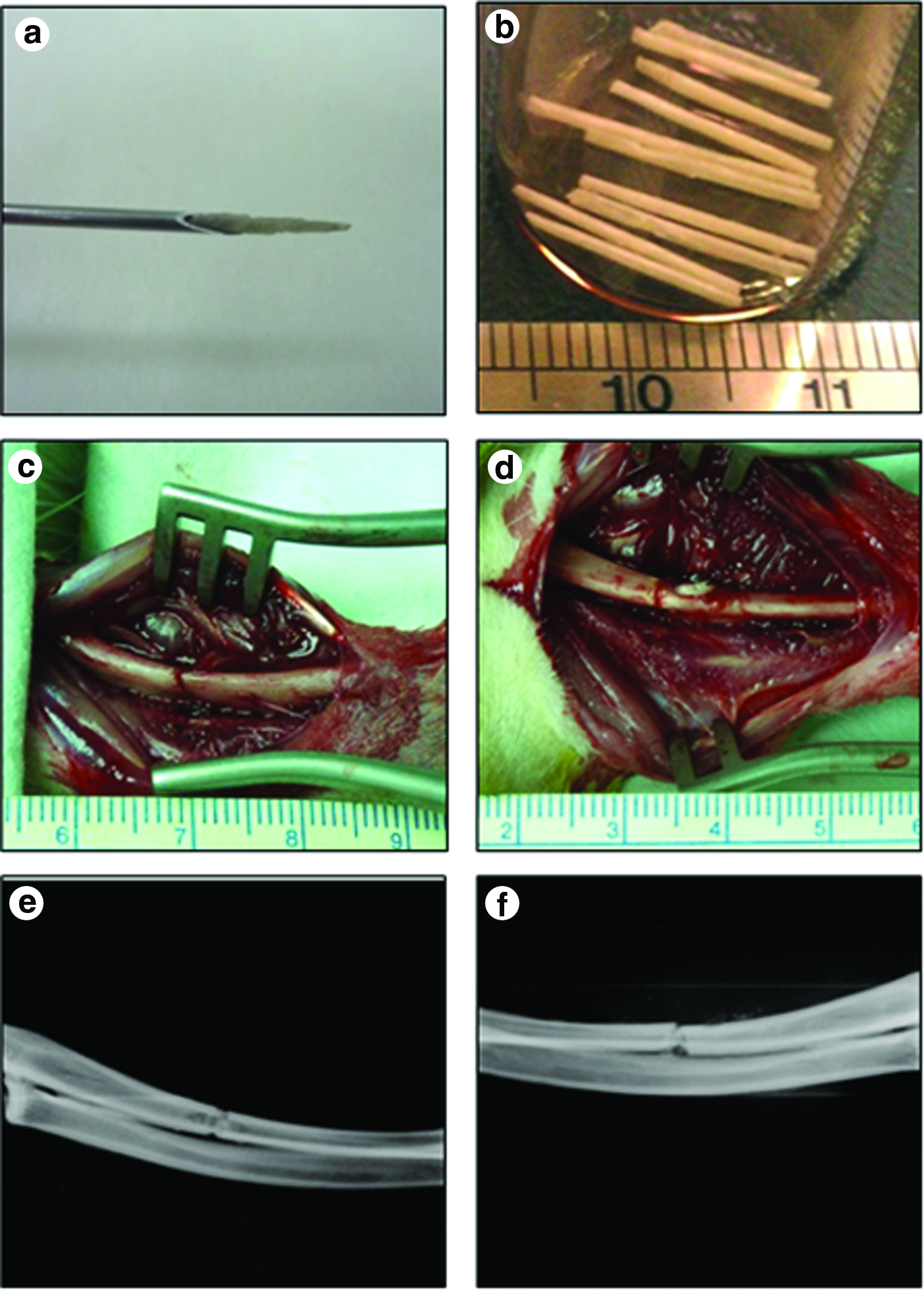
Fish scale pin implantation.
Regular radiological examination was performed at the predetermined intervals. Based on the radiographic analysis, the progress of fracture healing was determined for limbs implanted with a bone pin (Fig. 6). For the control group, nonunion was noticed. Although the stiffness of the fish scale bone pin was much lower than that of a traditional metallic pin, the immobilization of the forelimb fracture was kept well with the aid of the fish scale bone pin. Fracture union was achieved 12 weeks after surgery for all defects that were fixed with the fish scale bone pin.

Bone healing analyzed by X-ray photography and histology. Regular radiological examination was undergone at 4, 8, 12, and 24 weeks postoperatively. Fracture union was observed at 12 weeks postsurgery for defects fixed with the fish scale bone pin. The bone implantation was also harvested at predetermined intervals. The samples were prepared for H&E and Masson's Trichrome (MT) staining. Inflammatory cells were found around the bone pin at week 4. Mild-to-moderate inflammatory reaction could be seen and the bone pin was integrated with the surrounding tissue at week 8. The surrounding cells grew into the bone pin and new extracellular matrix was also formed around the implant. The degradation of the fish scale bone pin and amount of ingrowth of fibrovascular tissues were more evident with new bone formation at week 12. Gradual integration of the fish scale bone pin at week 24 was obvious. MT staining showed that the newly secreted collagen around and within the matrix increased with time. The results showed the bone tissue ingrowth with new collagen synthesis and fish scale degrading with time. H&E, hematoxylin and eosin. Arrows indicate the implantation site. Dotted red squares indicate the fish scale bone pin in fracture site. Red square boxes indicate high magnification image of H&E and MT from fracture site with bone pin. Color images available online at www.liebertpub.com/tea
At weeks 4, 8, 12, and 24, the animals were sacrificed, and the forelimbs were harvested for histological examination (Fig. 6). For gross inspection of the specimens from the experimental animals, the structures of both forelimbs were intact and the fracture was healing well. Moreover, Supplementary Figure S1 (Supplementary Data are available online at www.liebertpub.com/tea) shows that the restoration of the fracture with bone pin fixation was better than that of the untreated fracture. Based on histological analysis, the bone pin showed good biocompatibility using H&E staining. At 4 weeks postimplantation, inflammatory cells were found around the bone pin. However, no evidence of infection or rejection was observed. After 8 weeks, only a mild-to-moderate inflammatory reaction could be observed, and the bone pin was integrated with the surrounding tissue. The surrounding cells grew into the bone pin, and new extracellular matrix was formed around the implant. After 12 weeks, degradation of the fish scale bone pin and the ingrowth of fibrovascular tissues were more evident, and new bone formation was noted around the implant. After 24 weeks, an obvious, gradual integration of the fish scale bone pin was observed, and degradation of the bone pin and newly formed osteoid tissue around the degraded matrixes were simultaneously shown. The surrounding osteoprogenitors and osteoblasts invaded the matrix, forming new osteoids adjacent to the ingrowth of macrophages. In contrast, the bone fracture of some rabbits of the control group recovered spontaneously, even in the absence of bone pin fixation; however, delayed union or nonunion was found in these rabbits at the same intervals. Serial radiographic images showed that delayed union of the right limb was found at 6 and 12 weeks (Supplementary Fig. S1). Using Masson's Trichrome staining, the collagen in the tissue was stained blue, and nuclei were stained black. With increasing time, the newly secreted collagen around and within the matrix also increased.
Discussion
Accurate reconstruction and secure fixation are critical for the repair of fractured bones, while unstable fixation may result in nonunion. To achieve better fracture fixation, fixation devices made of bioabsorbable materials, such as poly-L-lactide (PLLA), have been shown to have various clinical advantages. However, several disadvantages of PLLA, including delayed foreign-body reaction, have been reported in the past.25,26 It has also been reported that the ideal biomaterials for constructing fixation devices should be able to help fracture healing and promote bony tissue integration/immobilization. 27 Compared with PLLA, HA-collagen hybrids that mimic the bone composition may be a better material for bone repair. 11
Collagen used for clinical purposes can be derived from various sources and prepared through different processes, such as extraction from fish origin.13,14 It has been found that higher collagen-matrix density confers higher tensile strength with a slower absorption rate. 28 Moreover, the pore size, porosity, and surface pattern of a bone filling material have been shown to influence the bone remodeling process. In collagen-based, biodegradable bone implants, crosslinking is often used to improve mechanical properties, and the degradation rate is consequently modulated by the level of crosslinking.29,30 To evaluate the mechanical properties of fish scale in this study, the chosen area of the fish scale had to be the adequate size and a complete and homogenous, collagen-layered structure. Therefore, fish scale with high-density collagen distribution will reveal better mechanical properties. 31
Decellularization is a critical process in the preparation of fish-scale-derived materials that eliminates any cellular residue of the fish scale. Incomplete decellularization may cause an acute immune response when the fish scale is implanted. An ideal decellularization process should be able to thoroughly remove the fish cells and maintain the native mechanical strength. In our preparation procedure, the cell layer that is located on the surface of the fish scales was completely removed after decellularization treatment without altering the specific pattern of the fish scales.
Cell growth on the top of the decellularized fish scales was observed to be similar to cell culture on a tissue culture plate (Fig. 3c), which showed that the acellular fish scales provided a biocompatible surface for cell growth. In addition, we observed that the cells were elongated and distributed along the surface of the fish scale (Fig. 3a). High-resolution SEM images further showed that the cells adhered to the collagen fibrils of the specific layer and secreted abundant extracellular matrices (Fig. 3b). These results revealed that decellularized fish scales provided an original structure with stratified layers of collagen fibrils for cell attachment.
Numerous clinical studies support the use of controlled, low-temperature irradiation of allograft implants. Sterilization is another preparation step that may affect the mechanical property of fish scales. We chose gamma-ray irradiation to sterilize fish-scale-derived materials because that irradiation has been shown to cause much less disruption to collagen fibrils compared with autoclaving.32,33 The low, 10–20-kGy dosage that was used for terminal sterilization deactivated 99.9% of any remaining, low-density HIV in allograft tissues. 34 Low-dose gamma irradiation tissue-processing procedures allow for thorough bactericidal treatment while maintaining intrinsic biomechanical properties and ensuring successful clinical performance. 35 Additionally, the fish-scale-derived bone pin in this study had passed sterilization validation of gamma irradiation from SGS Taiwan Ltd. In another study, gamma-ray irradiation resulted in a slight dose-dependent reduction of the elasticity of a hydroxyapatite/collagen implant. Moreover, the bone conductivity at skeletal implantation sites was not impaired after gamma-ray irradiation. 36
Moreira-Silva et al. have reported that the naturally occurring patterns on fish scales can guide human adipose-derived stem cells to grow and support cell adhesion and cytoskeletal organization. 37 Hence, in vivo bone repair may be promoted by better cell growth and adhesion. In addition, fish scales display good mechanical properties because of their layered/oriented collagen fibers with HA. 38 Therefore, fish scales possess outstanding pliable and tensile strength to be used as bone pins for fracture fixation.
The thermal properties of different scales can be evaluated for molecule stability. The grass carp fish scale was evaluated using TGA/DTA and showed a bound water loss lower than 200°C, while the collagen shrinkage temperature was at a low temperature of 49.08°C. 39 These results showed that collagen fragmentation at ∼320.26°C was higher than 311.43°C for the decalcified scale. The higher fragmentation temperature for the acellular collagen-HA composition could be attributed to the more stable thermal property of the decellularized fish scale. A high decomposition temperature occurring at ∼721.56°C was observed for the decalcified scale. 40 In the native fish scale, decomposition occurred at a broader temperature range of 600°C to 1000°C. These results showed a more stable composition of decellularized fish scale collagen with HA than decalcified fish scale, which indicated that natural HA could help with stability. The major collagen within fish scale is type I; however, its molecular weight is ∼270 kDa, which is smaller than the mammalian collagen of 320 kDa.41,42 In addition, the content of hydroxyproline of fish scale is less than that of mammals and could affect the denaturation temperature of type I collagen within fish scale. 42 The denaturation temperature of fish scale collagen is ∼25°C, which is slightly higher than its living temperature. 40 Once the thermal property was more stable for the decellularized fish scale structure than the decalcified scale, the grass carp fish scale showed a good thermal property as a material for bone pin.
In the rabbit fracture model, a bone pin constructed of decellularized fish scale showed an ability to immobilize fractured bones and stabilize fixation (Fig. 6), resulting in good bone repair. According to our histological analysis, inflammatory cells were initially observed around the bone pin. However, previous studies showed that these macrophages decreased over time, and collagen was found to be secreted by osteoblasts in the defect site. 43 Although the fish scale graft consisted of dense collagen, it still allowed cells to grow/migrate inside and secret extracellular matrix. We have found that the tissue adjacent to the graft integrated well with gradual graft degradation over time. Such bone repair processes may be promoted by the osteoconductive HA in the graft.44–46 Therefore, the HA in the fish scale graft may contribute to bone repair. The degradation time was at least 6 months, as shown in this study. For fractures or defect remodeling, the required time for bone repair is ∼4–6 months; therefore, degradation of the graft should satisfy this requirement for bone repair progression. The results showed characteristics of good biocompatibility and degradation properties in bone fracture of this rabbit model after 6 months.
This preliminary study can provide an animal model and promising material properties for bone repair or regeneration in future studies. The native structure of fish scale possesses good mechanical properties and biocompatibility. Further, fish-scale-derived materials can be used as scaffolds or implants for tissue regeneration.
Conclusions
We have shown the responses of cell morphology, cell viability, and bone implantation when using fish scale. Our results demonstrated that cells adhered onto the fish scale surface along collagen fibrils with a specific pattern. The fish scale also displayed a biocompatible surface for the C2C12 cell line in terms of the edge as well as lateral wall of an acellular fish scale. In addition, the in vivo results of a rabbit model showed that a fish-scale-derived bone pin can provide good biocompatibility for tissue ingrowth and integration. The role of implant composition in tissue reactions would require further elucidation. Fish scale provides a native structure composition that could be a promising implant material for bone repair. This study suggests that decellularized fish scale could be a potential material for bone healing that should be further studied for the regeneration of other tissues and clinical applications.
Footnotes
Acknowledgments
The authors thank Wan-Ching Lee at Mackay Memorial Hospital for her great contribution in histological staining and cell culture and Shiang-Huan Hou of Body Organ Biomedical Corporation for his excellent examination of mechanical properties.
Disclosure Statement
No competing financial interests exist.
References
Supplementary Material
Please find the following supplemental material available below.
For Open Access articles published under a Creative Commons License, all supplemental material carries the same license as the article it is associated with.
For non-Open Access articles published, all supplemental material carries a non-exclusive license, and permission requests for re-use of supplemental material or any part of supplemental material shall be sent directly to the copyright owner as specified in the copyright notice associated with the article.