
Abstract
Scaffold architecture and composition are crucial parameters determining the initial cell spatial distribution and consequently bone tissue formation. Three-dimensional poly-ɛ-caprolactone (PCL) scaffolds with a 0/90° lay-down pattern were plotted and subjected to (1) an oxygen plasma (PCL O) or (2) a postargon plasma modification with gelatin and fibronectin (PCL Fn). These scaffolds with an open pore structure were compared with more compact scaffolds fabricated by conventional processing techniques: oxidized polylactic acid (LA O) and collagen (COL) scaffolds. Human adipose tissue-derived stem cell/scaffold interactions were studied. The study revealed that the biomimetic surface modification of plotted scaffolds did not increase the seeding efficiency. The proliferation and colonization was superior for PCL Fn in comparison with PCL O. The plotted PCL Fn was completely colonized throughout the scaffold, whereas conventional scaffolds only at the edge. Protein-based scaffolds (PCL Fn and COL) enhanced the differentiation, although plotted scaffolds showed a delay in their differentiation compared with compact scaffolds. In conclusion, protein modification of plotted PCL scaffolds enhances uniform tissue formation, but shows a delayed differentiation in comparison with compact scaffolds. The present study demonstrates that biomimetic PCL scaffolds could serve as a guiding template to obtain a uniform bone tissue formation in vivo.
Introduction
A
The hypothesis of this study is that cell density is a crucial factor in the differentiation process and tissue formation in 3D scaffolds and can be regulated by the scaffold architecture and composition. The influence of scaffold architecture (plotted vs. conventional scaffold) and composition/surface chemistry (oxidized polyester vs. protein-based surfaces) on adipose tissue-derived stem cell (ADSC) adhesion, proliferation, colonization, and differentiation was studied. Two modification strategies (1) an oxygen plasma modification and (2) a double protein coating by means of postargon plasma AEMA grafting followed by gelatin immobilization and Fn physisorption, on plotted PCL scaffolds were compared. These plotted scaffolds with an open pore architecture were compared with conventional scaffolds based on oxidized poly D,D-L,L-lactic acid (LA O) and COL having a more compact architecture with an irregular pore network.
Materials and Methods
Three-dimensional PCL scaffold fabrication
PCL pellets (MW=80 000 g.mol−1) were obtained from the Sigma-Aldrich Company. Porous cylindrical PCL scaffolds with a height of 3 mm and a diameter of 4.5 mm were produced using the Bioscaffolder® device (Sys-Eng). The scaffolds were designed in Inventor, while PrimCam (Sys-Eng) was used to create the final structure. The needle was 27 gauge, the pressure was maintained at 5 bar, and the temperature was set to 120°C. The selected lay-down pattern was 0/90° and the anticipated pore size was 300 μm.26,27
Scaffold surface modification
Oxygen plasma modification
Scaffolds were subjected to an oxygen plasma treatment (dielectrical discharge plasma reactor, Model Femto, version 3; Diener Electronic) for 60 s. 27 These scaffolds will be denoted as PCL O.
Multistep gelatin-Fn modification
Scaffolds were subjected to a multistep procedure involving a double protein coating of gelatin and Fn.24–26 Briefly, PCL scaffolds were preactivated by Ar plasma treatment followed by exposure to the atmosphere. These scaffolds were immersed in a 1 M AEMA solution and subsequently subjected to UV irradiation. After thoroughly rinsing with deionized water, the scaffolds were immersed in 1 mg/mL of gelatin type B (gelB) solution in distilled water. Subsequently, 1 mg/mL of water soluble 1-ethyl-3(-3-dimethylaminopropyl) carbodiimide hydrochloride solution was added. After this immobilization step, several cleaning cycles were performed using deionized water followed by an overnight incubation at 37°C. In the last modification step, the scaffolds were coated with Fn by immersion in a 0.1 mg/mL Fn solution for 60 s followed by drying at an ambient atmosphere. These scaffolds will be denoted as PCL Fn.
PCL O and PCL Fn scaffolds were sterilized using ethylene oxide (UZGhent) for the cell culture experiments.
Reference scaffolds
BD™ three dimensional OPLA® scaffolds (LA O) (Cat. No. 354614) and BD three dimensional COL scaffolds (Cat. No. 354613) (Becton Dickinson) were used as reference materials. LA O is synthesized from D,D-L,L polylactic acid and oxidized by an atmospheric plasma treatment. COL comprised a mixture of soluble and fibrillar COL (type I and type II COLs). These sponge-type 3D reference scaffolds have a diameter of 4.7 mm and a height of 2.25 mm.
Scaffold characterization
Scanning electron microscopy (SEM) analysis was performed on a JEOL JSM-5600 (Jeol) instrument. The apparatus was used in the secondary electron mode (SEI). Different dimensions of the scaffold were measured: strut diameter, interstrut distance, height of the struts, and pore size (mean and SD of triplicate values). The porosity of 3D plotted scaffolds was calculated. 28 Bioplotted PCL and the conventional scaffolds were evaluated using the inverted contrast light microscope (Olympus inverted Research System Microscope; CellM Software).
Cell culture and cell seeding onto PCL and conventional scaffolds
ADSCs (Cryo-Save) were plated at a density of 5000 cells/cm2 in MesenPRO (Invitrogen) and expanded until P3-6 that was used for all experiments performed in our study. Cells were cultured at 37°C (5% CO2). For the cell/scaffold experiments, cells were cultured in α-MEM glutamax (Gibco Invitrogen) supplemented with 10% fetal calf serum (Gibco Invitrogen) and 0.5 vol% penicillin–streptomycin (10,000 U/mL-10,000 μg/mL; Gibco Invitrogen) named as standard medium.
Before cell seeding, the scaffolds were immersed in a serum-free standard medium in Eppendorf tubes. Air was removed from their pores by generating vacuum with a 20-mL syringe equipped with a 18-gauge needle. The scaffolds were left in the medium on a gyratory shaker (37°C, 70 rpm). After 24 h, the scaffolds were placed into 96-well tissue culture dishes (for suspension culture). Cells were seeded at a density of 0.75×10⁶ cells/40 μL/scaffold and were allowed to adhere for 4 h. Medium (160 μL) was added to each well and the seeded scaffolds were further incubated overnight. After 24 h, cell/scaffold constructs were placed in a 12-well plate. Three milliliters of osteogenic culture medium (standard medium supplemented with 100 μM L-ascorbic acid 2-phosphate [Sigma], 100 nM dexamethasone [Sigma], and 10 mM β-glycerophosphate [Sigma]) was added and the cell/scaffold constructs were cultured for 28 days (5% CO2/95% air, 37°C) with a culture medium change twice a week.
Characterization of cell/scaffold constructs
Cell seeding efficiency, adhesion, proliferation, colonization, and differentiation were evaluated at different time points (1, 7, 14, and 28 days postseeding).
Seeding efficiency
Twelve hours postseeding, the scaffolds were removed and the remaining cells in the wells were counted using a counting chamber. The seeding efficiency was calculated using the equation:
Phase-contrast and fluorescence microscopy
To visualize cell adhesion and colonization on the scaffolds, cell/scaffold constructs were evaluated using inverted contrast light microscopy and fluorescence microscopy (Olympus inverted Research System Microscope, type U-RFL-T; CellM Software) after calcein acetoxymethylester/propidium iodide staining as described previously. 21
Histology
Cell/scaffold constructs were rinsed with phosphate-buffered saline (PBS), fixed with 4% phosphate (10 mM)-buffered formaldehyde (pH 6.9) (4°C, 24 h), dehydrated, and embedded in paraffin. Five- to 7-μm sections were stained with hematoxylin and eosin and Masson's Trichrome and mounted with a mounting medium (Cat.No. 4111E; Richard-Allan Scientific).
Immunohistochemistry
Immunohistochemistry, using antibodies directed against collagen I (COL I) (polyclonal rabbit anti-human, Acris R1038; Acris) and osteocalcin (OCN, polyclonal goat anti-human, V-19 sc-18319; Santa Cruz Biotechnology), was performed on the tissue sections as described previously. 21
Protein assay and alkaline phosphatase activity
Cell/scaffold constructs were lysed into 0.5 mL of a 1% Triton X-100 containing Tris HCl buffer, homogenized by two freeze–thaw cycles and sonicated on ice for 3×10 s (amplitude of 40%) (Vibra Cell™ SONICS [ANALIS]). The protein content and alkaline phosphatase (ALP) were determined as previously. 29 The ALP activity was expressed as mM p-nitrophenol/mg protein.
Real-time RT-PCR analysis
ADSC cultures were trypsinized, collected, and centrifuged at 1000 rpm, for 5 min. After removal of the supernatant, the TRI Reagent was added. Cell/scaffold constructs were rinsed in PBS and the TRI Reagent was added. RNA was isolated followed by DNA treatment and RNA was transcribed to cDNA. Real-time PCR was performed on the ABI 7500 Fast Real Time PCR device with TaqMan probes for the following genes: runt-related transcription factor (RUNX2), collagen type I α1 (COL1A1), and bone γ-carboxyglutamate protein (BGLAP) (OCN) as described previously. 21 Relative quantification (Rq, n fold expression) values were calculated using the equation 2−ΔΔCt.
Statistical analysis
A Mann–Whitney test using SPSS 19.0 was performed to compare the differences among groups. Differences among groups were considered as statistically significant when p≤0.05. Mean and SD are reported in each Figure.
Results
Scaffold characterization
In Figure 1, a photograph of the bioscaffolder (Fig. 1a) and a plotted PCL scaffold with a 0/90 lay-down pattern (Fig. 1a, insert) are presented. SEM images of a top view (c) and a cross section (d) are presented. The scaffold properties are presented in Table 1, according to the scaffold dimensional parameters (Fig. 1b). Phase-contrast micrographs of the scaffolds are shown in Figure 2. The plotted scaffolds (Fig. 2a, d) consist of repeating structural units, well controlled interconnected pores with a pore size of±300 μm and a porosity of 60–78%. This is in contrast to LA O (Fig. 2b, e) and COL (Fig. 2c, f) scaffolds, which have a more compact architecture, an irregular structure, and pore morphology (pore size±100–200 μm) and a porosity of 90–98%. The fibrillar network of both scaffolds is presented in Figure 2c and f.

Fabrication and characterization of bioplotted poly-ɛ-caprolactone (PCL) scaffolds.
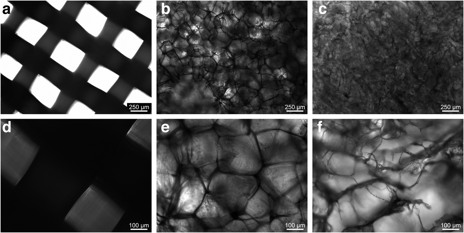
Phase-contrast images.
PCL, poly-ɛ-caprolactone; LA O, oxidized polylactic acid; COL, collagen; n.a., not applicable.
The characterization of surface modified PCL scaffolds by direct oxygen plasma (PCL O) and a multistep procedure involving postargon plasma AEMA grafting, gelB immobilization, and Fn physisorption (PCL Fn) was reported earlier.24,25,27,30
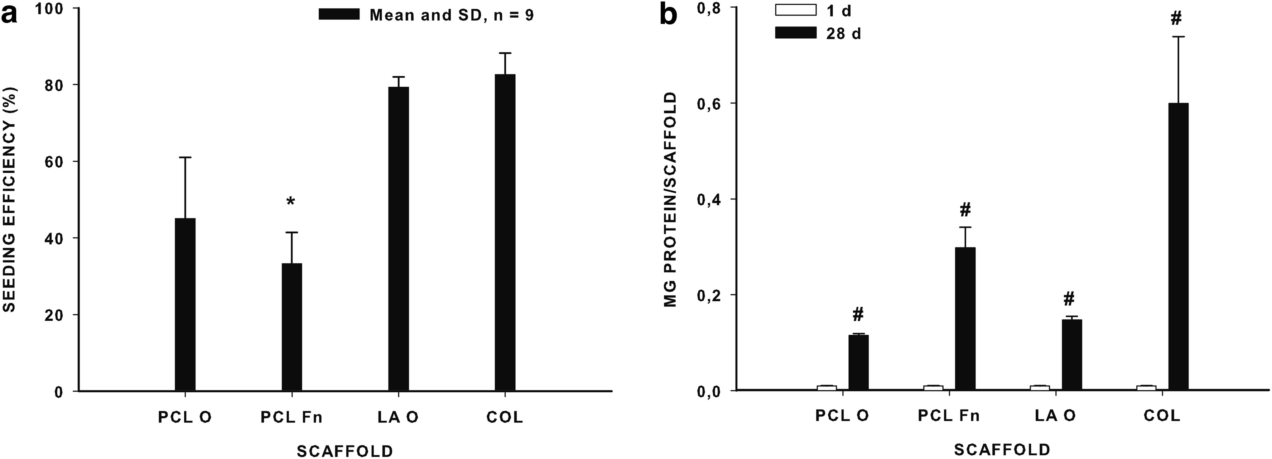
Cell seeding efficiency
The seeding efficiency is presented in Figure 3a. Low-seeding efficiencies were obtained for plotted PCL scaffolds: PCL O (45.1%±15.9%) and PCL Fn (33.3%±8.1%). Conventional LA O- and COL-based scaffolds reach seeding efficiencies of 78.6%±2.6%, respectively, 82.7%±5.5%.
Cell proliferation and colonization
The results of cell imaging for plotted and conventional scaffolds as a function of time are depicted in Figure 4. After 4 h, a lot of round, nonadherent cells between the polymer struts of the plotted scaffolds are visible by phase-contrast microscopy (Fig. 4a, a′, b, b′). Round cells can also be detected on the conventional scaffolds (Fig. 4c, c′, d, d′). After 7 days, the polymer struts of the plotted scaffolds are completely covered with viable cells, independent of the surface modification (Fig. 4e, f). The cells are bridging the pores of the plotted scaffolds (elongated cells) (Fig. 4e′, f′). The surface of LA O and COL is completely covered with viable cells (Fig. 4g, h). After 14 days, the surface of the plotted scaffolds is completely covered with viable cells and bridging of the pores continues (Fig. 4i, i′, j, j′). The cellular appearance and the amount of viable cells on the surface of the conventional scaffolds remain unchanged (Fig. 4 k, k′, l).
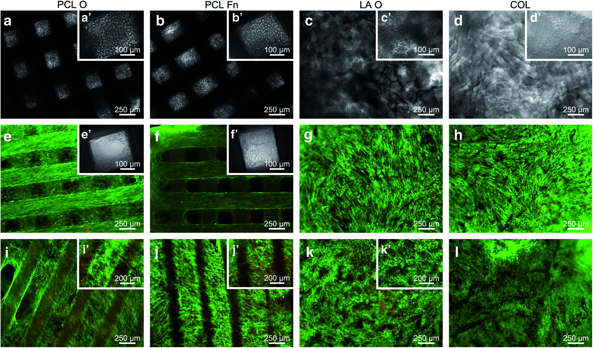
Influence of surface modification and scaffold design on the colonization of the scaffolds. Phase-contrast
In a next step, we were interested to study cellular colonization in the scaffolds by looking at cross sections as shown in Figure 5. After 7 days, PCL O scaffolds are colonized only at the edge of the scaffold. In the center of the scaffold, no cells are present (Fig. 5a). PCL Fn scaffolds show a better colonization in the center of the scaffold. The pores are completely filled with cellular material (Fig. 5b). The LA O and COL scaffolds show only cell layers at the edges of the scaffolds (Fig. 5c, d). After 28 days in culture, the colonization of PCL O scaffolds is still limited mostly to the edge of the scaffold (Fig. 5e′). In the center of the scaffold, almost no colonization is detected (Fig. 5e). In contrast, plotted PCL Fn scaffolds are homogeneously colonized throughout the scaffold (Fig. 5f). This is in contrast to the conventional scaffolds where the cellular colonization did not proceed toward the center of the scaffold (Fig. 5g, h). More cell layers can be observed in COL (Fig. 5h) than LA O (Fig. 5g) scaffolds.

Influence of surface modification and scaffold design on the colonization of the scaffolds. Histological analysis of 3D scaffolds after 7
The protein content of cell/scaffold constructs after 28 days is shown in Figure 3b. The protein content is highest for COL followed by PCL Fn, LA O, and PCL O. The protein content of blanc protein-based scaffolds (PCL Fn and COL) was negligible. Oxidized (PCL O and LA O) protein-based scaffolds (PCL Fn and COL) have a comparable proliferation ratio (Fig. 3b).
Extracellular matrix formation
The extracellular matrix (ECM) formation is visualized in Figure 6. After 7 days, a dense layer of cells can be seen on the periphery of LA O (Fig. 6c) and COL (Fig. 6d) scaffolds. The initial ECM formed at the edge of the conventional scaffolds already has a dense appearance. In contrast, plotted PCL scaffolds are colonized at the edge (PCL O) or throughout the scaffold (PCL Fn) (Fig. 6a, b). For both plotted scaffolds, the ECM formation is loose (Fig. 6e, f). For PCL Fn scaffolds, the ECM is uniformly distributed throughout the scaffold (Fig. 6b, f).

Influence of surface modification and scaffold design on the extracellular matrix formation. Histological analysis of 3D scaffolds after 7
After 28 days, the ECM remains loose in the plotted scaffolds (Fig. 6i, j, m, n). Sometimes, a more dense ECM can be observed at the edge (Fig. 6n′) or between the narrow space of 2 struts (artefact) (Fig. 6m′). In the conventional scaffolds, the ECM is dense and situated parallel to the surface of the scaffold (Fig. 6k, l, o, p). In the center, almost no ECM can be observed.
Differentiation
In the last part of our work, we assessed cell differentiation by evaluating the levels of ALP, RUNX2, COL1A1, BGLAP (OCN) phenotypic expression, and (immuno) histochemistry.
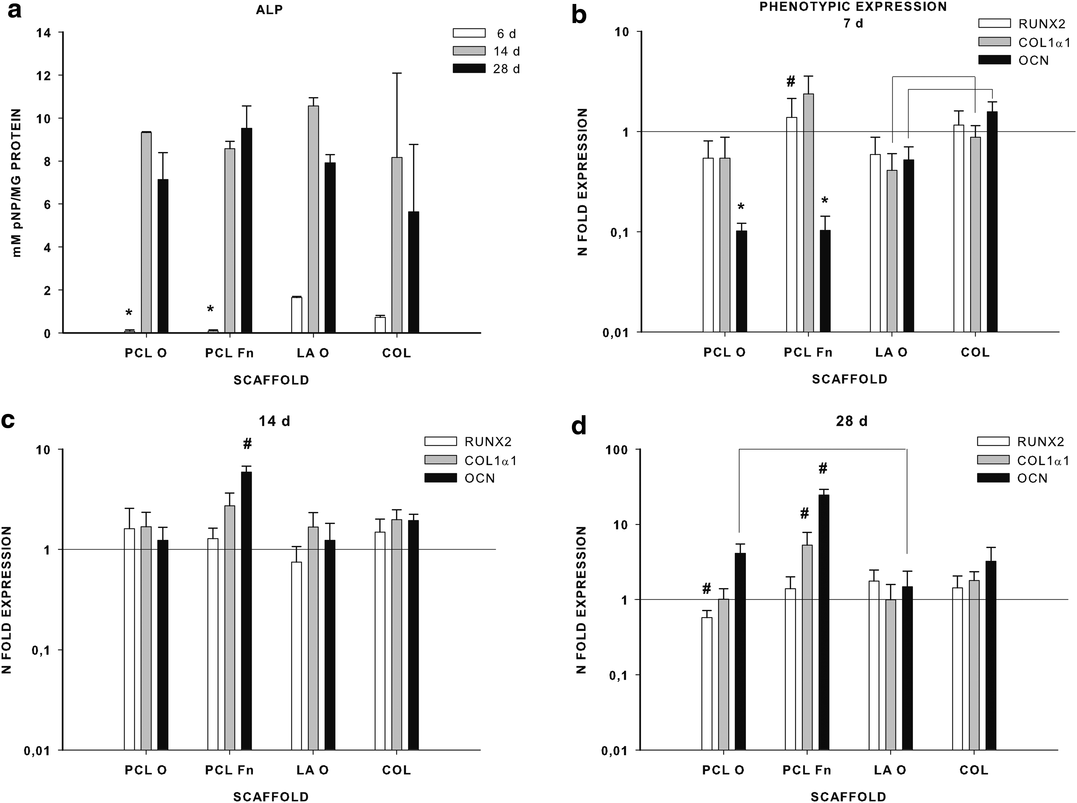
ALP was undetectable for the plotted scaffolds after 7 days, compared to the conventional scaffolds. However, after 14 and 28 days, the ALP activity increased drastically for these structures and reached similar values as the conventional scaffolds (Fig. 7a). The phenotypic expression of RUNX2 is upregulated after 7 days in COL and PCL Fn scaffolds. An upregulation of COL1A1 can be observed for the PCL Fn scaffold. An upregulation of BGLAP (OCN) can already be noticed for the COL scaffold. For all plasma-treated scaffolds (PCL O and LA O), the three osteogenic markers are downregulated. After 14 days, RUNX2 is upregulated for all the scaffolds except LA O. Also, COL1A1 is upregulated in all scaffolds, but highest for PCL Fn and COL. In addition, BGLAP (OCN) is upregulated in all scaffolds, but highest in the protein-based scaffolds (PCL Fn and COL). After 28 days, COL1A1 is still upregulated in protein-based scaffolds (PCL Fn and COL). BGLAP (OCN) upregulation is more pronounced in PCL Fn, followed by PCL O, COL, and LA O (Fig. 7d).
Influence of surface modification and scaffold design on the differentiation of ADSCs.
Immunohistochemical analysis of the osteogenic differentiation of ADSCs in plotted PCL compared with conventional scaffolds is presented in Figure 8. The formation of an ECM by ADSCs after 28 days in culture was observed in both plotted and conventional scaffolds. However, it must be noted that the conventional scaffolds (LA O and COL) show an intense staining of the ECM by Trichrome Masson (Fig. 8c, d, g, h) in contrast to plotted scaffolds where the stain is less intense (Fig. 8a, b, e, f). Only at the edge (Fig. 8f′) or between the narrow space of 2 artefact struts, the ECM is stained more intensely (Fig. 8e′). The ECM is staining positive for COL I. COL I was observed predominantly at the periphery of the conventional scaffolds (Fig. 8k, l) and almost throughout the plotted scaffolds (Fig. 8i, j). Again, the COL I immunostaining is more intense on the conventional than plotted scaffolds. In addition, OCN can be detected in both plotted and conventional scaffolds. The ECM stains of plotted scaffolds are less intense for OCN than the conventional scaffolds. It must be reported that OCN already could be detected by immunostaining after 7 days (photograph not shown), which was also detected by qRT-PCR.
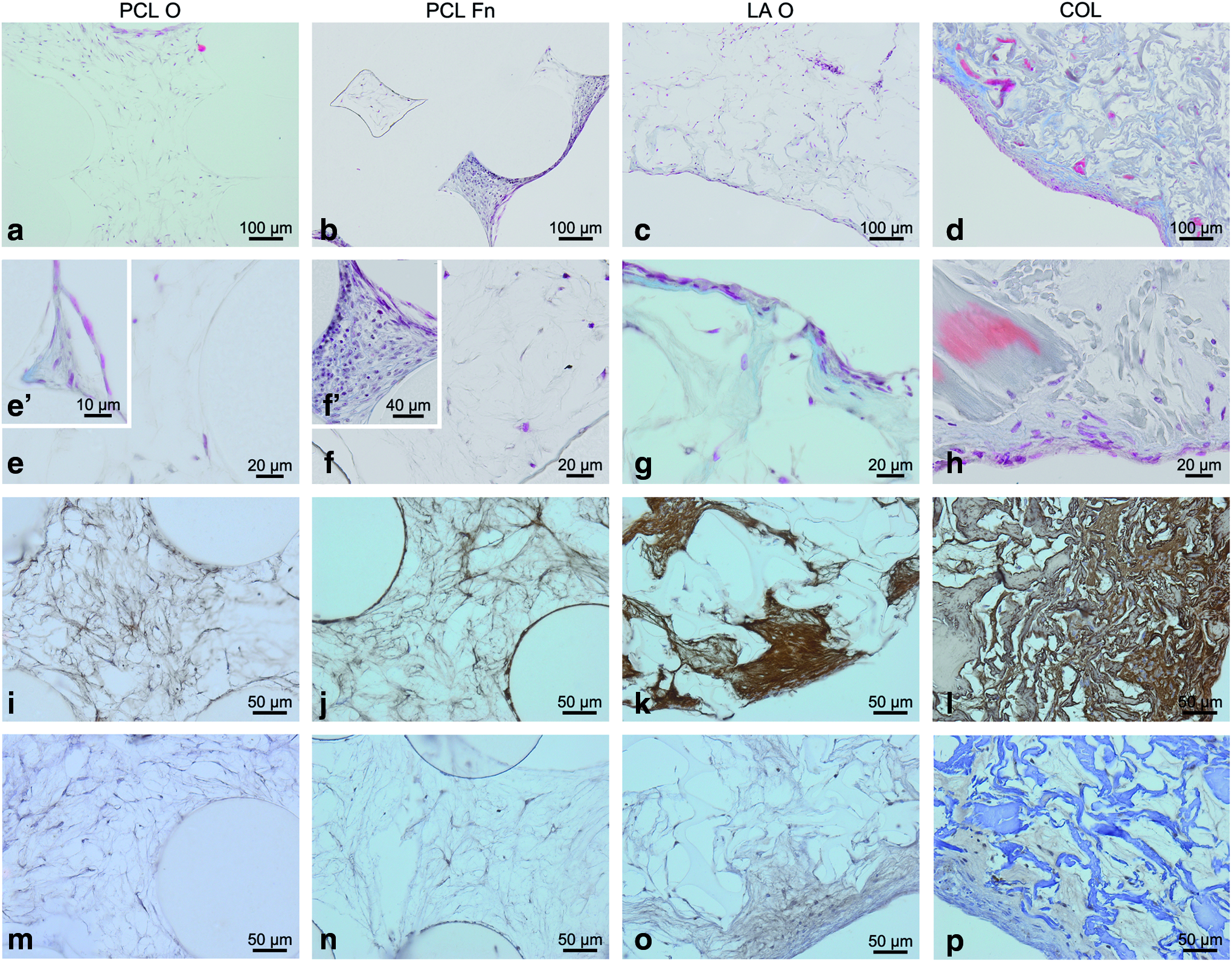
Differentiation of ADSCs after 28 days in plotted 3D scaffolds compared with conventional scaffolds. Histological analysis.
Discussion
In TE, the scaffold plays an important role as temporary support for the development of new tissue either through in vivo cell invasion or through in vitro culturing cells on the scaffold before implantation. In this regard, tissue formation in 3D scaffolds is influenced by the scaffold design and composition. Hence, the architecture and physicochemical properties of the scaffold that support cell colonization, growth, and differentiation need to be considered. Scaffold architecture influences passive cell distribution while seeding as well as active cell movement and tissue formation. 4 The scaffold composition should elicit certain biological cues to direct the cells toward colonization and tissue formation.
In the present study, we analyzed the influence of scaffold architecture and composition on mesenchymal stem cell adhesion, colonization, and differentiation. Two bioplotted PCL scaffolds (surface modified with, respectively, an oxygen plasma and a gelatin/Fn coating) with an open pore and uniform structure were compared with more compact, irregular LA O- and COL-based scaffolds.
The seeding efficiency has been associated with the surface area available for cells to attach to and the surface characteristics influencing the ability of cells to adhere. In addition, also the scaffold architecture and pore structure could play an important role. 31
In our study, no increase in the seeding efficiency of double protein-coated PCL (PCL Fn) compared with plasma modification of PCL scaffolds (PCL O) was found. In addition, the conventional plasma-treated LA (LA O) scaffold had a comparable seeding efficiency as the COL scaffold. Cell seeding onto plotted PCL scaffolds was significantly lower (39.2%±8.3%) compared to the conventional scaffolds (81.9%±1.0%). The two scaffold types (plotted vs. conventional scaffolds) on which cells were seeded on the surface clearly showed the effect of scaffold architecture. The conventional scaffolds (COL and LA O) are the densest and retain the cells on the surface. In contrast, plotted scaffolds have an open pore network and are highly geometrical leading to cell loss during seeding. The biomimetic surface modification of the 3D PCL plotted scaffold did not contribute to a higher cell seeding efficiency. Consistent with other studies, higher seeding efficiencies were reported for more compacted architectures.10,31,32 From these results, we can suggest that 3D scaffold architecture has a more profound influence on the seeding efficiency than surface chemistry, at least with the pore size geometry combination evaluated in the present work.
Although a biomimetic coating (PCL Fn) or scaffold (COL) did not increase the seeding efficiency compared to oxidized scaffolds (PCL O and LA O), protein-based surfaces (PCL Fn and COL) did have a clear benefit on the proliferation and colonization of ADSC. PCL O and LA O showed little proliferation, whereas PCL Fn and COL showed a high increase in cell/protein content. Histology demonstrated that only the edges of the PCL O scaffolds were colonized, but not the center of the scaffolds, while for PCL Fn the complete structure was colonized after 28 days. COL and LA O scaffolds were only colonized at the edges, but the peripheral tissue layer was denser on COL scaffolds.
We can thus conclude that both surface modification and scaffold architecture have a clear influence on the cellular proliferation and colonization. The influence of a protein-based surface modification was also reported by Yildirim et al. who observed an increased cell amount in an oxygen plasma/Fn modification of PCL compared to plasma-only modified PCL. 14 Consistent with other reports,4,33,34 a limited colonization at the edge of the conventional scaffolds was observed resulting in a strong gradient of cell density from the surface to the inner scaffold region.
As tissue formation is often preceded by high cell densities, the observed difference in seeding efficiency, proliferation, and colonization between the scaffolds, varying in architecture and composition, will inevitably lead to differences in ECM formation and differentiation. In conventional scaffolds (LA O and COL) with a compact design, a dense ECM is formed at the edge of the scaffold already after 7 days in culture. In contrast, the ECM formed in plotted scaffolds was loose even after 28 days in culture. This is reflected in the osteogenic differentiation: the ALP activity and late-stage osteogenic markers BGLAP (OCN) in conventional scaffolds could already be detected after 1 week. Kumar et al. described that the scaffold structure was more influential than the scaffold composition on the cellular gene expression of human bone marrow stromal cells. 35 Nevertheless, during further culture periods, the osteogenic markers in plotted scaffolds reached levels competing with the conventional scaffolds. Cells on protein-based surfaces (PCL Fn and COL) did show increased osteogenic marker expression throughout the whole culture period. Also, Yildirim et al. reported that a combined plasma/Fn modified scaffold led to a higher ALP activity compared to plasma-only modified scaffolds. 14 In general, it can be concluded that protein-based surfaces stimulate the differentiation of ADSCs toward bone cells. However, it should be noticed that the cell differentiation on uniform, open pore structured scaffolds produced by RP is delayed compared to the differentiation on compact, irregular pore structured scaffolds processed by conventional techniques. It can be hypothesized that this is due to the fact that cells need a longer proliferation period on the plotted scaffolds, to colonize the complete structure. Therefore, the required cell density to differentiate is reached at a later stage compared to the conventional scaffolds where the cells remain on the edge.
Cipitria et al. reported that plotted scaffolds acted as a guiding substrate to enable the formation of a fibrous network as a prerequirement for later bone formation. The fibrous network morphology, which in turn is guided by the scaffold architecture, influences the microstructure of the newly formed bone. A structured fibrous tissue across the entire defect was formed, which acted as a secondary supporting network for cells. 36
The double protein modified PCL scaffold, combining the advantages of the bioplotter technology and the biomimetic properties, is a promising scaffold serving as a guiding template during the bone regeneration process. Future directions in guided tissue regeneration will focus on optimization of scaffold design by tailoring the scaffold structure or by combining multiple processing techniques to create hybrid scaffolds.35,37,38
Conclusions
In the present study, PCL scaffolds were plotted applying the 3D Bioscaffolder technology and successfully modified with two surface modification strategies: an oxygen plasma modification and a double protein biomimetic coating. Uniform but loose tissue formation was obtained in both plotted scaffolds in contrast to conventional scaffolds, where the tissue formation is dense but non-homogeneous. The biomimetic coating of PCL resulted in an increased osteogenic differentiation compared with oxygen plasma-only modified PCL scaffolds, but a delayed differentiation compared with scaffolds with a more compact architecture. In conclusion, the present study successfully demonstrates that biomimetic PCL scaffolds can serve as a guiding template to obtain a uniform bone tissue formation.
Footnotes
Acknowledgments
The authors would like to thank David Schaubroeck for the SEM images. A special thanks to Leen Pieters and Toke Thiron for the excellent technical assistance. This work was supported by the FWO research grant G.0006.10 and the Ghent University by providing Dr. H. Declercq a postdoc mandate (BOF IV1-I/0002/03).
Disclosure Statement
No competing financial interests exist.