
Abstract
Tissue-engineered small-diameter vascular grafts have been developed as a promising alternative to native veins or arteries for replacement therapy. However, there is still a crucial need to improve the current approaches to render the tissue-engineered blood vessels more favorable for clinical applications. A completely biological blood vessel (3-mm inner diameter) was constructed by culturing a 50:50 mixture of bovine smooth muscle cells (SMCs) with neonatal human dermal fibroblasts in fibrin gels. After 30 days of culture under pulsatile stretching, the engineered blood vessels demonstrated an average burst pressure of 913.3±150.1 mmHg (n=6), a suture retention (53.3±15.4 g) that is suitable for implantation, and a compliance (3.1%±2.5% per 100 mmHg) that is comparable to native vessels. These engineered grafts contained circumferentially aligned collagen fibers, microfibrils and elastic fibers, and differentiated SMCs, mimicking a native artery. These promising mechanical and biochemical properties were achieved in a very short culture time of 30 days, suggesting the potential of co-culturing SMCs with fibroblasts in fibrin gels to generate functional small-diameter vascular grafts for vascular reconstruction surgery.
Introduction
W
Using purified proteins as scaffolds to engineer small-diameter vascular grafts can eliminate the limitation associated with synthetic polymers. Fibrin gels, whose precursors (fibrinogen and thrombin) can be obtained allogeneically or from a patient's own blood, 14 have been shown to promote the deposition of collagen and other extracellular matrix (ECM) molecules.15–17 Several groups have created small-diameter vascular grafts using fibrin gel scaffolds, with some engineered vessels showing good mechanical properties.18–20 Recently, the Tranquillo group generated fibrin gel-based small-diameter vascular grafts using human dermal fibroblasts with the potential to be used for arterial reconstruction. 21 However, their approaches did not achieve sufficient suture retention strength in the grafts, which also lacked elastin.
Elastic fibers are large ECM complexes that include an elastin core formed by the cross-linking of tropoelastin, and an outer layer of fibrillin-rich microfibrils used as a template for tropoelastin deposition. 22 Although human skin fibroblasts have been shown to produce microfibrils in engineered vascular tissues in some studies, 23 dermal fibroblasts are less capable of making mature elastin as compared with smooth muscle cells (SMCs). 24 In most of the studies where mature elastin deposition in engineered vascular grafts has been reported, SMCs from different species have been used25,26 (also see review in Bashur et al. 27 ). Previously, we have shown that bovine SMCs were capable of making large amounts of collagen matrix and providing strong mechanical properties in engineered vascular grafts. 28 In this study, we tested the hypothesis that a combination of bovine SMCs and human dermal fibroblasts, when cultured in fibrin, will not only improve collagen production, but also stimulate elastin deposition in fibrin gel-based vascular grafts.
Materials and Methods
Cell culture
Neonatal human dermal fibroblasts (nHDFs; Invitrogen) were grown in Medium-106 with Low Serum Growth Supplement (Invitrogen) according to the manufacturer's instructions and passaged at confluency for three passages, after which they were grown for an additional passage in a 50:50 mixture of Medium-106 with Low Serum Growth Supplement and bioreactor basal medium [Advanced Dulbecco's modified Eagle's medium (DMEM)/F-12 (Invitrogen) supplemented with 20% fetal bovine serum (FBS), 100 U/mL penicillin, 100 μg/mL streptomycin, and 2 mM
Bovine SMCs were harvested fresh from the descending aorta of adult cows as described previously. 29 The aorta was stripped of its adventitia and cut into 1 cm squares, which were placed in tissue culture flasks with DMEM supplemented with 20% FBS, 500 U/mL penicillin, and 500 μg/mL streptomycin. SMCs migrated out of the tissue sections and grew to confluency, after which they were passaged and grown in bioreactor basal medium. Culture medium was changed twice weekly and cells were passaged once a week. SMCs were harvested for use in the bioreactor at passage 5.
Bioreactor construction and setup
The bioreactor (Fig. 1) was modified from Miller et al. 30 Briefly, three 4-cm long segments of 3-mm outer diameter silicone tubing are positioned to run through two parallel PTFE discs (Fig. 1B). Where the tubing passes through the disc, shallow circular depressions allow two halves of a cylindrical PTFE mold to be held in place coaxial with the silicone tubing. The mold produces a 7-mm diameter cavity around the silicone tube, which can be filled with the fibrin-cell mixture through an injection port at one end. When the gel is solidified, the halves of the molds are removed and a 2-mm thick fibrin gel vessel forms around the silicone tubes (Fig. 1B). Externally threaded fittings at each end of the gel grip it and prevent it from contracting axially.
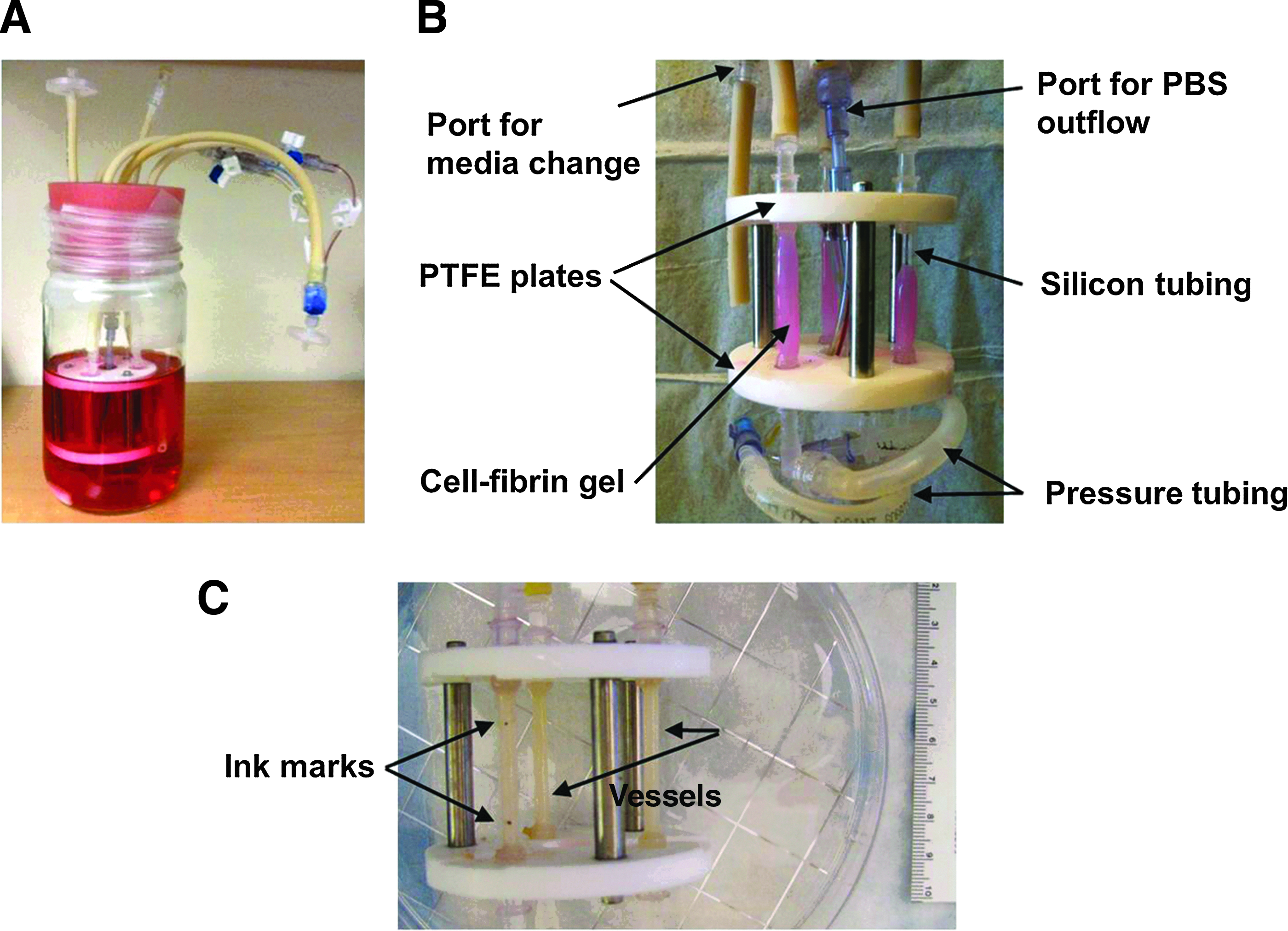
The whole apparatus is placed in a glass jar sealed with a silicone stopper (Cole-Parmer) (Fig. 1A). Two 0.2 μm disc air (PTFE) filters on the lid allow for gas exchange. A peristaltic pump (Cole-Parmer) draws phosphate-buffered saline (PBS) from a reservoir and pushes it past an in-line pressure transducer and through the silicone tubing to produce cyclic distention in the engineered vessels. An adjustable valve and an elevated reservoir downstream of the bioreactor provide back-pressure.
Engineered vessel culture in bioreactor
Cell-seeded fibrin gels were similarly formed as described previously. 21 Briefly, fibrinogen from human plasma (Sigma) was diluted in advanced DMEM/F-12 to 10 mg/mL and kept on ice. SMCs and nHDFs at passage 5 were collected in bioreactor complete medium (bioreactor basal medium containing 3 ng/mL CuSO4, 50 g/mL proline, 20 g/mL alanine, and 50 g/mL glycine, modified from Niklason et al. 28 ) at 2.4×10⁶ cells/mL each. Thrombin from human plasma (Sigma) and CaCl2 were added to the cell suspension to a final concentration of 10 U/mL and 10 mM, respectively. The cell/thrombin mixture was also kept on ice until the moment of seeding.
Five milliliters of each component was combined and the mixture was quickly injected into the three molds of the bioreactor via the injection ports. When all the molds were filled, the bioreactor was moved to an incubator at 37°C for an hour to allow gelation. The bioreactor was placed on its side and manually rotated about the vessel axis 120° every 10 min to prevent the uneven accumulation of gel on one side of the silicone tubing. After an hour, the molds were gently removed from the fibrin gel and vessels were cultured in 400 mL of bioreactor medium at 37°C and 5% CO2 for 30 days.
Pulsatile stretching of the vessels began on day 4 and was maintained between 160 and 180 beats per minute as previously described 28 with a peak to peak pressure amplitude of about 300 mmHg. Half the bioreactor medium (200 mL) was replaced twice weekly except for day 7 when all 400 mL was withdrawn and replaced. The amount of FBS in the culture medium was reduced from 20% to 10% on day 7 and maintained at 10% for the rest of the culture time to increase ECM production. 31 The culture medium was supplemented with ascorbic acid (50 μg/mL) every other day to stimulate collagen synthesis, 32 and with aprotinin from bovine lung (Sigma) (20 μg/mL) every time the medium was changed to slow down the process of fibrin gel degradation.
Measurements of vessel length
At the time of harvest, vessels were marked with ink near their ends (Fig. 1C) and the distance between was measured and designated as the “extended length.” Vessels were then cut from the bioreactor and the silicon tubing was removed. The distance between the two ink dots was measured again and designated as the “contracted length.” The contracted length was divided by the extended length to determine the contraction ratio of the engineered vascular graft after harvesting from the bioreactor.
Measurements of vessel mechanics
Vessel mechanical characterization, including burst pressure, suture retention, and compliance was performed as described previously.
28
Vessels were stretched to their original length as in the bioreactor (i.e., the distance between the two ink dots was at the extended length), and attached using sutures to a perfusion system that allowed application of intraluminal fluid at controlled pressures. Vessels were preconditioned by infusing with PBS at 1 mL/min and were cycled from 0 to 120 mmHg five times. Subsequently, the vessel was infused with PBS at 0.5 mL/min and images were taken to determine the vessel outer diameter at 10 mmHg intervals from 0 to 120 mmHg. After 120 mmHg, the vessel was infused with PBS at 5 mL/min until failure. Physiological compliance (C) was calculated by the equation:
which gives compliance as a % diameter increase/100 mmHg, and where D120 is the diameter at 120 mmHg, D80 the diameter at 80 mmHg, and ΔP the pressure difference of 40 mmHg. 21
Suture retention was determined as described previously 28 by hanging weights on a loop of 6-0 Prolene suture that was threaded through one side of the vessel wall, 2 mm from the end, with force exerted in the axial direction of the vessel. The weights were increased from 10 to 20 g and then in 5 g increments until failure.
Histological analysis
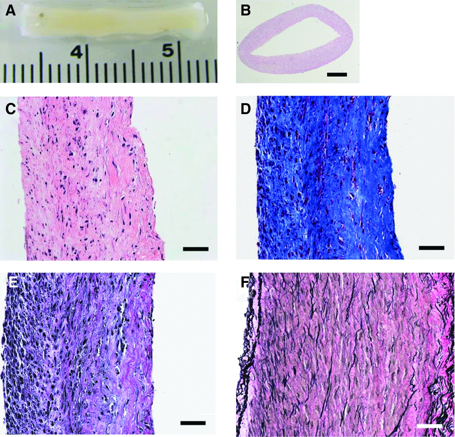
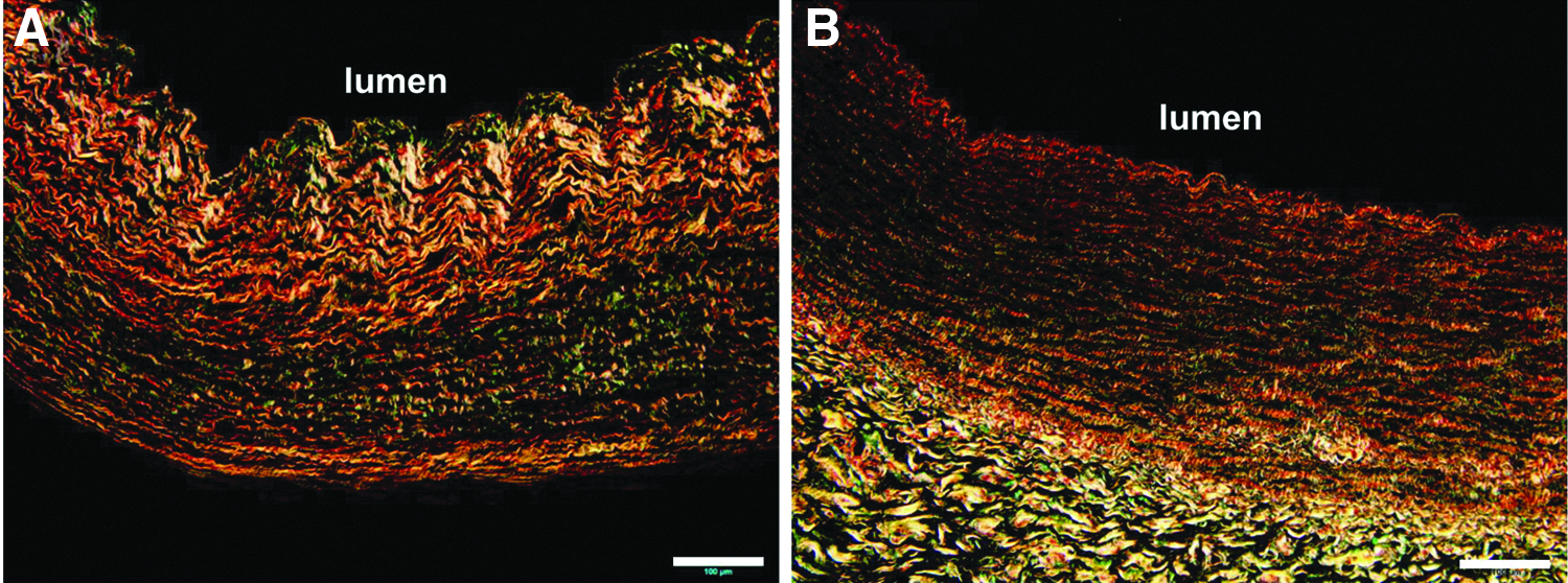
Vascular grafts were fixed in 10% neutral-buffered formalin, embedded in paraffin, cut into 5-μm sections, and stained with hematoxylin and eosin (H&E), Masson's Trichrome for collagen, or Elastic-Van Gieson (EVG) stain for elastin. Paraffin-embedded sections were also stained with picrosirius red, which exploits the birefringence of collagen fibers in the tissue. 33 Images were obtained under polarized light, where mature, thick collagen fibers stain red, orange, and yellow; immature, thin fibrils stain green. 34
Collagen quantification
The collagen content of vascular graft samples was determined by measuring the levels of hydroxyproline as described previously. 35 Collagen was calculated as 10 times the amount of hydroxyproline. 36 Collagen content was calculated as the mass per dry weight of tissue sample.
Desmosine quantification
Desmosine, a cross-link specific to elastin, was used to quantify the elastin content of vascular graft samples. Briefly, tissue samples were hydrolyzed in 6 N HCl at 110°C for 24 h, lyophilized, redissolved in water, and the protein concentration was measured. 37 The amount of desmosine in tissue samples was measured using the radioimmunoassay as described previously. 38 Desmosine content was expressed as picomoles of desmosine per milligram protein in the tissue sample.
Transmission electron microscopy
Vascular grafts were cut into 5×5 mm pieces and fixed in 4% glutaraldehyde in 0.1 M sodium cacodylate buffered fixative (pH 7.4) at room temperature for 30 min followed by 30 min at 4°C. Fixed tissues were rinsed thrice in 0.1 M sodium cacodylate buffer and postfixed in 1% osmium tetroxide for 1 h, then en bloc stained in 2% uranyl acetate in maleate buffer (pH 5.2) for a further hour. Then, the samples were rinsed, dehydrated through a graded ethanol series and infiltrated with epon resin and baked overnight at 60°C. Hardened blocks were cut using a Leica UltraCut UCT and 60 nm sections were collected on nickel grids and stained using 2% uranyl acetate and lead citrate. Samples were viewed on a FEI Tecnai Biotwin transmission electron microscope (TEM) at 80 kV. Images were taken using a Morada CCD digital camera using iTEM (Olympus) software.
Immunofluorescence staining
Immunofluorescence staining of vascular graft samples was performed as described previously. 39 Primary antibodies were used at 1:50 dilution and include rabbit anti-elastin (ab21610; Abcam), rabbit anti-calponin (ab46794; Abcam), mouse anti-α-smooth muscle actin (α-SMA) (ab18147; Abcam), and mouse anti-β-actin (A1978; Sigma).
Statistics
Data are expressed as mean±standard deviation of at least six samples. Statistical significance was determined by t-test. Difference (p-value)<0.05 was considered significant.
Results
Vessel morphology and cellular properties
Tissue-engineered vascular grafts generated from co-culturing nHDFs and bovine SMCs in fibrin gel scaffolds in a pulsatile bioreactor for 30 days had an appearance similar to a native artery (Fig. 2A, B). H&E staining showed that cells were uniformly present throughout the whole tissue (Fig. 2C). To determine the cell phenotype in the engineered vessels, cells were stained for both SMC markers and a fibroblast marker using immunofluorescence. Almost all cells stained positive for α-SMA, while some were also stained positive for calponin (Fig. 3), indicating the contractile phenotype of SMCs. 31 Interestingly, there was no detectable staining for fibroblasts (anti-Fibroblast Surface Protein staining, data not shown) in the engineered vessels, possibly due to the differentiation of nHDFs to myofibroblasts (characterized by the neo-expression of α-SMA 40 ) during the 30 days' culture under pulsatile stretching. Mechanical tension has previously been shown to contribute to myofibroblast induction. 41
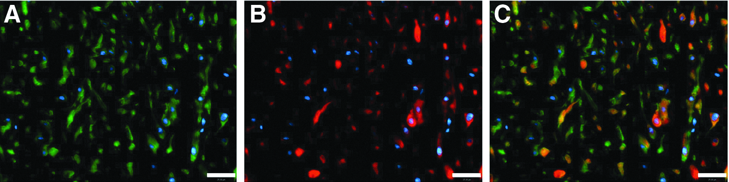
Representative images of engineered vascular grafts stained for α-smooth muscle actin (α-SMA)
The total length of the engineered vascular grafts in bioreactor was 4 cm, same as the distance between the two PTFE plates (Fig. 1C). When cut from the bioreactor and removed from the silicon tubing, engineered vessels contracted to 71.4%±11.3% (n=6) of their original length as in the bioreactor, mostly due to the presence of contractile SMCs and collagen matrix (Fig. 2C). The harvested engineered vessel measured ∼2 cm in total final length (Fig. 2A). The average wall thickness of engineered vascular grafts was 548.9±108.8 μm (n=6), while the original cell-containing fibrin gel had a thickness of about 2 mm (Fig. 1B), suggesting the remodeling of fibrin scaffolds during the 30 days' culture.
Collagen and elastin
Masson's Trichrome staining indicates that collagen was the major ECM component present in the engineered tissues (Fig. 2D). Average collagen content (calculated from hydroxyproline) in the engineered vessels as a fraction of dry weight was 67.2%±12.1%, which is greater than in native bovine arteries (45%±9%) 28 or in native human vessels (35.6%±11.6%, Table 1).
nHDF, neonatal human dermal fibroblast; SMC, smooth muscle cell.
Picrosirius red stain indicates that collagen fibers were circumferentially aligned in the engineered vessels, similar to native arteries (Fig. 4) and other engineered grafts. 21 In addition, mature thicker collagen fibers were mainly localized near the lumen, while newly formed and thinner collagen fibers were primarily found toward the abluminal surface (Fig. 4A). This distinct pattern might reflect the sequential deposition and maturation of collagen fibers in the engineered vessel wall over the culture period, as has also been observed in other tissue engineered grafts (A. Huang, et al., unpublished data). In other words, collagen fibers near the lumen may be “older,” more mature, and hence of larger diameter, than those near the abluminal surface. For purposes of comparison, native coronary artery had collagen fibers that stained yellowish-orange to red, suggesting mature collagen fibers 42 were present throughout the whole wall thickness in native vessels (Fig. 4B).
Elastic fibers, in the form of both microfibrils and mature elastic fibers, were also detected in engineered grafts by ultrastructural analysis using TEM (Fig. 5). Average desmosine content, used as a means for elastin quantification, was 126.7±42.4 pmol/mg in the engineered vessels (Table 1), about 15.6% of that in native bovine arteries (813.3±30.6 pmol/mg).
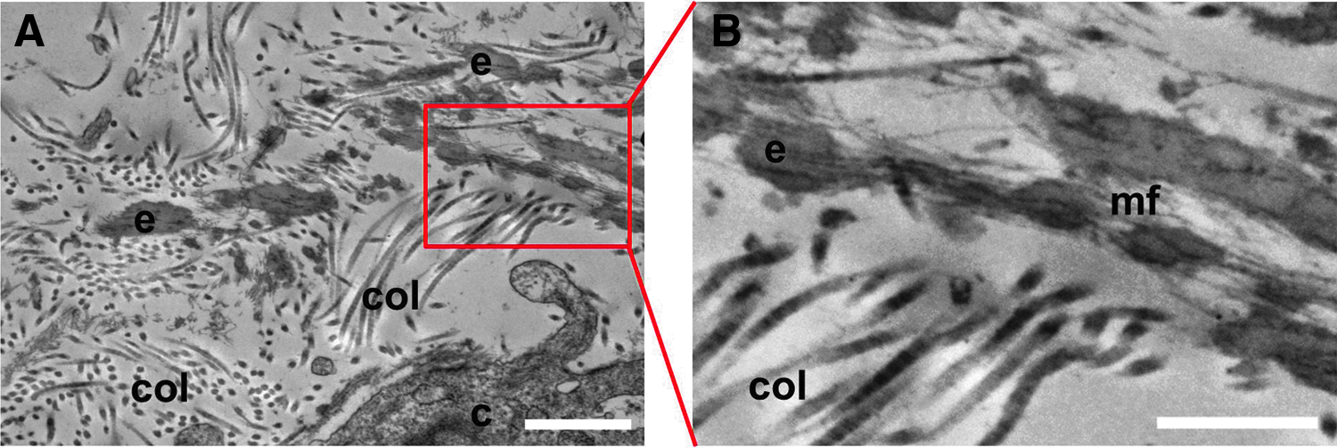
Transmission electron microscopy images of tissue-engineered vascular graft showing collagen fibers (col) and elastin fibers (e) surrounding cells (c). Elastin cores are integrated within a bundle of microfibrils (mf). Scale bars, 1 μm in
Immunostaining for elastin showed that elastin was present throughout the tissues, but was more concentrated near the luminal side (Fig. 6A), where mature thick collagen fibers were also localized (Fig. 4A). These results suggest a gradual deposition and maturation of the elastin matrix along with the collagen matrix during culture in the fibrin substrate. Consistent with EVG staining (Fig. 2F), native coronary artery showed elastin staining throughout the tissue (Fig. 6B).

Immunostaining for elastin in engineered vascular graft
Vessel mechanical properties
The engineered vascular grafts made from co-cultures of nHDFs and bovine SMCs in fibrin scaffolds for 30 days had an average burst pressure of 913.3±150.1 mmHg (Table 1), which is comparable to human umbilical arteries (969.7±154.4 mmHg, p=0.59), and is much higher than engineered grafts cultured from nHDFs in fibrin scaffolds for total 5 weeks (598±28 mmHg). 21 The compliance of the engineered grafts was 3.1%±2.5% per 100 mmHg (Table 1), also comparable to the human umbilical arteries (5.8%±3.1% per 100 mmHg, p=0.27) or engineered grafts cultured from nHDF in fibrin scaffolds for a total of 7 weeks (4.4%±1.6% per 100 mmHg, Table 1). In addition, the suture retention of the engineered grafts was 53.3±15.4 g, sufficient for surgical handling and much higher than other fibrin gel-based vascular grafts cultured for 7 weeks (19.4±5.1 g, Table 1). In light of the short culture time (30 days), co-cultures of nHDFs and bovine SMCs in fibrin scaffolds have greatly improved vessel mechanical properties as compared with cultured nHDFs alone. Future studies, however, are required to further improve vessel strength and compliance to match those of native vessels, 43 and to understand the interplay of these two cell types during culture.
Discussion
In this study, we describe the development of completely biological small-diameter vascular grafts that are grown in a pulsatile bioreactor by co-culturing bovine SMCs and nHDFs in fibrin gels for 30 days. The engineered vascular grafts contained circumferentially orientated collagen fibers and mature SMCs in the vessel walls, and exhibited good mechanical strength and adequate compliance suitable for implantation. In addition, these engineered vessels contain mature elastic fibers, which have not been previously observed in polymer-based vascular tissue engineering.11,28,44
An ideal tissue engineered vascular graft should possess similar vasoreactivity and biomechanics to native vasculature, and should include a confluent endothelium to resist thrombosis when implanted in vivo. SMCs provide vasocontractile function in native arteries. Starting with a 50:50 mixture of bovine SMCs and nHDFs, at the end of 30 days culture, most of the cells that populated the engineered vessel wall stained positive for α-SMA, indicating a smooth muscle or myofibroblast phenotype. Some of the SMCs also expressed calponin, an indication of a contractile SMC phenotype. 31 Because the culture medium used in our bioreactor contained high amounts of serum that would have supported fibroblast proliferation in addition to SMC proliferation, the absence of fibroblasts in the engineered vascular grafts is possibly due to the differentiation of dermal fibroblasts into myofibroblasts, a widely known characteristic of fibroblasts. 40 Pulsatile stretching of the engineered vessels during culture might have contributed to the transition of fibroblasts to myofibroblasts. 41
The fibrin gel-based vascular grafts, derived from co-culturing of bovine SMCs and nHDFs for 30 days, showed higher collagen content and mechanical strength as compared to engineered vessels generated from bovine SMCs cultured on polyglycolic acid (PGA) scaffolds for 5 weeks. 28 Previous studies suggest that SMCs might take a significantly longer time to make ECM as compared to fibroblasts. 45 Thus, the fibroblasts in the fibrin gels might have contributed to the early ECM deposition, before these cells were differentiated to myofibroblasts. Bourget et al. have shown that vascular grafts engineered from seeding SMCs on fibroblast-derived ECM possessed much higher mechanical strength as compared with SMCs alone. 46 Thus, it is possible that the initially deposited ECM contributed to the enhanced collagen matrix deposition in our vascular grafts. Future studies, though, are required to determine whether the increase in collagen content is also due to the differential effects of fibrin gels and PGA scaffolds for supporting matrix synthesis and deposition by SMCs. On the other hand, the engineered vascular grafts derived from co-culturing of bovine SMCs and nHDFs showed an approximately threefold increase in suture retention, compared with those engineered from nHDFs only. 21 These results thus strongly suggest the synergistic roles of bovine SMCs and nHDFs in enhancing ECM deposition, although mixing cells of the same species, in particular, human, will be more clinically relevant. Future studies could be designed to determine whether the limitations of human vascular SMCs 47 can be minimized by co-culturing human SMCs with human neonatal or adult dermal fibroblasts 23 to engineer vascular grafts.
Vessel mechanical properties are mainly determined by ECM, wherein collagen matrix is responsible for vessel strength and mature elastin contributes to vessel compliance.48,49 Early studies assigned compliance mismatch as one of the main contributors to graft failure due to neointimal hyperplasia.50–53 During elastogenesis, soluble tropoelastin is deposited into the extracellular space onto a fibrillin-rich microfibril template to form mature, insoluble elastic fibers. 22 As such, fibrillin-1 and tropoelastin have routinely been used for identifying elastin in the engineered tissues.25,26 In this study, we showed the deposition of elastin into the extracellular space in engineered vascular grafts, which represents a significant improvement to the previous approach where fibroblasts alone were cultured in fibrin gels. 21 Further, we demonstrated the presence of mature elastic fibers in our engineered vessels via TEM, which is a more dispositive method than immunostaining. Lee et al. recently showed 19% elastin content in their engineered grafts as compared with native vessels, 25 however, there was little direct evidence of mature, cross-linked elastin in that report. In contrast, we used desmosine, a cross-link specific to elastin for elastin quantification, and have shown a 15.6% desmosine content in the engineered grafts as compared with native arteries. Compared to using fibroblasts alone, 21 the addition of SMCs in fibrin gels not only improved the production of collagen matrix and thus the mechanical strength of the engineered vessels, but also may have induced the deposition of mature elastin. However, there were obvious differences in the distribution and organization of collagen and elastic fibers between the engineered grafts and the native vessels, which might indicate the less optimal mechanical properties of the engineered grafts as compared with the native vessels (Table 1). In the future, we may further increase mature elastin deposition in the engineered vessels with longer culture time, and/or by adding biochemical stimulation as described by us and others.26,54–56 In addition, as a confluent endothelium is highly desired prior to implantation, in the future we will also seed endothelial cells in the lumen of the engineered vascular grafts as described previously.23,28,57
In conclusion, we have shown that co-culturing of bovine SMCs with nHDFs in fibrin gel scaffolds under pulsatile stretching produced engineered arterial conduits having strong mechanical properties, and substantial amounts of both collagen and elastin. Using this approach, we have significantly decreased the culture time to produce robust ECM, thus simplified the handling to generate a mechanically strong vessel that is completely biological.
Footnotes
Acknowledgments
This work was supported by R21 HL107768-01A1 (Humphrey, Niklason co-I), R01 HL083895-06A1 (Niklason), and 1P01HL107205-01A1 (Simons).
Disclosure Statement
L.E.N. has a financial interest in Humacyte, Inc., a regenerative medicine company. Humacyte did not fund these studies, and Humacyte did not affect the design, interpretation, or reporting of any of the experiments herein.