
Abstract
The mesenchymal stem cell (MSC) supernatant is well known as a rich source of autologous cytokines and universally used for tissue regeneration in current clinical medicine. However, the limitation of conditioned medium used in open-wound repair compels the need to find a more sophisticated way to take advantage of the trophic factors of MSCs. We have now fabricated a three-dimensional membrane from freeze-dried bone marrow mesenchymal stem cells-conditioned medium (FBMSC-CM) using a simple freeze-dried protocol. Scanning electron microscopy images showed the microstructure of the FBMSC-CM membrane (FBMSC-CMM) resembling a mesh containing growth factors. ELISA was used to test the paracrine factors retained in the FBMSC-CMM, and the results indicated that FBMSC-CMM withheld over 80% of the paracrine factors. Live/dead assays were adopted to test the toxicity of the FBMSC-CMM on cultured rat dermal fibroblasts, and the results confirmed its biological safety with low toxicity. Moreover, the FBMSC-CMM could significantly accelerate wound healing and enhance the neovascularization as well as epithelialization through strengthening the trophic factors in the wound bed as determined by immunohistochemical staining. Thus, the ability to maintain paracrine factors and enhance the effectiveness of these growth factors in the wound as well as the simple procedure and economical materials required for production qualifies the FBMSC-CMM to be a candidate biomaterial for open-wound regeneration.
Introduction
T
Wound healing requires complex biomolecular processes, including cell migration, proliferation, and angiogenesis, as well as extracellular matrix remodeling. 4 MSCs have long been demonstrated to be involved in the regeneration of damaged tissues in the wound, and isolated MSCs may have a broader potential (i.e., plasticity) than was previously thought.5–7 They not only have the capacity for self-renewal and generation of differentiated cells, but also are able to produce a broad repertoire of secreted trophic factors, growth factors, cytokines, and chemokines, as well as immunomodulatory cytokines, which can provide considerable promise for the treatment of refractory wound diseases. 8 Furthermore, the potential to prepare and apply stem cells as an allogeneic graft enables their development as an extraordinary therapeutic tool.
The traditional MSC-conditioned medium (MSC-CM) has been one of the most frequently used form of MSC secretome in preclinical application.9–11 However, the efficacy of such conditioned medium has been debated. Previous studies have argued that injection of the MSC supernatant could mediate only the early steps of the tissue repair process due to its massive, uncontrollable, and short-term effects after transplantation in vivo.12,13 Thus, harnessing the power of the MSC secretome for meaningful therapeutic outcomes remains a challenge. Recently, the MSC secretome was modulated in some preclinical studies through different methods, including growth factor preconditioning and physiological, pharmacological, cytokine and genetic manipulations,14–18 before transplantation, as well as by combining MSCs with functional extracellular matrices.19–21 However, thorough in vivo examinations and the demonstration of effective modulation of the MSC secretome, which seem essential for rational therapy design and improvement of existing therapies, are still lacking.
During the last several years, platelet-rich plasma (PRP), which is highly enriched in autogenous growth factors, has been well established to be successfully freeze dried into a powder and to exert a significant therapeutic role in wound repair and tissue regeneration.22–24 Comparably, the MSC-conditioned medium may also be a panacea as it is enriched with many types of cytokines. However, whether the paracrine factors of stem cells can also be made into a solid membrane or powder without losing their therapeutic function has not been reported.
Enlightened by the usage of freeze-dried PRP, we considered that the freeze-dried bone marrow mesenchymal stem cells-conditioned medium membrane (FBMSC-CMM) may also preserve the function of paracrine factors and provide stronger and more durable effects on cellular proliferation, survival, and wound regeneration compared with fresh medium or other types of synthetic growth factors. Therefore, the potential of FBMSC-CMM as a bioscaffold was first evaluated in our study through scanning electron microscopy (SEM) observations of the microstructure. Paracrine factors maintained in the FBMSC-CMM were detected by rehydrating it at 4°C first and then analyzing the extract by ELISA. The toxicity of FBMSC-CMM also was evaluated by examining the proliferative and survival rates of rat dermal fibroblasts (RDFs) cultured in rehydrated FBMSC-CMM. We further performed in vivo tests of the FBMSC-CMM on wound skin regeneration as well as explored the potential mechanisms for the FBMSC-CMM-mediated tissue healing by western blot and immunofluorescence analyses.
Materials and Methods
Experimental design
We hypothesized that freeze drying of BMSC-CM may not only be useful for the storage of proteins in a conditioned medium, but also as a new biomaterial that can benefit wound healing. Therefore, we designed both in vitro and in vivo experiments to test the proteins preserved in FBMSC-CMM and evaluated the biological function of the membrane. BMSCs were cultured to prepare the conditioned medium, which was either stored in −20°C or freeze dried to formulate the FBMSC-CMM. SEM and ELISA were adopted to observe the structure and protein reservoir of FBMSC-CMM. Apoptosis and survival of RDFs cultured within FBMSC-CMM were examined to test its toxicity and biocompatibility. Cells cultured in fetal bovine serum (FBS), BMSC-CM, serum-free medium (SFM), and freeze-dried biochemical stabilization buffer (FBSB) served as control groups. For evaluating the regenerative function, rats with (full-thickness) acute skin wounds were used as animal models and divided into groups treated with FBMSC-CMM, BMSC-CM, SFM, or FBSB. Nontreated animals served as a negative control group. Skin wound healing and regeneration, including epithelialization, angiogenesis, as well as related mechanisms, were evaluated to verify the biological function of the novel biomaterial, and the results were analyzed by statistical methods.
Animals
All Sprague Dawley (SD) rats, weighing 300±20 g, 8–12 weeks old (certificate No. 2007-0005) were purchased from the Military Academy of Medical Sciences of PLA, Laboratory Animal Co., Ltd. and housed in the Veterinary Service Center of the First Affiliated Hospital, General Hospital of PLA (Guangzhou, China) at least 1 week before the experiment in accordance with the National Institutes of Health (NIH) and the Institution-Approved Animal Care Guidelines. All procedures were approved by the Administrative Panel of the General Hospital of PLA on Laboratory Animal Care (Guangzhou, China).
Isolation and expansion of rat bone marrow MSCs
SD rat bone marrow MSCs (BM-MSCs) were isolated as previously described. 25 Briefly, bone marrow was isolated from the tibias and femurs of male SD rats into phosphate buffered saline (PBS; Invitrogen, Carlsbad, CA). Cells were then cultured in plastic dishes in high glucose Dulbecco's modified Eagle's medium (DMEM, containing 4.5 g/L glucose; Invitrogen), supplemented with 10% FBS (Gibco, Carlsbad, CA) and antibiotics (100 U/mL penicillin G, and 0.1 mg/mL streptomycin; Invitrogen). The medium was changed 48 h after initial plating to remove all nonadherent cells and thereafter changed every 2–3 days. Cells were detached with trypsin-EDTA (1:250) and passaged at 80% confluency. Cells were used at passages 3 to 6 for subsequent experiments.
The potential of multilineage transdifferentiation of BM-MSCs was determined by Alizarin Red staining (osteogenesis) and Oil Red O staining (adipogenesis). The superficial markers of BM-MSCs, including CD34, CD44, CD45, CD90, and CD11b, were analyzed by flow cytometry.
Evaluation of FBMSC-CMM and its effect on RDFs in vitro
Preparation of rat BM-MSC-CM and FBMSC-CMM
BM-MSCs of passage 3 were detached after treatment with trypsin-EDTA (1:250) (PAA, Linz, Austria) and seeded in six-well plates at the density of 3–5×10⁵ cells per well in a DMEM medium supplemented with 10% FBS and antibiotics. The cells were cultured until reaching 80% confluency, and then the attached cells were washed three times with PBS. Subsequently, they were continued to be incubated with 1 mL serum-free DMEM for 24 h to generate BM-MSC-CM, which were either used to generate FBMSC-CMM or cultured RDFs.
After 24 h, conditioned medium was collected and centrifuged at 1500 g for 10 min and then the concentration (10%, 10 mL buffer B was added to resuspend the proteins) was adjusted with a physiological buffer (buffer B; 136 mM NaCl, 11.9 mM NaHCO3, 5.6 mM glucose, 5 mM HEPES, 2.7 mM KCl, 2.0 mM MgCl2, 0.42 mM NaH2PO4; pH 7.4) after passing through a 0.22-μm filtration unit (Millipore, Bedford, MA). One milliliter of this medium was obtained to test the concentration of the major factors, whereas the rest was concentrated 10-fold by tangential flow dialysis (Bio-Rad, Berkeley, CA) and stored at −80°C until use.
To prepare the FBMSC-CMM, we first thawed 10 mL of the 10× medium, and then it was combined with 80 mL of buffer B, 10 mL buffered biochemical stabilization solution [250 mM amilioride, 100 mM adenosine, 50 mM sodium nitroprusside, 1% (v/v) dimethyl sulfoxide, 4% (w/v) polyvinyl pyrrolidone (Plasdone C-15; International Specialty Products, Wayne, NJ)], as well as 5 g mannitol. Thus, the concentration of proteins in this diluted conditioned medium was identical to that of the fresh medium (10%). One milliliter of the gel-like medium was added into each well of the 24-well plates, which were then placed in a LyoStar freeze dryer (FTS Kinetics Systems, Stone Ridge, NY).
The biochemical stabilization buffer with mannitol alone (0.5 mg mannitol/mL buffer) also was prepared to obtain the FBSB, which was used as a positive control for FBMSC-CMM. All procedures were performed under sterile conditions and were identical to those described previously for the preparation of freeze-dried PRP. 23 The FBMSC-CMM and FBSB were packed under dry nitrogen and stored at −80°C until use.
ELISA for paracrine factors in MSC-CM and FBMSC-CMM
To verify the obtained cell supernatants and investigate the differences in protein preservation between fresh MSC-CM and FBMSC-CMM, we first rehydrated the FBMSC-CMM and adjusted the final concentration of FBMSC-CMM to the same level as that of fresh BMSC-CM (10%) by dilution with buffer B as described above. Levels of various bioactive proteins were measured, including proliferative and antiapoptotic factors such as hepatocyte growth factor (HGF), vascular endothelial growth factor (VEGF); chemokines such as stem cell-derived factor-1α (SDF-1α); monocyte chemoattractant protein-1 (MCP-1), inflammatory cytokines such as tumor necrosis factor-α (TNF-α) and interleukin-6 (IL-6); and adipokines such as leptin and plasminogen activator inhibitor-1 (PAI-1) (VEGF, TNF-α; R&D Systems, Minneapolis, MN, HGF; Institute of Immunology, Tokyo, Japan, IL-6; Pierce, Rockford, IL, Leptin; AdipoGen, Seoul, Korea, PAI-1; Oxford Biomedica Research, Oxford, CT, SDF-1, and MCP-1; R&D Systems).
Scanning electron microscopy
For SEM observation, the prepared FBMSC-CMM and FBSB were fixed in 4% paraformaldehyde for 2 h at 4°C, followed by dehydration through increasing concentrations of ethanol. The dried specimens were then gold sputter coated and examined with an environmental scanning electron microscope (TM-1000; Hitachi, Tokyo, Japan) with an accelerating voltage of 15 Kv. 26
Cell survival and proliferation assay
RDFs purchased from a commercial source (catalog No. R106K-05n, cryopreserved RDFs; Cell Applications, San Diego, CA) were plated at 5000 cells per cm2 in DMEM containing 10% FBS and trypsinized when they reached 70–80% confluency. Passage 3 fibroblasts were seeded into a 96-well plate at a density of 4000 cells per well and cultured with DMEM with FBS (10%), MSC-CM, SFM (DMEM), FBSB solution (rehydrated as FBMSC-CMM), or FBMSC-CMM rehydrated solution. The medium was replaced every 3 days. A live/dead assay was used to assess cell viability of RDFs for 1, 3, 7, and 14 days per the manufacturer's instructions (Live/Dead Double Staining Kit; Calbiochem, Gibbstown, NJ). Images were obtained with a fluorescence microscope (Olympus, Tokyo, Japan) with band-pass filters set to detect FITC and Texas Red. At the same time, identical cell samples were labeled with MTT (Sigma-Aldrich, St. Louis, MO), and absorbance was determined at 570/630 nm with a microplate reader (Bio-Rad). Results were normalized as a ratio of each type of medium (10% FBS). Every experiment was repeated three times with five samples for each group.
Rat skin wound model and FBMSC-CMM application
A total of 15 male SD rats were divided into five groups: FBMSC-CMM-treated group (F, 10×40-fold), MSC-CM-treated group (C, 10×40-fold), FBSB-treated group (B), medium (M) alone-treated group, and nontreated group (N). The cells were examined at 4 days (n=5), 12 days (n=5), and 20 days (n=5) postwounding, which were appropriate intervals to evaluate the initial phase, the late phase of the proliferation stage, and the remodeling stage, respectively. As described in a previous report, 27 surgical procedures were performed under standard aseptic conditions. After anesthesia with 10% chloral hydrate, two circular full-thickness skin defects (diameter=10 mm) were prepared in each rat, and 30 min later the wounds were treated or not with the above agents. One milliliter of medium was added all at once by dropping directly into the wound bed with a 1-mL syringe. The FBMSC-CMM membrane or FBSB (both of which were identical in volume before being freeze dried) was placed onto the wound bed directly. To prevent the diffusion of the medium and possible infection, the wounds were further covered by a polyurethane film (Department of Plastic Surgery, Guangzhou General Hospital, GuangDong), which was changed every 3 days to monitor the wound condition and measure the area.
The rate of wound healing was calculated by the following formula: percentage of the initial wound area=[wound area at each time point]/[initial wound area]×100%.
Histological processing and skin regeneration
At 7 or 14 days postwounding, rats were euthanized, and full-thickness skin samples, including subcutaneous tissue were obtained with surgical scissors at the center of the wound and surrounding connective tissue. Fixed samples were embedded in paraffin, cut into sagittal sections of about 5 μm thickness, and then directly stained with Masson's trichrome or probed with the primary antibodies rabbit anti-rat CD31 (1:100; EPITOMICS; Abcam, Cambridge, MA) overnight at 4°C. Subsequently, the samples were incubated with horseradish peroxidase-coupled goat anti-rabbit IgG (1:1000; Santa Cruz Biotechnology, Santa Cruz, CA) as the secondary antibody for 2 h at room temperature followed by analysis under a light microscope (KS400; Zeiss, Munich, Germany).
Immunohistochemical staining of frozen samples was performed by embedding the tissue in OCT compound (Tissue-Tek 4583®; Sakura Finetek USA, Inc., Torrance, CA) and cut into 10-μm-thick sections at−22°C. To stain the regenerated arterioles and epidermis as well as trophic factors in the wound bed, sections were incubated with rabbit monoclonal anti-CK5, anti-HGF, anti-TGFβ1 (1:100; Abcam) and mouse monoclonal anti-SMA, anti-VEGF or anti-bFGF (1:100; Abcam) primary antibody, and then probed with FITC-conjugated donkey anti-rabbit IgG (catalog No. A21206 and No. A31571; Invitrogen) or Alexa Fluor 488-conjugated goat anti-mouse IgG (1:100; Invitrogen) at 37°C for 2 h. Thereafter, sections were counterstained with DAPI (Sigma-Aldrich) and examined by fluorescence microscopy (Olympus).
Western blotting
Protein expression of specific trophic factors were further analyzed by using immunoblotting. Protein lysates were obtained from skin tissue samples (untreated or treated with MSC-CM and FBMSC-CMM) using lysis buffer (Sigma-Aldrich) and separated by 12% sodium dodecyl sulfate–polyacrylamide gel electrophoresis. Transferred membranes were blocked and labeled with rabbit monoclonal anti-HGF, anti-TGFβ1 (1:1000; Abcam) and mouse monoclonal anti-VEGF and anti-bFGF (1:1000; Abcam), and β-actin (1:1000 dilution; R&D Systems) primary antibodies for 3 h at room temperature, followed by staining with goat anti-rabbit or anti-mouse biotin-conjugated secondary antibodies for 1 h (1:2000 dilution, WesternDot™ Goat Anti-Rabbit or Anti-Mouse Western Blot Kit; Life Technologies, Carlsbad, CA) at room temperature. Peroxidase activity was detected using the Western Blot Luminol reagent per manufacturer's instructions (Santa Cruz Biotechnology). Protein band intensities were normalized to β-actin, which served as the loading control.
Statistical analysis
In the in vitro proliferation study, results are expressed as the mean±standard deviation of four samples from representative single experiments. Statistical significance of differences between groups was analyzed by the Student's t-test and Kruskal–Wallis one-way analysis of variance of ranks (SigmaPlot version 8.0; Systat Software, San Jose, CA). Dunn's method was used to analyze multiple comparisons versus the control group. p-Values <0.05 were considered significant.
Results and Discussion
Identification of BM-MSCs
Cultured cells were 99.05% pure for CD90 and 99.23% for CD44. The contaminating population of hematopoietic stem cells positively expressed the markers CD11b, CD45, and CD34 at 0.230%, 0.15% and 0.23%, respectively. Cultured BM-MSCs had a strong ability to proliferate, form colonies, and differentiate into multiple mesenchymal lineages (data not shown).
Quantification of growth factors and chemokines in frozen MSC-CM and FBMSC-CMM
In previous reports, MSCs were shown to have cell protective effects and induce angiogenesis through secretion of various cytokines, including VEGF, HGF, and SDF-1α.28–30 To compare the proteins secreted by cultured MSCs before and after the freeze-dried process, ELISA was used to investigate the production of several growth factors and cytokines. As compared with frozen MSC-CM and FBMSC-CMM released all types of factors more slowly (most factors were collected at 24 h after dehydration). Not only was over 75% of HGF and VEGF, which are antiapoptotic and angiogenic factors, preserved, but also SDF-1α and MCP-1, which are cell migration-related chemokines, were maintained in FBMSC-CMM. However, FBMSC-CMM released significantly lower levels of the inflammatory cytokines TNF-α and IL-6. There was no significant difference in several secreted adipokines, such as leptin and PAI-1 between frozen MSC-CM and FBMSC-CMM (Fig. 1).
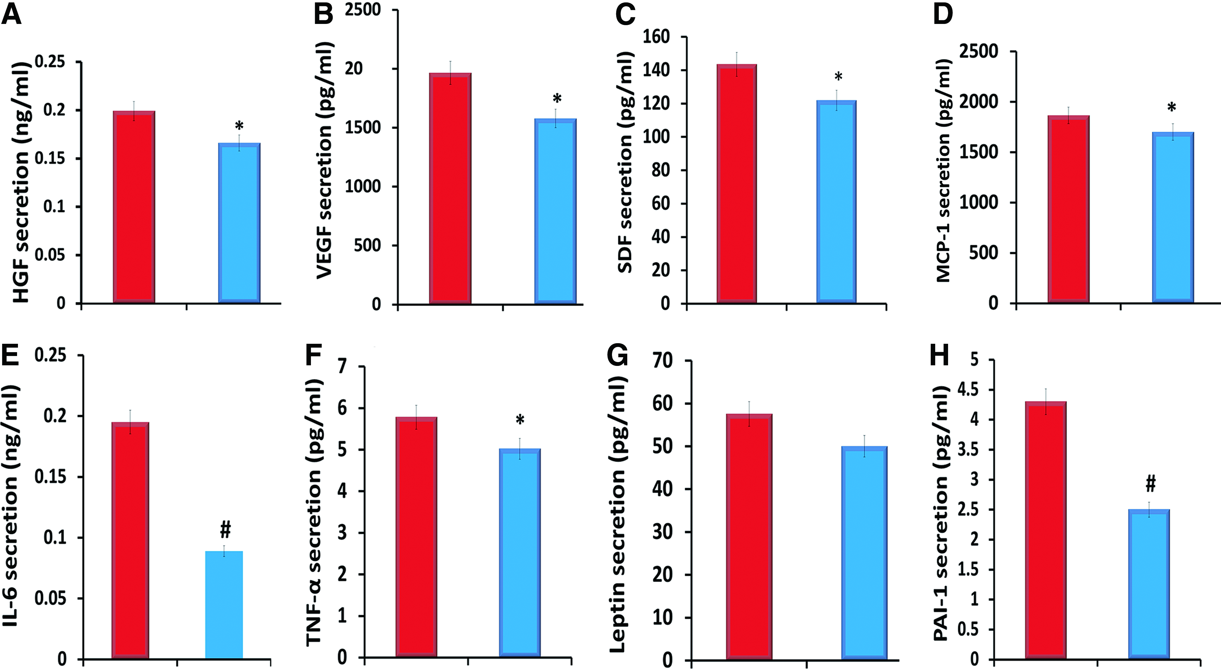
Secretory proteins from frozen BMSC-CM (red) and rehydrated freeze-dried bone marrow mesenchymal stem cells-conditioned medium membrane (FBMSC-CMM) (blue). Fresh conditioned media from bone marrow mesenchymal stem cells (BM-MSCs) were collected after incubation for 24 h in Dulbecco's modified Eagle's medium. Hepatocyte growth factor (HGF)
Morphological characteristics and biocompatibility of MSC membrane
To assess the feasibility of FBMSC-CMM as a novel material for wound regeneration, we evaluated the cell morphology, viability, and proliferation capacity of cultured RDFs within the rehydrated FBMSC-CMM. Proteins or minerals appeared to be attached to the mesh and conformed to the three-dimensional topography of the scaffold. The majority of the proteins or minerals in the membrane exhibited a rounded morphology and clustered around the mesh pores. FBSB only showed small pores (Fig. 2A). The results assayed by the live/dead kit on the 1st, 3rd, 5th, 7th, and 14th day suggested that a higher death rate was present in the FBMSC-CMM compared with frozen MSC-CM and FBS on days 1, 3, and 7. The cells then survived well within the rehydrated FBMSC-CMM from day 7 and a greater than 84% of viable cells remained for up to 14 days in vitro (Fig. 2C, D), implying that the FBMSC-CMM acts as a functional growth factor drug for the cell population. Proliferation of RDFs seeded within FBMSC-CMM was compared with those in frozen MSC-CM and FBS (Fig. 2B). RDFs cultured within FBMSC-CMM supplemented with DMEM showed a lower proliferation rate during the first 7 days compared with those in FBS and MSC-CM (Fig. 2B), whereas they became identical in these three groups after day 7 (data not shown). RDFs cultured both in FBSB and SFM showed lower survival rates and higher death rates compared with other groups at each time point due to the lack of trophic factors, especially in the FBSB. Thus, we can conclude that no specific effects were exerted by the stabilization solution on the therapeutic potential of FBMSC-CMM.

Biocompatibility of rat dermal fibroblasts (RDFs) within a biomimetic FBMSC-CMM.
Wound closure and histological healing
We utilized a rat model of normal wound healing to assess the therapeutic efficacy of FBMSC-CMM in vivo (Fig. 3A). On day 1, 3, 7, 10, 14, 18, and 22, the macroscopic wound areas were quantified by tracing the wound margin and calculating the pixel area in relation to a ruler with a fine resolution computer mouse. A significant acceleration of wound healing was observed at each time point in both the local FBMSC-CMM and BMSC-CM groups compared with the nontreated group (e.g., % original wound area: 40.8%±3.2%, p=0.01 in FBMSC-CMM group; 57.8%±3.3%, p=0.05 in BMSC-CM group vs. 66.2%±4.3% in nontreated group on day 7) (Fig. 3B). Wounds healed significantly faster when treated with FBMSC-CMM scaffolds as observed on days 5, 7, 10, and 14 compared to that with a local injection of BMSC-CM, but the differences disappeared gradually after day 14. However, FBMSC-CMM-treated wounds healed nearly 4 days faster compared with those treated with BMSC-CM (day 18.2±0.5 vs. 22.0±0.4, p=0.003) (Fig. 3B). Moreover, FBMSC-CMM-treated wounds demonstrated a greater regenerative healing capacity with improved skin architecture, including the enhancement of epithelialization with the highest density and best-organized collagen deposition compared with other groups (Fig. 3C). The results also showed that the wound healing rates of both the FBSB and medium alone-treatment groups were identical to that of the normal control group. No specific benefits to the wound healing rate were conferred by FBSB or medium alone.
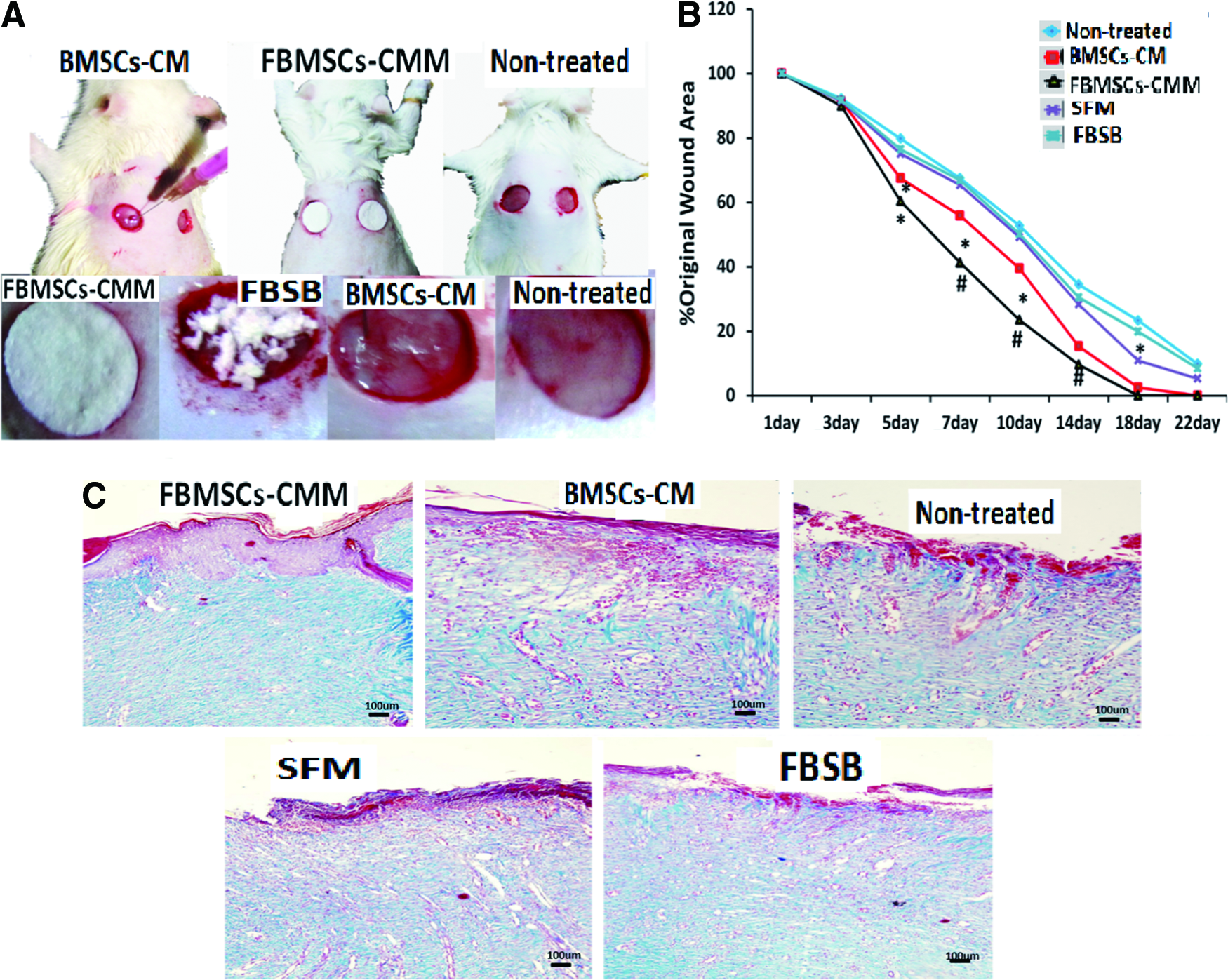
Effects of FBMSC-CMM on wound closure.
Skin vascularization and epithelialization
Excisional wounds treated with either BMSC-CM or FBMSC-CMM appeared grossly more vascular than wounds in other groups (Fig. 4A). To characterize wound vascularization, the number of dermal microvessels was counted in CD31-stained immunohistochemical sections of healed day 14 skin wounds (Fig. 4B). There was a moderate increase in the number of microvessels in wounds treated with a local injection of BMSC-CM (8.8±0.4 vessels per high-powered field [hpf] vs. normal control 5.6±0.5, p=0.03), but neovascularization was further augmented when FBMSC-CMM was applied (15.7±0.6 vessels/hpf; vs. untreated p=1.0×10⁻⁶; vs. BMSC-CM p=2.5×10⁻⁴). The increase in vascularization seen in FBMSC-CMM-treated wounds was further confirmed by immunofluorescent staining of α-SMA. A significantly higher staining intensity of α-SMA was seen in wounds treated with the local BMSC-CM injection compared with the other three control groups. However, α-SMA staining was further increased in wounds treated with FBMSC-CMM (Fig. 4C, D). Not surprisingly, both the α-SMA and CD31 expression levels in the FBSB and medium alone-treatment groups were similar to that in the normal group.
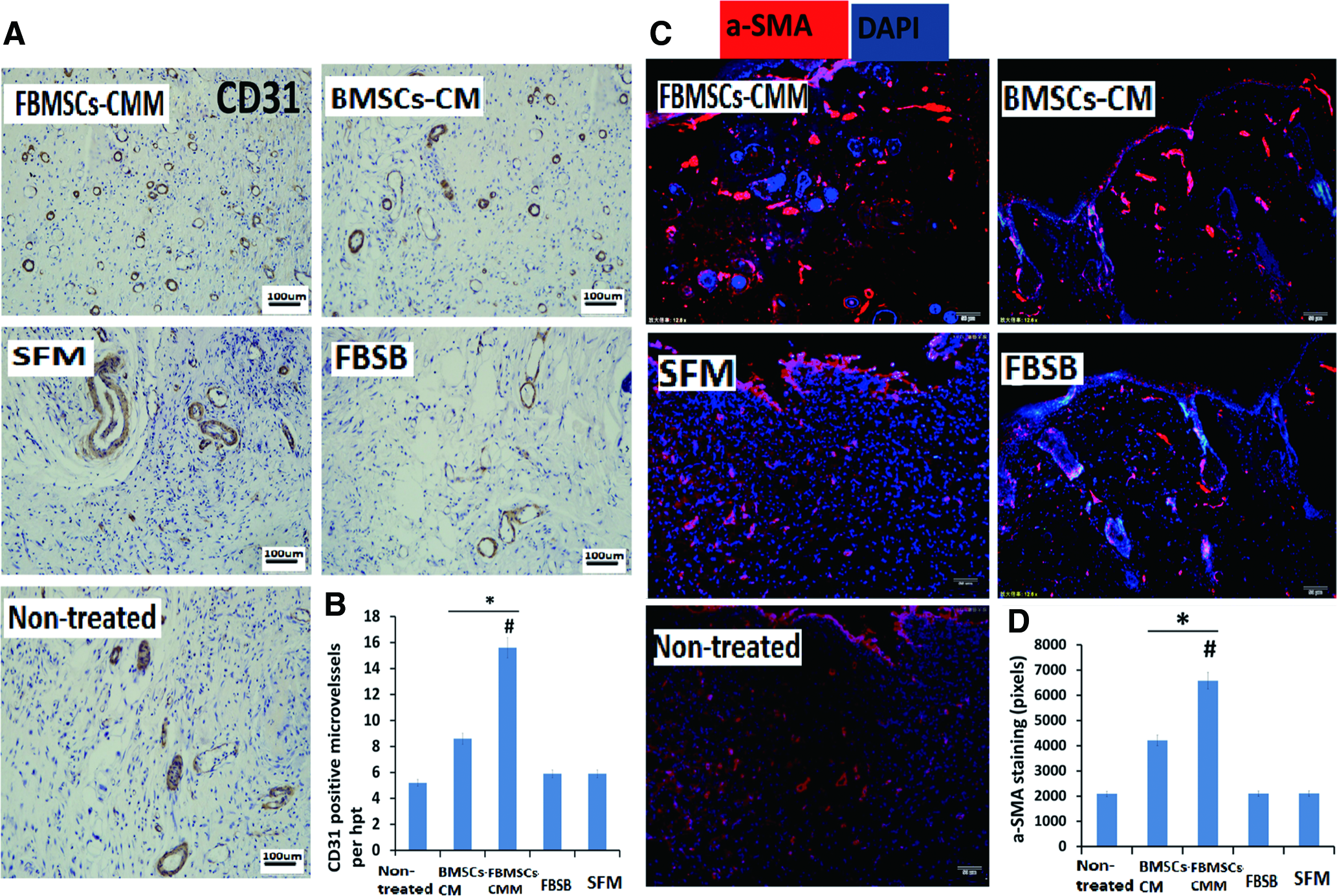
FBMSC-CMM enhances wound vascularization.
To assess the epithelialization in the FBMSC-CMM-mediated improvements in animal wounds, samples from wounds at day 7 were assayed by CK5 immunofluorescent staining. A significant difference in CK5 expression was found in wounds treated with the local BMSC-CM injection compared with that in untreated wounds (0.45±0.04 vs. 0.28±0.04 relative pixel density, p=0.03). Wounds treated with FBMSC-CMM had further increased levels of CK5 expression (0.58±0.10 vs. normal controls 0.28±0.04, p=0.001) (0.58±0.10 vs. BMSC-CM 0.45±0.03, p=0.01) (Fig. 5A, B). Medium alone-treatment groups showed slightly higher CK5 expression compared with the FBSB and normal groups. No additional effects were detected in the FBSB group (stabilization solution).
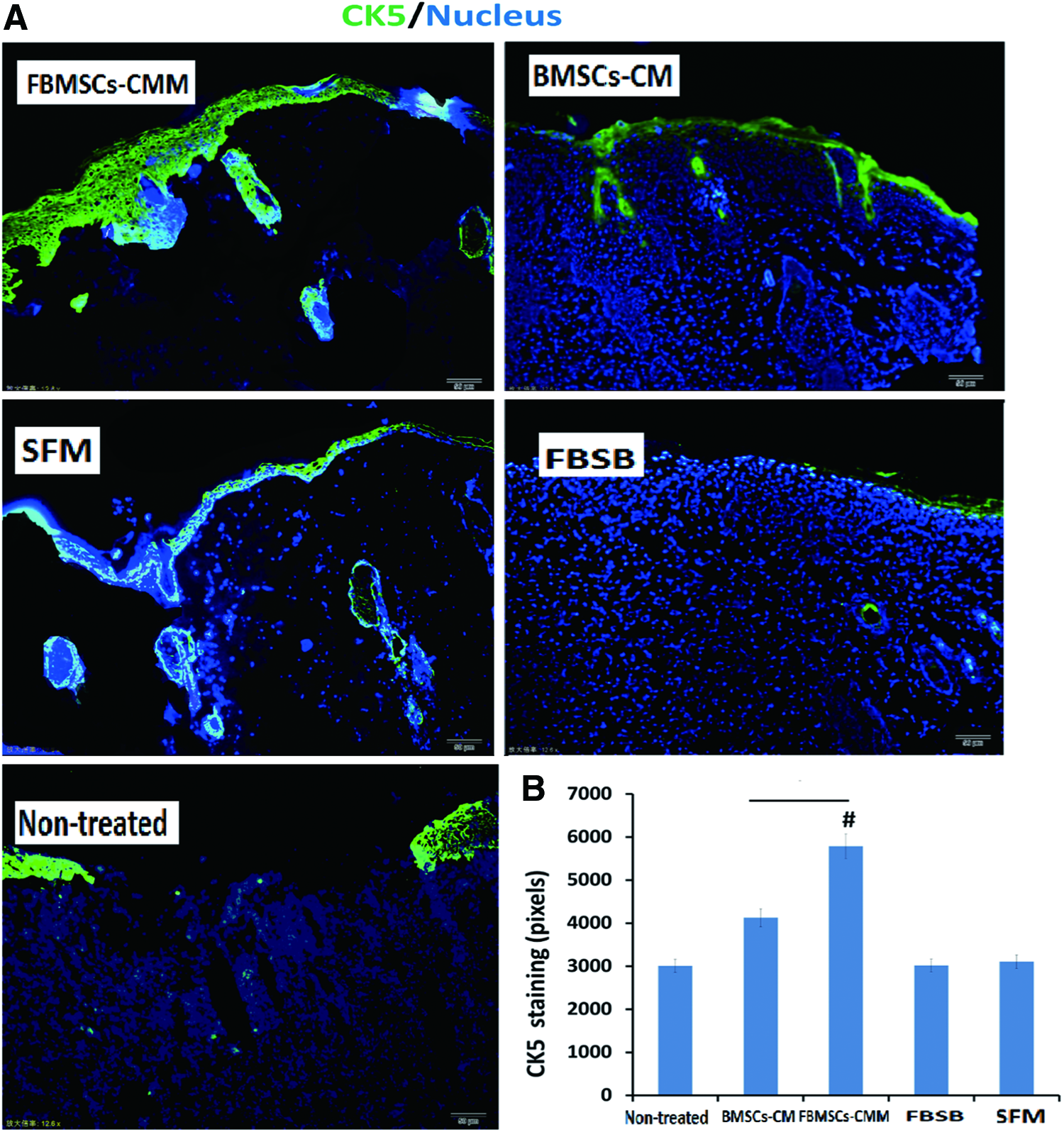
Evaluation of wound epithelialization in wound bed.
Enhanced tissue trophic factors by FBMSC-CMM
To detect the mechanisms that account for the therapeutic effects of FBMSC-CMM, trophic factors in the wound bed at day 7 were evaluated by immunofluorescent staining and western blot analysis. Both the MSC-CM injection and FBMSC-CMM-treated groups showed enhanced trophic factors in the wound bed compared with those in the normal control groups. However, trophic factors of the FBMSC-CMM clearly functioned better in promoting regeneration of the wound bed compared with those of the BMSC-CM (Fig. 6A–C).
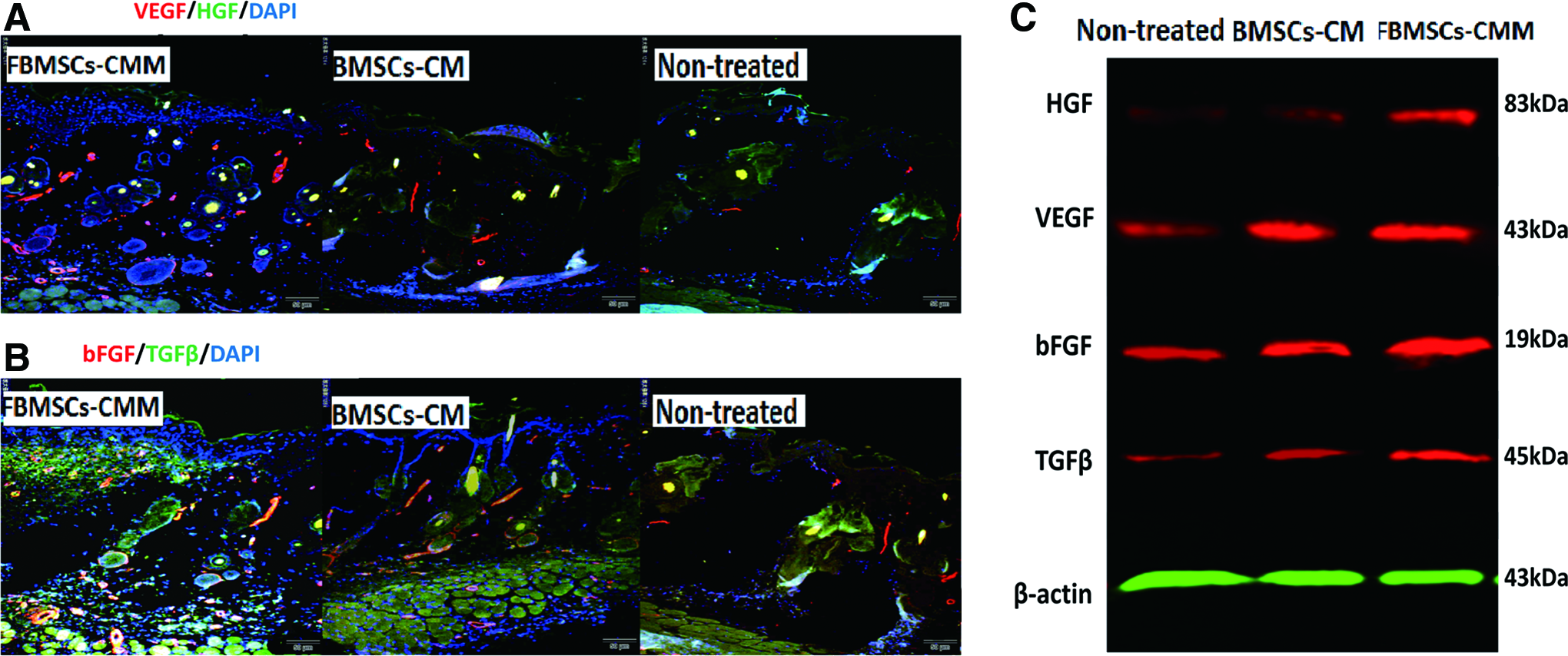
Detection of trophic factors secreted in wound area.
Discussion
The great potential of MSCs for facilitating wound repair and skin degeneration has been well established to rely on several key properties, including the precise recruitment to the injured site, multipotential differentiation function, immune modulation capability, and the ability to secrete soluble factors. However, the contribution of transdifferentiation of stem cells to specific cells in tissue repair is still controversial because only a limited number of donor-derived cells can be detected in vivo, and the incidence of differentiation of implanted MSCs in the target tissue is quite low.31,32 Furthermore, evidence for the differentiation potential of MSCs in patients is rare or even lacking. 33
Thus, previous research has shown increasingly that the greatest therapeutic impact of MSCs in vivo may depend on the potency of their trophic, paracrine, and immunomodulatory functions. MSCs are known to secrete a broad variety of cytokines, including VEGF-1, IGF-1, EGF, NO, HGF, keratinocyte growth factor, angiopoietin-1, SDF-1, MCP-1, and erythropoietin.30,34,35 In fact, the therapeutic effects of these trophic factors have been well documented in the clinic to improve cardiac function through increasing the capillary density and decreasing the infarct size.35,36 More recently, paracrine factors of MSCs were reported to have intriguing effects in brain cancer treatment. 37 Simultaneously, other studies also demonstrated that the administration of conditioned medium of MSCs could act as a chemoattractant for recruiting macrophages and endothelial cells as well as local stem/progenitor cells to enhance wound healing. 38 However, cytokine-based approaches cannot be adequately performed in clinical trials, in general, due to the inherent limitations in tissue transport, pharmacokinetics, and protein stability in vivo. Therefore, harnessing the MSC secretome for wound repair seems, in principle, to have significant clinical potential.
Our study is the first to investigate the biological characteristics of the FBMSC-CMM and reveal its ultrastructure by SEM. Its effects on RDF survival and proliferation in vitro and the repair function after application in the skin wound area were also evaluated. A significant outcome of this study was that the 3D FBMSC-CMM preserved over 80% of the soluble factors secreted by MSCs after rehydration in DMEM. However, levels of inflammatory cytokines, including TNF-α and IL-6, in FBMSC-CMM were significantly lower than those in the BMSC-CM. This result may be due to the fact that the TNF-α and IL-6 secreted by MSCs were extremely low. On the other hand, with an inherited short half-life both of these molecules may be more susceptible to degradation after the freeze drying and rehydration processes compared with other cytokines. The RDFs survived well in the rehydrated membrane after the seventh day, in spite of the fact that their proliferation rate was lower than those in FBS and frozen conditioned medium at the early stage. Perhaps time is needed for the cells to adapt themselves to a new niche. Furthermore, the requirement for a significant increase of various growth factors for angiogenesis and skin cell migration, proliferation, and survival in wound beds may account for the emerging therapeutic effects of FBMSC-CM in skin regeneration. However, whether stem/progenitor cells were recruited into the wound beds and increased cytokine secretions in the wound area were not determined in this study.
The potential interference of the stabilization solution and SFM on the effects of FBMSC-CMM were excluded in both in vitro and in vivo studies. Thus, our results were validated and demonstrated, for the first time, that the FBMSC-CMM fabricated by a selective physical and chemical method, which biologically preserved the main function of fresh conditioned medium, could stimulate RDF survival and contribute to wound skin turnover. The ability of FBMSC-CMM to maintain cytokines over an extended period of time and gradually release them during treatment may account to some degree for such advanced effects on wound healing. Altogether, this study demonstrated that the simple freeze-dried membrane with good biocompatibility is economically feasible to produce and may serve as a potential tool for therapeutic treatments aimed at enhancing wound regeneration.
Conclusion
Currently, the increased aging population and the ongoing epidemic of type II diabetes39–41 means that the search for more effective therapeutics to treat recalcitrant wounds should be ongoing. Our results suggest that the freeze-dried BMSC-CM represents a promising biomaterial that can be used to stimulate quality tissue formation in chronically refractory wounds. Moreover, the paracrine factors may be used in the clinic (e.g., during bone or alveolar surgery) as an emergency drug if prepared in other forms such as a powder. Since the paracrine factors can be readily obtained, FBMSC-CMM may further alleviate the limitation of clinical use of stem cells due to their extremely low survival cell number in vivo. However, our study is limited in the sample size, and comparison of FBMSC-CMM with other scaffolds. Moreover, we only placed our focus on the difference in treatment potential between the BMSC-derived medium and freeze-dried membrane. The specific factors responsible for the regeneration and the other immunomodulatory functions in FBMSC-CMM should be studied in the future. The optimal standards for the production of FBMSC-CMM are needed in particular. Finally, whether formation of such a biomaterial can support trophic factors in the wound bed and reduce wound reopening in the long run remains to be evaluated.
Footnotes
Acknowledgments
This study was supported by grants from the Science and Technology Project of Guangzhou, China (2012J4100044), the National Natural Science Foundation of China (81171812), and the National Basic Science and Development Program, China (973 Program 2012CB518105).
Disclosure Statement
The authors declare no funding or conflicts of interest.