
Abstract
Delayed re-endothelialization is one of the major disadvantages in synthetic vascular grafts, especially in small-diameter grafts (inner diameter <6 mm), leading to thrombosis and stenosis of the grafts. Simvastatin, a serum cholesterol-lowering drug, has promotional effects on endothelial progenitor cell (EPC) mobilization from bone marrow and recruitment to sites of vascular injury exhibiting acceleration of re-endothelialization. In this study, we prepared double-layer vascular patches from Thai silk fibroin/gelatin with gelatin hydrogel incorporating simvastatin-micelles (SM) for sustained release of simvastatin to recruit circulation EPCs. To enhance simvastatin solubility, simvastatin was entrapped in micelles of
Introduction
T
Simvastatin (3-hydroxy-3-methyl-glutaryl-CoA reductase inhibitors), a hydrophobic drug widely used for lowering serum cholesterol levels, was reported to be involved in mobilizing and recruiting endothelial progenitor cells (EPCs) from bone marrow (BM) to the injured vascular sites and accelerating re-endothelialization of vascular defects in animal models.4–6 Simvastatin can also induce EPC migration, augment EPC chemotaxis, and inhibit EPC apoptosis.7,8 Moreover, it has been reported that simvastatin induced angiogenesis with the increasing of capillary density.9,10
We have previously reported the application of gelatin hydrogels as an effective sustained release carrier for many bioactive agents such as basic fibroblast growth factor, transforming growth factor-β1, bone morphogenetic protein-2 (BMP-2), and stromal cell-derived factor-1 (SDF-1) by means of degradation control.11–15
However, their interactions with hydrophobic drugs were limited. Polymeric micelles are amphiphilic block copolymers that are formed through the self-assembly in an aqueous environment. They generally have a nanospherical core/shell structure. In an aqueous medium, the inner hydrophobic core acts as a microreservoir for encapsulation of hydrophobic drugs, while the hydrophilic shell interfaces with the circumstances. The potential applications of polymeric micelles in the drug delivery system are enhancement of hydrophobic drug solubilization, controlled drug release, and drug targeting.
16
Protein-polymeric micelles,
Silk fibroin (SF) is a natural fibrous protein from silk cocoons. SF provides inherently excellent properties as a biomaterial such as mechanical properties, due to its β-sheet structure, bio-blood compatibility, low immunogenicity, and biodegradability.18–22 Therefore, it has been applied as substitutes for various tissues such as blood vessel, bone, ligament, cornea, and wound dressing patch.23–32 As artificial vascular grafts, SF has been fabricated into a tube form by various techniques such as dip-coating technique, electrospinning, fibroin fiber weaving and dip-coating with SF solution, and high-concentration SF solution injection.27,28,33,34 It has been reported that SF vascular grafts had comparable mechanical properties to those of native blood vessels.27,32,35,36 Thai SF, Bombyx mori Nangnoi Srisaket 1, has been studied in biomedical applications such as bone tissue engineering and wound dressing.23,24,29,30 Unlike some wild-type silkworms such as Antheraea pernyi, Bombyx mori silkworm SF has no RGD (arginyl–glycyl–aspartic acid) sequence, which is one of the cell adhesion recognition motifs. 37 Therefore, gelatin, a natural polymer derived from collagen containing RGD motif, was additionally used to improve cell adhesion on the patch and also improve its flexibility.
In this study, double-layer vascular patches from Thai SF and gelatin were fabricated. The outer structural layer was made of Thai SF/gelatin, which can provide mechanical strength to withstand the physiological blood pressure. The inner layer of the patches was gelatin hydrogel incorporating SM designed for controlled release of simvastatin. If simvastatin, a chemoattractant of EPCs, can be controlled released from gelatin hydrogel incorporating SM, then circulating EPCs will be recruited to the vascular patches to accelerate re-endothelialization. We evaluated the ability of this bioactive layer in recruiting EPCs on the luminal surface of vascular patches. In vitro releases of the drug from the gelatin hydrogel incorporating SM and the responses of EPCs to the bioactive layer were investigated. In vivo recruitment of EPCs and re-endothelialization were also examined in rat models.
Materials and Methods
Materials
Yellow Bombyx mori silk cocoons, Nangnoi Srisaket 1, were a kind gift from the Queen Sirikit Sericulture Center, Nakorn Ratchasima and Srisaket Provinces, Thailand. Lithium bromide (LiBr) was purchased from the Sigma-Aldrich Co., LLC. Gelatin powder (MW 100 kDa) with isoelectric points of 5 and 9 were kindly supplied by Nitta Gelatin, Inc. Disuccinimidyl carbonate (DSC) and 4-dimethylaminopyridine (DMAP) were purchased from Nacalai Tesque, Inc. Simvastatin and other chemicals were purchased from Wako Pure Chemical Industries and used without further purification.
Preparation of LAo- grafted gelatin
LAo (MW 1000 Da) was synthesized from
Critical micellar concentration of LAo-grafted gelatin
The conventional fluorescence technique using pyrene as a fluorescence probe was used to determine the critical micellar concentration (CMC) of LAo-grafted gelatin micelles. The micelle solution at the concentration of 1000 μg/mL was serially diluted to 1 μg/mL and added to 6×10⁻⁶ M pyrene powder. The CMC of the micelle solution was determined from emission spectra at an excitation wavelength of 333 nm. Emission spectra were scanned from 350 to 500 nm. The CMC was measured from the drastic changes in slope of graphs plotted between the intensity ratio (II/IIII) of pyrene and polymer concentration. 39
Preparation of gelatin-LAo micelles entrapping simvastatin
SM were prepared using a dialysis method as previously described.9,17 LAo-grafted gelatin (1 mg/mL) was dissolved in DMSO at 37°C for 3 h and mixed with simvastatin solution (0.6 mg/mL) in DMSO at 37°C for 5 h. The solution was dialyzed against DDW for 3 days and centrifuged at 8000 rpm, 4°C for 10 min. The supernatant was collected and freeze-dried to obtain SM. All micelle preparation steps were performed in dark conditions to prevent drug destabilization. To measure the amount of simvastatin entrapped, the SM were dissolved in acetonitrile before absorbance measurement at the wavelength of 236 nm using a UV spectrophotometer. Drug concentrations were obtained from a standard curve of known concentration. 40
Size and surface charges of SM
To measure the average size and surface charges of the micelles, dynamic light scattering (DLS) measurement (DLS-DPA-60HD; Otsuka Electronic Co. Ltd.) was performed. The micelle solution in DDW (1 mg/mL) was measured with a He–Ne laser at a detection angle of 90° at room temperature. 17
Preparation of gelatin hydrogel incorporating SM
Gelatin hydrogel incorporating SM was prepared through chemical crosslinking with glutaraldehyde (GA).14,17 The aqueous gelatin solution (pI 9, 6% w/w) was mixed with the SM solution (40 mg/mL). GA solutions (0.045%, 0.09%, and 0.18% v/v) were added for the crosslink reaction. The mixture was cast into a polypropylene dish (Bio Bik) and stored at 4°C for 12 h in darkness. The reacted hydrogels were then rinsed with 0.1 M glycine solution for 2 h to block the unreacted GA. They were washed repeatedly with DDW followed by air-drying for 1 day. The gelatin hydrogels (3% w/w) incorporating SM (20 mg/mL) were obtained. They were sterilized in ethylene oxide gas and kept from light for further experiments.
Isolation and characterization of bone marrow-derived EPCs
EPCs were isolated from (male, 4 weeks) F344 rat bones using a modified method as described previously.41,42 After scarifying animals, the femur and hip bones were cut into small pieces followed by soaking under 5 mM ethylenediaminetetraacetic acid in phosphate buffer saline (PBS). Histopaque 1083 solution (Sigma) was used to isolate mononuclear cells from cell suspension by a density gradient centrifugation method. Red blood cells (RBCs) were lysed in NH4Cl at 4°C for 15 min. The obtained mononuclear cells were cultured in M199 media supplemented with 10% (v/v) fetal bovine serum (FBS) and 1% (v/v) penicillin/streptomycin at 37°C, 5% CO2 overnight. The nonadhered cells were collected and seeded on 10 μg/mL ProNectin® F (Sigma)-coated RepCell® dishes (CellSeed, Inc.). They were cultured in endothelial growth medium (EGM-2-MV Bullet Kit; Clonetics) supplemented with 10% (v/v) FBS for 7 days to obtain the EPC-rich cell population.
Characterization of the EPCs was performed by flow cytometric analysis. EPCs were incubated in 1% (v/v) paraformaldehyde in PBS at room temperature for 15 min. After fixation, EPCs were incubated with mouse anti-rat CD32 antibody (1:50 dilution) at 4°C for 15 min to reduce nonspecific binding. Next, EPCs were incubated with primary antibodies, biotin mouse anti-rat CD31 (BD Biosciences), biotin mouse anti-rat CD90 (BD Biosciences), and rabbit anti-rat vascular endothelial growth factor receptor-2 (VEGFR-2; Abcam) (1:50 dilution) at 37°C for 1 h. 43 After washing with a buffer solution (PBS supplemented with 2% fetal calf serum and 0.02% NaN3), the cells were incubated with PE-Cy™5 Streptavidin (1:250 dilution) (BD Biosciences) and rabbit-Alexa 647 (1:1000 dilution) (Invitrogen) at 37°C for 30 min as secondary antibodies. After washing with the buffer solution, expression of cell surface markers was evaluated by fluorescence-activated cell sorting (FACS Canto II flow cytometer; BD Biosciences).
Migration assay
To evaluate the chemoattractive effect of EPCs toward SM comparing to nonmodified simvastatin (S), an in vitro modified Boyden's chamber migration assay was performed. 44 Due to its original inactive lactone form, simvastatin was activated by dissolving in ethanol, then hydrolyzed with 0.1 N NaOH, and neutralized with HCl (∼pH 7.2). 45 The EPCs suspended in endothelial basal media (EBM-2) supplemented with 0.5% (v/v) albumin were seeded at 5×10⁴ cells/well in the upper chamber of the transwell culture plates (24-wells, 8 μm pore size; Corning/Fisher Scientific). Simvastatin (S) or SM solutions at concentrations of 0.1, 1, 10, and 100 μM simvastatin were added to the lower chambers. The EBM-2 media supplemented with 0.5% (v/v) albumin were used as a negative control (C), while SDF-1α (100 ng/mL) was used as a positive control. The cells were incubated at 37°C, 5% CO2 for 16 h. The cells that migrated across the membrane to the lower chamber were fixed with 4% (v/v) paraformaldehyde and stained with crystal violet. The stained cells were counted under a light microscope at 20× magnification (n=5).
Releases of simvastatin from gelatin hydrogel incorporating SM
Gelatin hydrogels incorporating SM at various GA concentrations were immersed under 1 mL of PBS at 37°C. At each predetermined time, the hydrogels were moved to new PBS tubes. After 3 days, the solution was changed to PBS containing 0.125 U/mL of collagenase IA and further incubated at 37°C until the hydrogels were completely degraded. To measure the amount of released simvastatin, the PBS or collagenase/PBS was freeze-dried followed by dissolution in acetonitrile. After centrifugation, concentrations of simvastatin in the supernatant were determined as the method described in the section “Preparation of gelatin-LAo micelles entrapping simvastatin” (n=5).
In vitro adhesion and proliferation of EPCs cultured on the gelatin hydrogel incorporating SM
The gelatin hydrogel (G) and gelatin hydrogel incorporating SM at the drug concentrations of 100 μM (GSM100) and 400 μM (GSM400) were swollen in EGM-2 MV media. They were cut as the size of the well and placed in a 24-well culture plate. EPCs were seeded on the hydrogels at 2×10⁴ cells/hydrogel and cultured in EGM-2 MV media supplemented with 10% (v/v) FBS for 6 h, 1, 3, and 5 days. Cell viability was evaluated using the cell counting kit-8 as a manufacturer's protocol (WST-8; Dojindo, Inc.). WST-8 could be reduced by dehydrogenase in living cells resulting in the water-soluble formazan (orange color). At each predetermined time, the WST-8 solution was added to the cultured cells at the ratio of 1:10 (WST-8: media volume), followed by incubation for 3 h. Light absorbance of the cell culture solution was measured using a microplate reader (Molecular Devices) at 450 nm wavelength. The percentage of initial cell adhesion, specific growth rate (μ), and population doubling time (PDT) were calculated as the equations given below (n=4). 46
Percentage of cell adhesion
when X0 is the initial cell seeding number and Xt is the number of cells at 6 h of culture
Specific growth rate (μ) (unit; h)
when X1 is the number of cells at cultured time t1 and X2 is the number of cells at cultured time t2 (t1=24 h, t2=72 h)
PDT (unit; h)
Preparation of SF solution
The SF solution was prepared as previously described. 47 Water-soluble parts of silk cocoon were removed with 0.02 M Na2CO3 solution. The fiber was dissolved in 9.3 M LiBr solution at 60°C for 4 h. The SF solution was then dialyzed against DDW for 3 days. After dialysis, the SF solution was centrifuged at 8000 rpm, 4°C for 20 min twice to remove impurities. The fibroin solution was concentrated to 15% (w/v) using Amicon® Ultra-15 centrifugal filter devices (Millipore) at 5800 rpm, 20°C for 30 min.
Fabrication of double-layer vascular patches
Tubular vascular grafts were fabricated using the dip-coating technique (Fig. 1). Polyethylene terephthalate (PTFE) tubes (outer diameter=1.67 mm; Chukoh Chemical Industries, Ltd.) were used as templates. The PTFE tube was dipped into the gelatin solution (3% w/v) with or without SM (20 mg/mL) and 0.09% (v/v) GA and then immediately dipped into liquid nitrogen. The coated tubes were stored at 4°C for 12 h in the dark to allow the crosslinking reaction. They were rinsed with 0.1 M glycine solution to block the unreacted GA and washed repeatedly with DDW. After air-drying overnight, the coated tubes were dipped into SF/gelatin solution (75:25, 15 wt% total solid weight) and immersed under 70% (v/v) ethanol at −20°C for 1 h followed by air-drying overnight. The double-layer tubes having gelatin hydrogel incorporating SM as an active inner layer and SF/gelatin as a reinforce outer layer were removed from the PTFE tube and sterilized in ethylene oxide. Before surgery, the tubular grafts were soaked in PBS and cut transversely into curve-shaped patches with the longitudinal length of 2 mm.
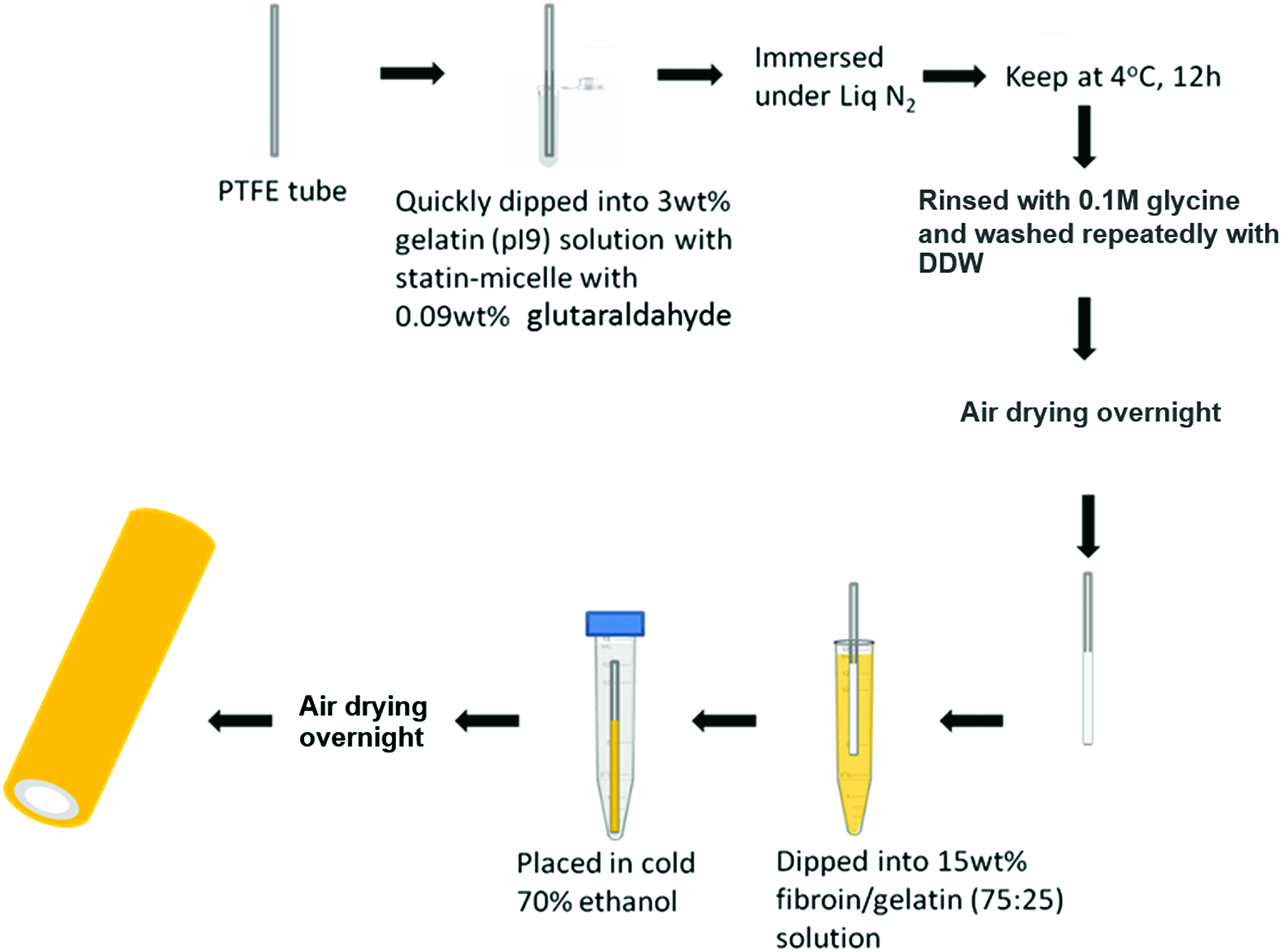
Fabrication process of double vascular tubes before punching into small patches. Color images available online at www.liebertpub.com/tea
In vivo evaluation of EPC recruitment
Double-layer patches from SF/gelatin with gelatin hydrogels (G-patches) (n=8) without or with SM at 100 μM of simvastatin (GSM100-patches) (n=9) were implanted in carotid arteries in male F344 rats. All the animal experiments were performed according to the animal experiment guidelines with permission of the Animal Experiment Committee of the Institute for Frontier Medical Science, Kyoto University. Male rats (500 g body weight) were anesthetized with isoflurane inhalation (3% v/v) followed by intraperitoneal (i.p.) injection with pentobarbital (50 mg/kg body weight). Left common carotid artery was exposed and blood flow was halted by clamping the distal side and proximal side of the artery with microclips. The arterial wall was cut and the patch was cropped out in oval shape using microscissors and a biopsy punch (2 mm diameter) (Kai Industries, Co. Ltd.), respectively. The oval-shaped patch was then placed on the opened artery and sutured with a 10-0 Prolene™ suture (Johnson & Johnson). A medical-grade glue (Aron alpha A) was applied to cover the anastomosis for prevention of blood leakage. Finally, the rats were heparinized (400 U/kg body weight, i.p.) to prevent acute thrombosis in sites of sutured patch.
The EPCs were stained with CellTracker™ CM-Dil (Invitrogen) according to the manufacturer's instructions and resuspended in EGM-2 MV media for tail vein injection (at 1×10⁶ cells in 100 μL of media) on day 1st and 5th after surgery. After 2 weeks of implantation, the rats were sacrificed, and the implanted patches were collected with neighboring arteries. EPC recruitment to the luminal surface of the patches was examined under a fluorescence microscope. The fluorescence intensity of the recruited EPCs on the hydrogels was evaluated using an ImageJ™ software and expressed as corrected total cell fluorescence (CTCF) value. Fluorescence double-immunostaining of the implanted patches was performed by staining with fluorescein isothiocyanate-isolectin B4 (Vector) for endothelial cell detection and staining with an anti-smooth muscle α-actin antibody (Abcam) and an Alexa 594 anti-rabbit immunoglobulin G antibody (BioLegend) for vascular smooth muscle cell (VSMC) detection. Nuclei were counterstained with the 4′,6-diamidino-2-phenylindole solution.
Statistical analysis
The experimental results are expressed as mean ± the standard deviation. Data were statistically analyzed by unpaired student's t test, and statistical significance was indicated by p-value<0.05.
Results
Characteristics of LAo-grafted gelatin and SM
The average size and zeta potential of LAo-grafted gelatin micelles and SM were 108.52 ± 55.98 nm and −4.49±0.28 and 256.42 ± 43.94 nm and −3.50 ± 0.21, respectively. The CMC of LAo-grafted gelatin was at 141.66 mg/L. The amount of simvastatin encapsulated in the micelles was at 4.1 ± 0.5 μg/mg micelles.
Releases of simvastatin from gelatin hydrogel incorporating SM
Figure 2 shows the time profiles of simvastatin released from GA-crosslinked gelatin hydrogel incorporating SM. When subjected in PBS solution, the initial burst release of simvastatin was observed for all hydrogels. After that, simvastatin was continually released from the hydrogels. The percentages of simvastatin released from the hydrogels crosslinked with 0.045%, 0.09%, and 0.18% GA reached 54.47 ± 0.7%, 40.79 ± 2.7%, and 29.10 ± 2.3%, respectively, at 3 days of incubation. Then, the solution was changed to the PBS solution containing collagenase. Different profiles of released simvastatin were observed. The hydrogels crosslinked with 0.045% GA completely released simvastatin within 4 days, while the hydrogels crosslinked with 0.09% and 0.18% GA gradually released simvastatin. Simvastatin was completely released from the hydrogels crosslinked with 0.09% and 0.18% GA after 11 and 14 days of incubation, respectively.
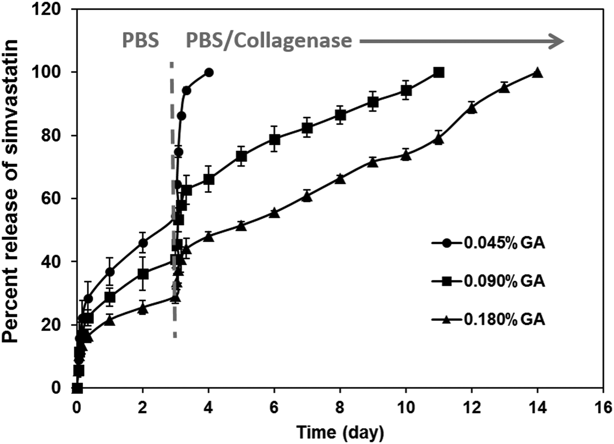
In vitro release profiles of simvastatin from the gelatin hydrogels. The gelatin hydrogel incorporating simvastatin-micelles was incubated in phosphate buffer saline (day 1–3) without or with 0.125 U/mL of collagenase (after day 3) over 14 days at different glutaraldehyde concentrations of 0.045%, 0.090%, and 0.180%.
Characteristics of BM-derived EPCs
After 7 days in culture, the BM mononuclear cells formed small multiple cell clusters (Fig. 3A, B). The population of cultured cells was positive for CD90 (48.8%; Fig. 3C), CD31 (30.7%; Fig. 3D), and VEGFR-2 (84%; Fig. 3E) as markers for EPCs.
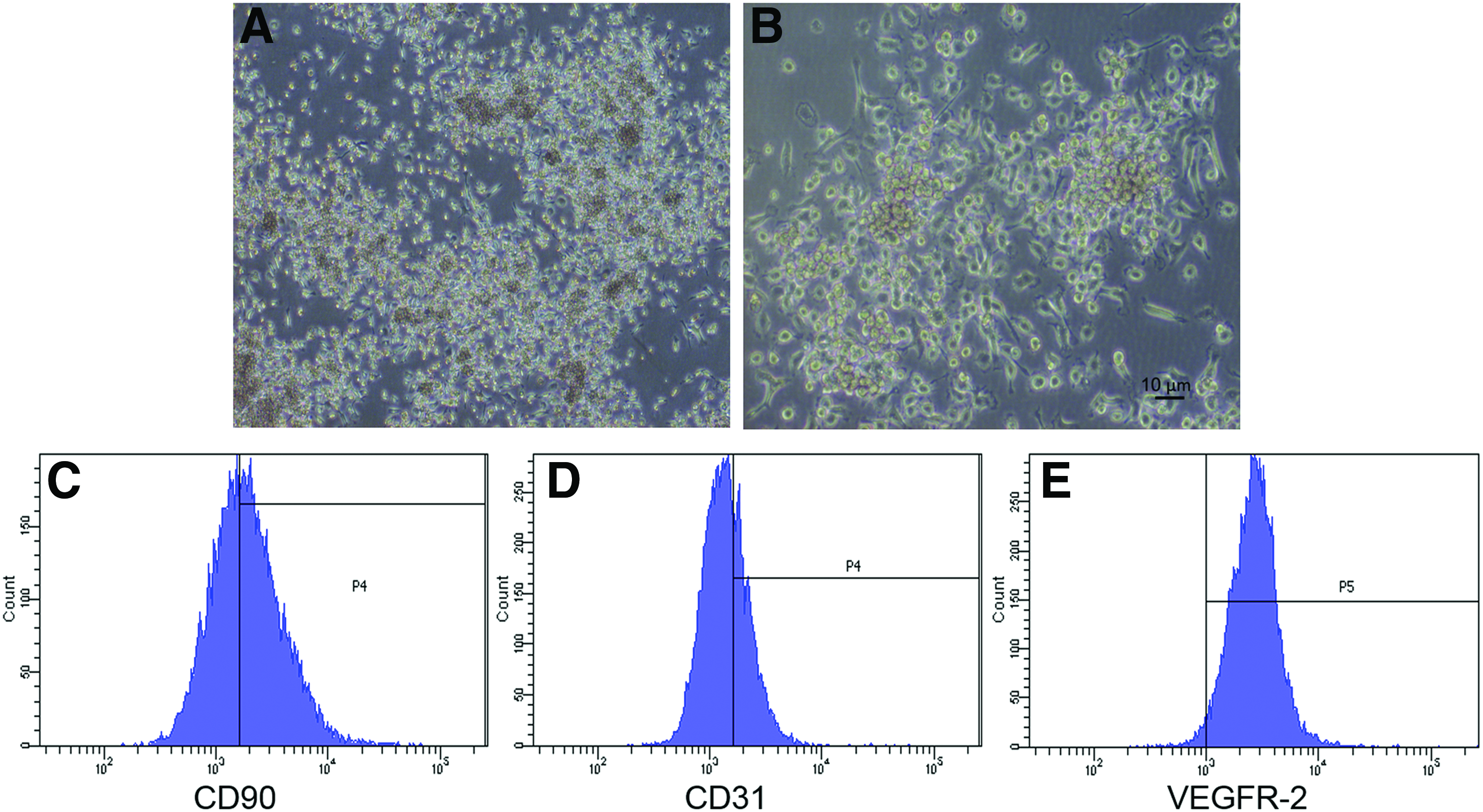
Characterization of bone marrow-derived endothelial progenitor cells (EPCs). The bone marrow mononuclear cells were cultured in the EPC differentiation medium for 6 days. The small cell clusters were formed at 4×
Migration of EPCs
Figure 4 shows the migration of EPCs toward the media containing simvastatin (S) or SM. The cells migrated dose dependently into the media containing S or SM at the drug concentrations of 0.1–100 μM. The number of migrated cells toward the media containing S or SM was higher compared with control media (the media containing 0.5% albumin). At a concentration range of 0.1–10 μM, the number of cells that migrated into the media containing S or SM was not statistically different. At a concentration of 100 μM, a high number of cells migrated into media containing S, while no cells migrated into media containing SM.
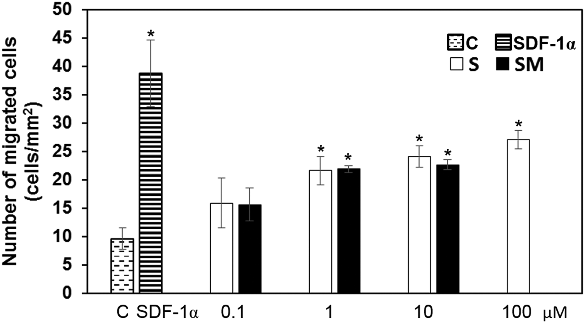
Migration of cultured EPCs. The EPCs were treated with simvastatin (S) and simvastatin-micelles (SM) at simvastatin concentrations of 0.1, 1, 10, and 100 μM. Endothelial basal media-2 media supplemented with 0.5% (v/v) albumin (C) was a negative control, and stromal cell-derived factor-1α (SDF-1α) was a positive control. There were no significant differences of migrated cells between the simvastatin (S) group and simvastatin-micelles (SM) group (*represents statistical significance at p<0.05 when compared to the negative control).
In vitro adhesion and proliferation of EPCs cultured on the gelatin hydrogel incorporating SM
Figure 5 shows the number of adhered and proliferated cells on the gelatin hydrogel (G) and the gelatin hydrogel incorporating SM at 100 (GSM100) or 400 μM simvastatin (GSM400). At 6 h, the number of cells initially adhered on the GSM100 hydrogel was significantly higher than those of G and GSM400 hydrogels. The higher number of proliferated cells was also observed for the cells cultured on the GSM100 hydrogel after 3 and 5 days.

In vitro adhesion and proliferation of endothelial cells. The EPCs were cultured on gelatin hydrogel (G), gelatin hydrogel incorporating simvastatin-micelles at simvastatin concentrations of 100 μM (GSM100) and 400 μM (GSM400) (*represents statistical significance at p<0.05 when compared to gelatin hydrogel films in each time point).
Table 1 shows the percentage of initial cell adhesion, specific growth rate (μ), and PDT of the cells cultured on the hydrogels. The cells cultured on the GSM100 hydrogel showed the highest percentage of cell adhesion (86%) and specific growth rate (15.33 h−1), but the lowest PDT (46.21 h).
Evaluated at 6 h of culture.
Evaluated at 1–3 days of culture (exponential phase) (* and ** represent statistical difference at p<0.05).
PDT, population doubling time.
In vivo evaluation of EPC recruitment
Figure 6 shows the implantation of the vascular patch in a rat's carotid artery. Figure 7 shows the implanted double-layer patches of SF/gelatin with gelatin hydrogel (G-patches) and the patches containing SM at 100 μM simvastatin (GSM100-patches) in carotid arteries after 2 weeks of implantation. Under a light microscope, macroscopic views of luminal surfaces of the implanted patches are shown in Figure 7A. The small dots of RBCs were found on three of the eight implanted G-patches and three of the nine implanted GSM100-patches (Fig. 7A[A]). Under a fluorescence microscope, a large number of CM-Dil-stained EPCs found on the luminal surfaces of GSM100-patches were higher than those on the G-patches (Fig. 7A[C, D]). The CTCF of the recruited EPCs on GSM100-patches was significantly higher compared with G-patches (Fig. 7B). Figure 8 shows the double-immunofluorescence staining of endothelial and VSMCs. Complete re-endothelialization was observed in the implanted GSM100-patches (Fig. 8E, F), whereas incomplete or no endothelial cell lining was observed in the implanted G-patches (Fig. 8B). In addition, the implanted G-patches were covered with a number of migrated VSMCs (Fig. 8A, C).

Macroscopic views of the double-layer patch of silk fibroin and gelatin hydrogel incorporating simvastatin-micelles. The patch was placed on a carotid artery beside an oval defect (a white dash)
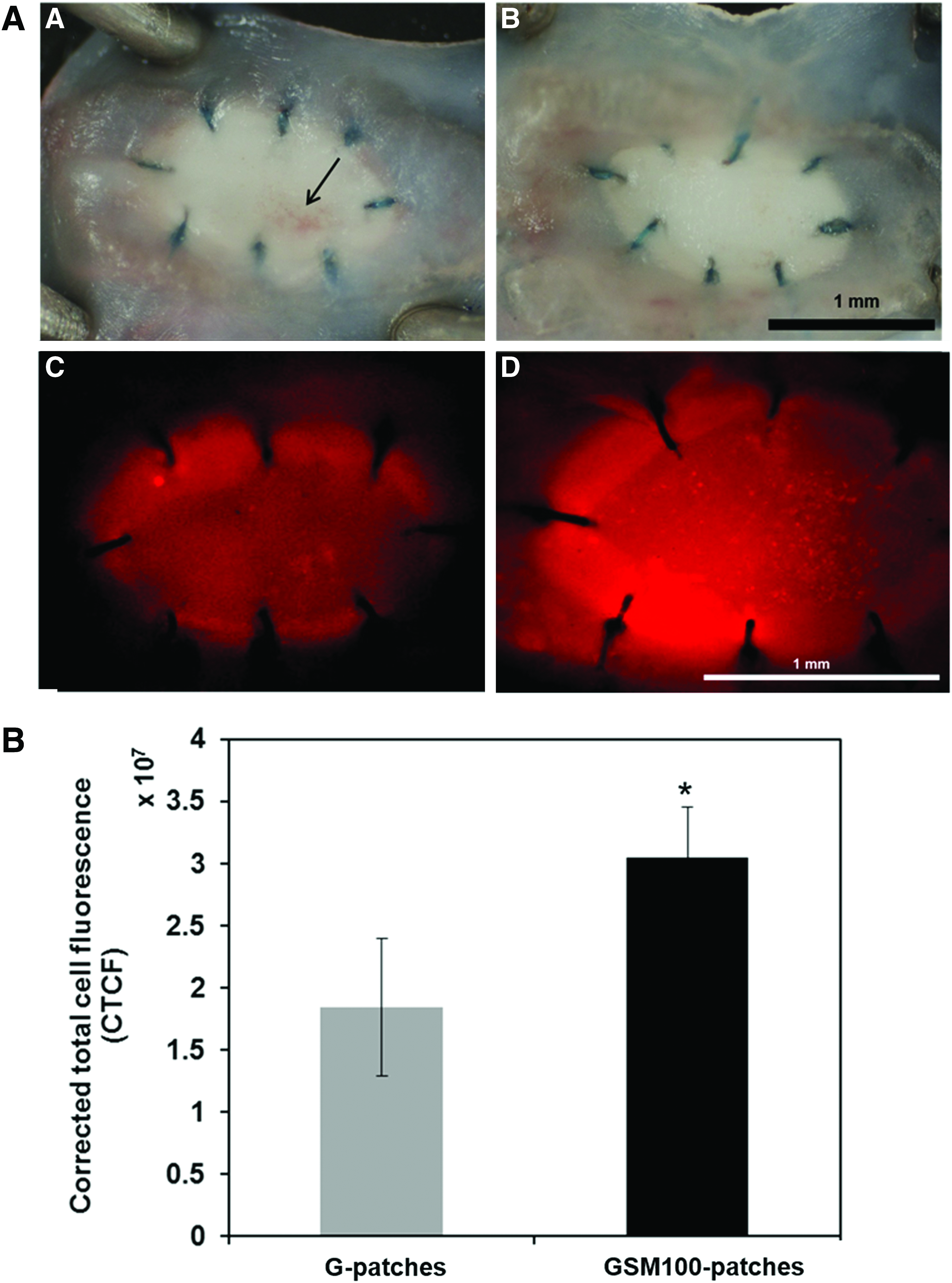
En face observation of the implanted patches in carotid arteries.

Double-immunofluorescence staining images of implanted patches in carotid arteries. The double-layer patches of silk fibroin and gelatin hydrogel (G-patches)
Discussion
The present study demonstrated the therapeutic potential of a vascular patch fabricated from Thai SF/gelatin with bioactive layer of SM. The dominant function of this patch is to recruit circulating EPCs to accelerate the re-endothelialization of vascular prostheses. We have shown that Thai SF has biocompatibility and tunable biodegradability.23,24 SF composited with gelatin and hydroxyapatite has been classified as “non-irritant” to “slight-irritant” according to the ISO 10993-6 standard. 48 The excellent mechanical properties of SF were due to its beta-pleated sheet structure, which resists human physiological enzymes. 18 The Thai SF-based double-layer vascular grafts we developed had suture retention forces similar to those of the native blood vessels. 32 In the design of double-layer vascular patches of this study, Thai SF/gelatin was used to produce the outer layer, which had flexibility and could withstand the normal physiological blood pressure. The gelatin hydrogel incorporating SM was produced as the bioactive inner layer of the patches to recruit EPCs and accelerate re-endothelialization.
Simvastatin, a hydrophobic drug, has limitation in homogenous distribution in gelatin hydrogels. To improve the distribution, LAo-grafted gelatin, which has both hydrophobic and hydrophilic sides, was used to entrap simvastatin in the form of SM. We showed that the SM had a chemoattractive effect similar to the nonmodified simvastatin (S) at the simvastatin concentration of 0.1–10 μM (Fig. 4). This indicated that the LAo-gelatin micelles could preserve the biological property of simvastatin. Noted that the micelles entrapped 100 μM simvastatin (SM100) did not have the same effect of migrating stimulation from our experiment due to high media concentration. This limitation resulted from the low drug loading nature of micelles. The limitation can be solved by distribution of SM in crosslinked gelatin hydrogels.
In vitro releases of simvastatin from the gelatin hydrogel incorporating SM were performed to experimentally confirm the release mechanisms of the hydrogel system (Fig. 2). This indicated that the release mechanisms depended on both diffusion and degradation of the hydrogels. The rate of releases was correlated with the concentrations of crosslinking agent. At the lowest GA concentration, the hydrogels showed the burst release and huge diffusion of simvastatin in PBS and rapidly destabilized in PBS/collagenase. Diffusion of the drug in PBS resulted from releasing of un-crosslinked gelatin out of the hydrogels due to too low crosslinking.
To evaluate the effect of simvastatin on EPC adhesion and proliferation, EPCs were cultured on gelatin hydrogel incorporating SM at two high concentrations of simvastatin (100 and 400 μM). The SM at 100 μM was chosen due to the best effect on EPC chemotactic activity in this study. In practice, the hydrogels with SM would be a drug reservoir to sustained release simvastatin to recruit EPCs from blood circulation. There were reports on successful mobilization of EPCs with high-dose simvastatin (20 mg/kg body weight) in systemic administration.49,50 Therefore, we used the high concentration of simvastatin (400 μM) to entrap in gelatin hydrogels. The gelatin hydrogels (G) without or with SM at 100 μM (GSM100) and 400 μM (GSM400) of simvastatin were tested for in vitro EPC adhesion and proliferation (Fig. 5; Table 1). GSM100 exhibited the highest promotional effects on initial cell adhesion of 86%, proliferation at specific growth rate (μ) of 15.33×10⁻³ h−1, and PDT of 46.21 h. Simvastatin has been previously reported to promote in vitro EPC adhesion and proliferation in serum-free M199 media at a dose of 1 μM. 51 In this study, it was found that GSM400 led to negative effects on EPC adhesion and proliferation. Similar to a previous study of Asakage et al., they cultured EPCs in the MCDB-151 medium containing 200–500 μM simvastatin and reported the negative effects on EPC proliferation by inducing cell cycle arrest. 52
In vivo implantation of the patches containing SM (GSM100-patches) in rat carotid arteries revealed successful EPC recruitment and re-endothelialization at 2 weeks after implantation (Figs. 7 and 8). The recruitment was attributed to pleiotropic effects of simvastatin on increasing VEGF, endothelial nitric oxide synthase, and integrin α5β1 in EPCs.4–6 Although gelatin has been reported to have blood hemostatic effects, 53 simvastatin has been claimed to reduce blood coagulation. 54 From the results of macroscopic observation of implanted-patch luminal surfaces under a light microscope, there were no differences of small RBC accumulation but not thrombus under the neoendothelium on G-patches and GSM100-patches. Further studies are therefore required to confirm the combination effects of gelatin and simvastatin on blood coagulation. In the results of immunohistological analysis, neointima was formed with migrated VSMCs in G-patches (Fig. 8A, C), while no neointimal formation was observed in GSM100-patches as we expected. The finding is consistent with previous reports in which an inhibitory effect of simvastatin on VSMC proliferation and migration was demonstrated.55,56
Comparing to one of the previous studies in which simvastatin was systemically administered for re-endothelialization, to achieve re-endothelialization in a balloon-injured carotid artery within 2 weeks, the animals had to undertake i.p. injection of high-dose simvastatin at 1.0 mg/kg body weight/day for 14 days (total 14 mg/kg). 55 In our case, only a small amount of simvastatin at 2.5 mg/kg body weight (or 100 μM simvastatin/500 g rat body weight) was contained in the hydrogels, and the drug was continuously and locally released for at least 14 days following implantation period. This system exhibited an advantage of the recruitment of transfused EPCs and accelerated re-endothelialization in artificial vascular patches with a low-dose simvastatin. Taken together, this study clearly indicated that the inner bioactive layer of gelatin hydrogel containing simvastatin entrapped in LAo-grafted gelatin micelles effectively sustained release of active simvastatin, which could promote EPC migration, adhesion, and proliferation leading to effective circulating EPC recruitment. Moreover, it should be noted that this hydrogel system can be applied to many other water-insoluble drugs.
For the current study, we utilized cultured EPCs isolated from BM and injected to syngeneic rats with vascular patch implantation to assess the recruitment of exogenously injected EPCs to gelatin hydrogel incorporating SM. In consideration of the assessment for endogenous BM-derived EPC recruitment to gelatin hydrogel incorporating SM in physiological settings, formally, BM transplanted immunodeficient nude rats with genetically labeled BM cells isolated from GFP-Sprague-Dawley rats should be used as a recipient of the patch implantation. However, it appeared to be technically pretty difficult to implant a tiny patch in a tiny carotid artery of nude rats. For this reason, we alternatively decided to use the current in vivo experimental design in this study. For future plans, we will further investigate the capabilities of the bioactive inner layer, gelatin hydrogel incorporating SM, on EPC recruitment and re-endothelialization with long-term vascular tissue ingrowth to the tissue-engineered vascular grafts.
Conclusion
We have demonstrated the potential of using double-layer vascular patches containing the inner bioactive layer of gelatin hydrogel incorporating SM for controlled release of hydrophobic drug, simvastatin. Release of biologically active simvastatin could be controlled correspondingly through diffusion and degradation of gelatin hydrogels. Sustained release of simvastatin could be achieved and resulted in EPC recruitment and accelerated re-endothelialization in vivo. The concept of multifunctional vascular grafts is therapeutically feasible, although further optimization of the system would be required.
Footnotes
Acknowledgments
This research was supported by grants under the program Strategic Scholarships for Frontier Research Network for the Join PhD Program Thai Doctoral degree from the Commission on Higher Education, Thailand, and the Ratchadaphiseksomphot Endowment Fund of Chulalongkorn University (RES560530060-AS). The Chulalongkorn University Centenary Academic Development Project (under Biomedical Engineering Development Program and Center of Innovative Nanotechnology, Chulalongkorn University) was gratefully acknowledged.
Disclosure Statement
The authors have no competing financial interests.