
Abstract
Objectives:
To investigate the efficacy of recombinant adeno-associated virus (rAAV)–human telomerase reverse transcriptase (hTERT)-transducted nucleus pulposus cells (NPCs) in disc degeneration process in a canine disc degeneration model.
Methods:
The intervertebral disc degeneration of lumbar (L) 1–2, L3–4, and L5–6, from 12 female mongrels was prepared with the 20-gauge biopsy gun. Four weeks after animal model preparation, intervention experiment with rAAV-hTERT-transducted NPCs was conducted: group A, L1–2, serum-free medium with rAAV-hTERT modified NPCs; group B, L3–4, serum-free medium with NPCs; group C, L5–6, serum-free medium alone. Canines underwent digital radiography and magnetic resonance imaging 1 day before intervention, and 4, 8, and 12 weeks after intervention to evaluate the change of disc height and hydration status of interventional intervertebral discs. Twelve weeks after intervention, histological, biomechanical, and biochemical studies were carried out.
Results:
The rAAV-hTERT-transducted NPCs were constructed successfully. The mRNA level of hTERT from rAAV-hTERT-transfected NPCs increased obviously. There was no significant change of disc height index observed between groups and within groups. The relative grayscale index (RGI) was maintained 8 weeks after the intervention in group A, whereas in group B and group C, the RGI decreased significantly (p<0.05). No significant differences of the angle of lateral bending and extension–flexion bending were observed in group A compared with other groups (p>0.05). The morphology of disc structure was preserved in group A. In group B, the structure of inner annulus was broken down and the jelly-like nucleus pulposus (NP) tissue transmitted into the fibrocartilaginous tissue. In group C, the jelly-like NP tissue was completely replaced by fibrocartilaginous tissue. In the NP, the content of proteoglycan (PG) and collagen II was higher in group A than in group C (p<0.05). The content of PG was 13, 8.9, and 15.6 times higher than the content of collagen II in group A, group B, and group C, respectively.
Conclusions:
In 12 weeks of observation, rAAV-hTERT-transducted NPCs could delay the degeneration process in the canine model which was superior than the capacity of NPCs in preserving structure integrity, content of extracellular matrix, and mechanical stability.
Introduction
I
The human telomerase, existing in a variety of stem cells in our body, cloned in 1995, 6 has the function of maintaining the capacity of cell proliferation. In our institute, we have transferred human telomerase reverse transcriptase (hTERT) into canine nucleus pulposus cells (NPCs) with recombinant adeno-associated virus (rAAV) successfully, and demonstrated that the natural aging progression of NPCs could be delayed by in vitro expression of the hTERT gene. 7 In this study, we attempted to investigate the efficiacy of rAAV-hTERT-transducted NPCs in IVDD treatment with the canine model.
Materials and Methods
Source of animals
Twelve female mongrels, 1 year old, weighing 10–12 kg, were purchased from a professional breeder, and placed in quarantine for at least 10 days before creating the IVDD model. The digital radiography (DR) (4 mA, 68 kV; GE) was performed to exclude animals with any abnormal structure (osteophyte formation, endplate calcification, Schmorl's node, and the variation of lumbar vertebral body number) in the spine. T2-weighted sections of magnetic resonance imaging (MRI) (1.5 T; GE) of lumbar discs in the sagittal plane with spine coil were obtained (spin echo sequence with repetition time of 2500 ms and echo time of 85 ms), and evaluated with Thompson classification (1, normal; 2, minimal decrease of signal intensity, but obvious narrowing of high signal area; 3, moderate decrease of signal intensity; and 4, severe decrease of signal intensity) to exclude animals with any lumbar disc degeneration sign (signal intensity reduction of NP and modic changes of endplate). The animal experiment protocols were approved by the local animal ethics committee (SYXK [Jun] 2002-011).
Animal models preparation
Computed tomography (CT)-guided NP biopsy technology was used to establish disc degeneration model in canine. 8 The canine intervertebral discs of lumbar L1–2, L3–4, and L5–6 were selected for disc degeneration preparation. In brief, general anesthesia with ketamine (10 mg/kg) and midazolam (0.5 mg/kg) was administered at the right forelimb of the canine. Skin preparation at the middle and left low back of the canine was conducted before operation. With right lateral recumbent position, the lumbar spine was scanned with CT (Brilliance iCT) in the following settings: scan time: 0.9 s, tube voltage: 140 kV, effective tube current: 300 mAs, and slice thickness: 3.0 mm. After puncture operation sites, depth and angle were ascertained with line markers, the skin of operation sites was sterilized with iodophor. In the left posterior route, the 20-gauge biopsy gun (Cook; Quick-Core) was punctured percutaneously into the NP area, and confirmed with CT scan. The stylet of the biopsy gun was pushed out to grab NP tissues. The preselected intervertebral discs of all canines were dealt with this method, respectively.
rAAV-hTERT modified NPCs preparation
NPCs were obtained from a 1-month-old male beagle dog, cultured and proliferated in vitro. 9 Briefly, NP of intervertebral disc was collected carefully to avoid other tissues. After washing twice with phosphate-buffered saline (PBS), the NP was cut into 1 mm2 pieces and digested overnight with 0.025% collagenase II (Sigma-Aldrich) solution in a serum-free medium at 37°C with 5% CO2. The digested cells were pelleted and collected by centrifugation at 400 g for 5 min with supernatant removed. Isolated NPCs were resuspended and cultured in a 25-cm2flask in Dulbecco's modified Eagle's medium (DMEM) with 10% fetal bovine serum (FBS) (Gibco, Invitrogen) at 37°C with 5% CO2. Culture medium was changed every 48–72 h. NPCs were subcultured when reached 80–90% confluence, and the second generation was used to construct rAAV-hTERT modified NPCs.
The procedure of rAAV-hTERT viral particles' preparation, purification, as well as transduction was performed according to our previous protocol. 7 The second generation NPCs were cultured in a six-well plate. When reached 70–80% confluence, the cells were washed with PBS, and rAAV-hTERT stocks were added into each dish with 1×10⁵ vg/cell multiplicity of infection and incubated at 37°C with 5% CO2 for 2 h for later use.
Intervention experiment
Four weeks after animal model preparation, intervention experiment was conducted. General anesthesia with ketamine (10 mg/kg) and midazolam (0.5 mg/kg), and skin preparation at the middle and right lower back of the experimental animal was performed. With left lateral recumbent position, a longitudinal skin incision, about 8 cm, was made along the transverse process of the vertebra from the second lumbar to the sixth lumbar. The subcutaneous tissues were separated. Retroperitoneal approach was applied to expose L1–2, L3–4, and L5–6 intervertebral discs, respectively. Microsyringe (50 μL; Fisher) was used to inject medium to each disc slowly: group A, L1–2, serum-free medium with rAAV-hTERT modified NPCs (50 μL, 2×10⁵ cells); group B, L3-4, serum-free medium with NPCs (50 μL, 2×10⁵ cells); group C, L5-6, serum-free medium alone (50 μL). As a radiology marker, a metal particle was placed at the fourth lumbar body. Wound was carefully washed with saline solution. The subcutaneous tissues and skin were closed in layers with degradable sutures. Blood loss, operation time, and intraoperative and postoperative complications were registered. All animals were handled under the same condition.
Canines underwent imageology assessment at 1 day preinjection and 4, 8, and 12 weeks postinjection, including DR and MRI, to evaluate the change of disc height and hydration status of interventional intervertebral discs. All experimental animals were executed 12 weeks postoperation. Histological, biomechanical, and biochemical studies were carried out to analyze the disc degeneration in each group.
DR and MRI analyses
The change of disc height was evaluated with three-centerline method.10,11 Briefly, on plain lateral film, four landmarks were chosen at the uppermost or lowermost corners of the vertebral bodies. Two straight lines were drawn using four landmarks. Three additional lines—A, B, and C—were drawn by dividing evenly the distance between the first two lines. Each disc height was measured with the computer software (Photoshop 7.0; Adobe) by averaging the distance at three different sites: (D+E+F)/3. The disc height at the experimental level was compared to the height of the seventh lumbar and expressed as a ratio for disc height index (DHI).
The hydration status of the NP in each group was reflected by the MRI grayscale on a T2-weighted sagittal scan, and determined with Photoshop (Version 6.0; Adobe). The grayscale of the target disc was normalized against that of the same level of cerebrospinal fluid, which was given an arbitrary value of 1. 12
Biomechanical analysis
Four disc segments in each group were used for biomechanical analysis. After disc segments were harvested, all residual musculature and ligaments (anterior longitudinal ligament, interspinal ligament, supraspinal ligament, and facet joint capsule) were removed. The whole disc segment with annulus fibrosus, NP, upper and lower endplates, and vertebral bodies was preserved. The vertebral bodies of disc segment in each group were embedded in denture base resin. The biomechanical test was conducted using a biomechanical machine MTS 858 Mini Bionix II (MTS). Before carrying out the test, a preliminary load was imposed on the segment to reduce the interference from the viscoelasticity of the disc segment. The multidirectional flexibility properties, including flexion, extension (±3 N·m), and horizontal torsion (±3 N·m), were determined. The range of motion (ROM) of each segment was measured three times for statistical analysis.
Histological study
Experimental intervertebral discs in each animal were harvested 12 weeks postoperation. Specimens were fixed in 4% formaldehyde for 1 week and decalcified using 20% EDTA for 1 month. After being decalcified and paraffin embedded, 6 μm thickness slides were achieved. Slides were stained with Hematoxylin–Eosin (HE) for cellular constituents and Safranin O for proteoglycans (PGs). 13
Biochemical study
hTERT mRNA detection in vitro
RNA extractions from NPCs and rAAV-hTERT-transducted NPCs were performed with TRIzol reagent (Invitrogen; Life Technologies) 7 days after transduction. Semiquantitative reverse transcription polymerase chain reaction (RT-PCR) was applied to detect the expression of hTERT and human glyceraldehyde-3-phosphate dehydrogenase (GAPDH, as internal control) in NPCs and rAAV-hTERT-transducted NPCs at the seventh day after transduction procedure in vitro. Briefly, RNA samples were reverse transcribed to cDNA using oligo-dT primer and AMV reverse transcriptase (Promega) at 42°C for 1 h, followed by specific amplification of hTERT and GAPDH genes. The reaction mixture was denatured at 95°C for 5 min, and the thermocycler performed 33 PCR cycles at 94°C for 45 s, 56°C for 45 s, and 72°C for 45 s, followed by 10 min incubation at 72°C. Primer sequences for hTERT were as follows: forward 5′-GCC AGC ATC ATC AAA CCC-3′ and reverse 5′-CCA CGA ACT GTC GCA TGT AC-3′; and primer sequences for GAPDH were as follows: forward 5′-GAA GGT CGG AGT CAA CGG-3′ and reverse 5′-GGA AGA TGG TGA TGG GAT T-3′.
The gel was scanned under UV light with the Densitograph System (ATTO Biotechnologies). Band intensities were quantified and normalized to GAPDH gene values using Quantity one (version, 4.62; Bio-Rad). HeLa cell lineage was chosen as the positive control group.
Enzyme-linked immunosorbent assay
Fresh NP tissue in each group was weighted and homogenized with homogenization buffer (100 mg tissue per mL RIPA lysis buffer) in a homogenizer with an ice bath. The homogenate was centrifuged at 120,000 g, 4°C for 20 min, and supernatants were collected for collagen I (Col I), collagen II (Col II), and PG measurements with ELISA Kits (BD Pharmingen), according to the instructions. Protein concentrations of all samples were measured using a BCA Protein Assay Kit, and equivalent amounts of proteins were used for the analyses. The concentrations of Col I, Col II, and PG were expressed as pg/mg.
Statistical analyses
Statistical analyses were performed using SPSS16.0. Statistical significance was set at α=0.05. All data were expressed as the mean±standard deviation. DHI was analyzed with one-way ANOVA (Mauchly's Test of Sphericity, p>0.05) and relative grayscale index (RGI) was analyzed with Repeated Measures. SNK (Student–Newman–Keuls) q test was conducted between groups and within each group. The biomechanical results and the content of Col I, Col II, and PG in each group were analyzed with one-way ANOVA.
Results
NPCs and rAAV-hTERT modified NPCs
NPCs were isolated from NP tissue of intervertebral disc in a young male mongrel and expanded as primary cultures. After attachment, the cells gradually spread out and assumed the fibroblastic morphology following treatment with 10% FBS-DMEM for 72 h. With continuous culture for 7 days, the NPCs appeared mostly spindle shaped (Fig. 1B). The rAAV-hTERT-transducted NPCs also showed spindle shape without any significant change compared with unmodified NPCs (Fig. 1C). The expression of hTERT was not detected in NPCs. Although slightly lower than the HeLa cell line, the mRNA level of the hTERT of rAAV-hTERT-transducted NPCs increased apparently compared with NPCs (Fig. 1A). The relative expression of hTERT in HeLa cell line was 90% approximately (compared with the GAPDH level); whereas 80% approximately in rAAV-hTERT-transducted NPCs.
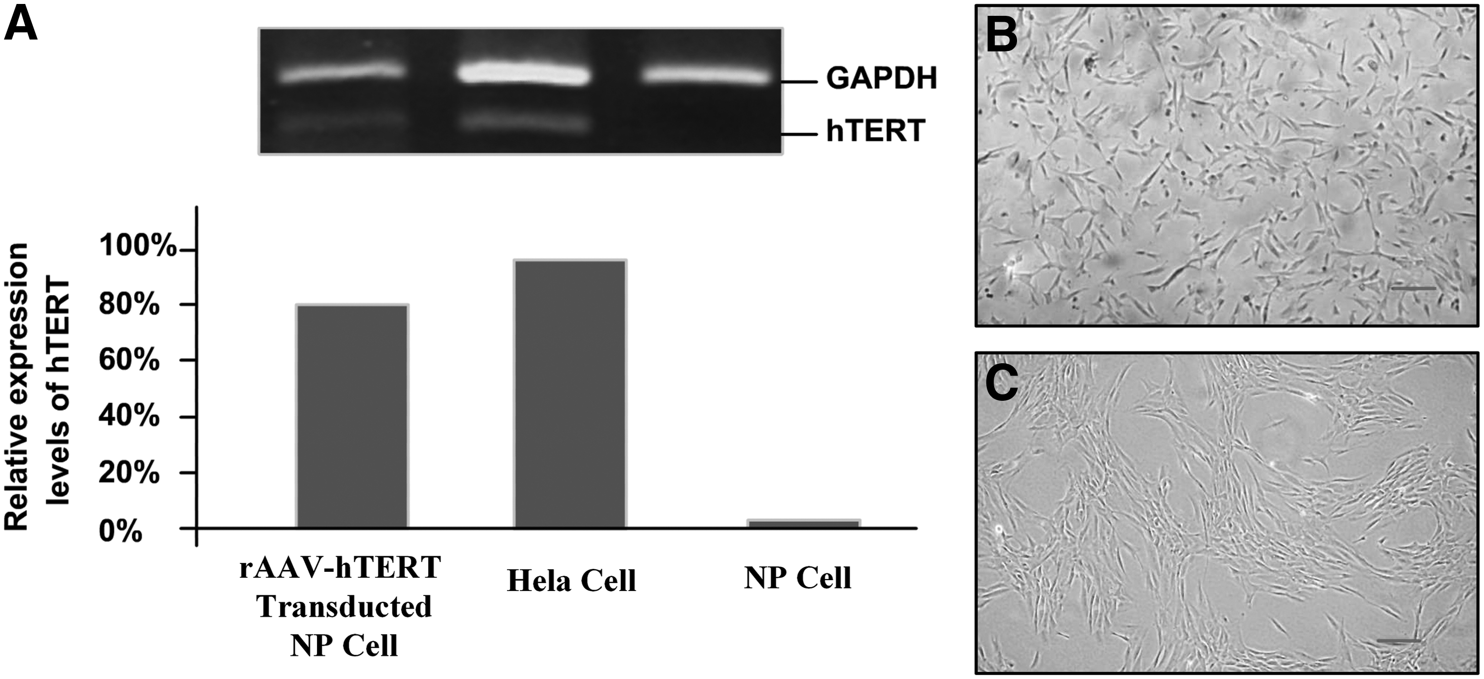
The characteristics of NPCs and rAAV-hTERT-transducted NPCs.
Animal model preparation and intervention procedure
One canine with endplate injury during preparation procedure was discarded because the vital function of endplate in nutrients supply and metabolic removal would interfere with the reliability of treatment outcomes. Two of the other 11 canines showed nerve irritation symptoms after animal model preparation procedure, and recovered within 2 months. The procedure of IVDD intervention in each animal consumed 1.5 h with 40 mL blood loss on an average. One canine showed inflammatory reaction at the surgery site, which dissipated in 1 week by enhancing the dose of antibiotics.
DHI analysis of DR
The DHI and RGI measurements were conducted by two independent observers for three times.
The data of DR were collected at four time points: 1 day before intervention and 4, 8, and 12 weeks after intervention (Fig. 2). The DHI of each group at the 1 day before intervention was similar (p>0.05), which implied that each group had the same baseline to compare with each other. Four weeks after intervention, the DHI in each group decreased compared with 1 day before intervention (p>0.05). Eight weeks after intervention, although DHI of cell intervention groups decreased sharply than the control group, there was no significant difference recorded (p>0.05). Twelve weeks after intervention, the DHI in group A and group B showed slight increase compared with the second month (p>0.05). The DHI in group C showed no significant change compared with 1 day before intervention and 4 weeks after intervention (p>0.05). All in all, although the changes of DHI were shown between groups and within groups, there was no significant change observed (Fig. 3).

Data of DR. Digital radiographs conducted at 1 day before intervention (T1), 4 weeks (T2), 8 weeks (T3), and 12 weeks (T4) after intervention. Group A: L1–2; Group B: L3–4; Group C: L5–6. The titanium metal particle was located at the fourth lumbar body as round highlight spot. DR, digital radiography.
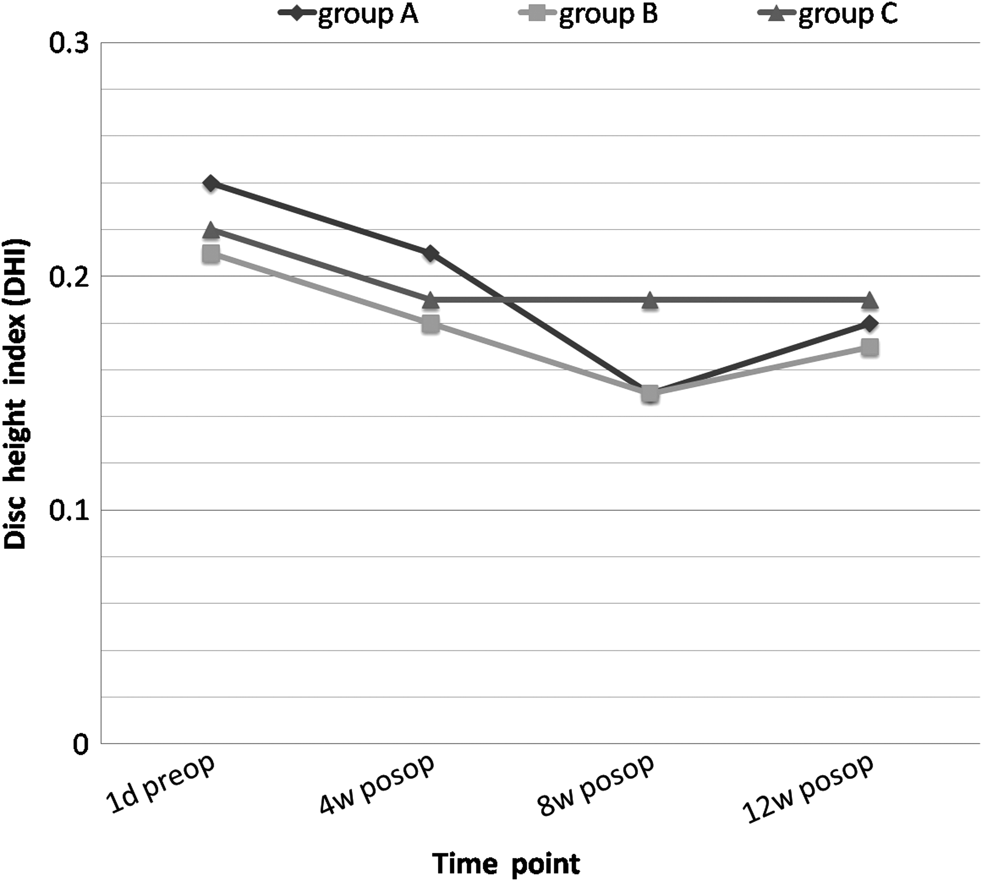
Relative disc index. The disc height of each group reduced gradually after intervention. DHI at T3 point, although DHI decreased sharply in group A and group B, no significant differences were observed compared with group C. DHI, disc height index.
RGI analysis of MRI
The data of MRI were also obtained at four time points as DR (Fig. 4). The RGI in each group was similar at 1 day before intervention. Four weeks after intervention, the RGI decreased in all groups. In group A, the RGI was maintained 8 weeks after intervention compared with the RGI 1 day before intervention; whereas in group B and group C, the RGI decreased significantly (p<0.05). Twelve weeks after intervention, still higher than other groups (p<0.05), the RGI in group A could not be maintained and decreased significantly compared with the RGI 1 day before intervention (p<0.05) (Fig. 5).

Data of MRI. MRI also conducted at four time points as DR. Group A: L1–2; group B: L3–4; and group C: L5–6. The transverse view of intervertebral disc of each group at T4 was provided. MRI, magnetic resonance imaging.
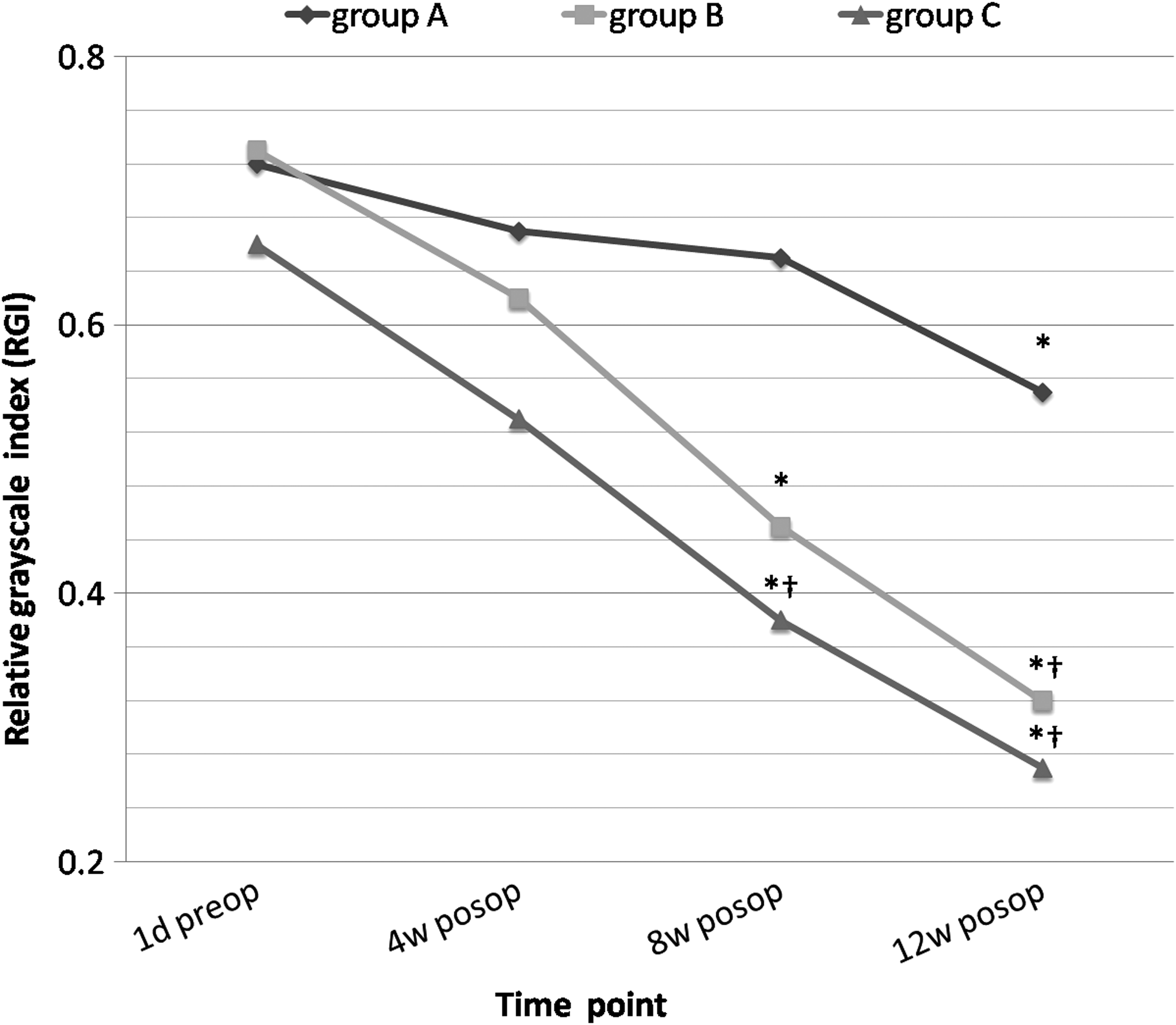
Relative grayscale index. In each group, the RGI reduced after intervention. In group A, the RGI reduced significantly at T4 when compared with the RGI at T1 (p<0.05), but still higher than other groups (p<0.05) at T4 (*compared with T1 within group, p<0.05; †compared with group A at the same time point, p<0.05). RGI, relative grayscale index.
Biomechanical test
The test of lateral bending and extension–flexion bending was conducted and the ROM was recorded (Fig. 6). Although the average flexible angle of lateral bending and extension–flexion bending was small in group A, no significant difference was observed when compared to group B and group C (p>0.05).

Range of motion in each group.
Histological study
The morphology of disc structure was preserved in group A (Fig. 7A). In HE staining, in the NP, amounts of chondrocyte-like cells could be observed which were surrounded by the pericellular matrix (PCM). Collagen fibers existed in the NP area which was stained pink (Fig. 7B). In Safranin O staining, the color of NP tissue was bright red (Fig. 7C), which indicated that PG took the major part in this area. In group B, the structure of inner annulus was broken down and the jelly-like NP tissue transmitted into fibrocartilaginous tissue (Fig. 7D). The volume of NP tissue and cellularity inside the NP area was reduced obviously in HE staining. Fibrous tissues could also be observed around the NP tissue (Fig. 7E). There were no evident differences in Safranin O staining compared with group A (Fig. 7F). In group C, the jelly-like NP tissue was completely replaced by fibrocartilaginous tissue. The structure failure of superior and inferior endplates could be observed (Fig. 7G). The round chondrocyte-like cell was diminished in the NP area, and replaced by spindle fibroblasts (Fig. 7H). In the Safranin O staining, the area of bright color was reduced (Fig. 7I).

Morphology and histology study. The scope of HE and Safranin O staining was indicated in the morphology image with black rectangular box. Group A was depicted in
Protein level of Col I, Col II, and PG level
In the NP, the PG and collagen II content was highest in group A, which was significantly higher than group B and group C (p<0.05). In group B the PG and collagen II content was also significantly higher than group C (p<0.05). The content of collagen I was highest in group B, which was significantly more than group A (p<0.05) (Fig. 8).
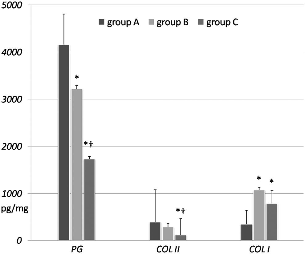
Content of PG, Col II, and Col I in NP in each group (*Compared with group A, p<0.05; †compared with group B, p<0.05). PG, proteoglycan.
Discussions
The etiology of disc degeneration is complex and multifactorial.14,15 IVDD reflects the imbalance between ECM synthesis and degradation in NP tissue, resulting in a gradual loss of disc ECM and, eventually, structural failure. The aim of biological treatments is to restore the balance between ECM synthesis and degradation in the NP. Among various biological strategies, cell-based gene delivery is an attractive option for treatment of moderate degenerative disc disease.16–18 In this study, we injected rAAV-hTERT-modified NPCs into a canine disc degenerative animal model to investigate the efficacy of hTERT in disc degeneration treatment.
In this study, the disc height reduced slightly in each group 12 weeks after injection, but significant difference was not apparent. This phenomenon could be attributed to the following two reasons. (1) In our previous study of disc degenerative animal model preparation, the disc height was not decreased significantly during 3 months observation time in the group of 20-gauge biopsy gun or even in the group of 18-gauge biopsy gun. In this study, the intervention was conducted 4 weeks after the animal model preparation and observed for 12 weeks, which suggested that a relatively short time of the observation may have contributed to the results. In the study of Ganey et al., 19 although the disc height decreased in 12 months, there were no significant differences between adipose tissue-derived stem cells' intervention group and the control group. In spite of promoting PG synthesis rate in vitro, TGF-β1 restored disc height inefficiently in in vivo study.20,21 (2) A certain volume of NP was removed during animal model preparation procedure and remaining NP tissue could not provide adequate space for cells to proliferate and secret ECM, which was considered to be a critical negative impact factor in cell therapy strategies. In this condition, it was possible that the disc height in both cell intervention groups decreased.
Fortunately, hTERT efficacy in disc degeneration was presented in MRI analysis. In the process of degeneration, along with the jelly-like NP replaced by fibrocartilaginous tissues, gradually the water content reduction and inhomogeneous stress distribution occurred inside the disc. In this study, RGI was applied to keep homogeneity between groups. In group A, RGI decreased slightly within 8 weeks postinjection (0.67±0.11, 0.65±0.17), and decreased significantly 12 weeks postinjection (0.55±0.15) compared with the baseline. In our previous in vitro study, gene level of hTERT declined gradually after gene transduction, almost 50% reduction within 3 months. In addition, in the degenerative intervertebral disc, cell proliferation and function was confined by low oxygen tension, poor nutrition supply, and even degradation and inflammatory factors.22–24 In the study of Liang et al., 25 the effectiveness of adenovirus-mediated growth and differentiation factor-5 (Ad-GDF5) had been demonstrated in reverse disc degeneration in mice disc degeneration model. These results might be related to the function of GDF5 in promoting cell proliferation and ECM secretion (type II collagen and aggrecan protein), and suppressing the expression of matrix metalloproteinase-3 (MMP-3), which was a vital degradation factor in disc degeneration process.26,27
In the NP area, cells were completely surrounded by a narrow region of tissue, the PCM, mainly consisting of collagen and PG. In group A, the PCM was clearly displayed in HE staining around chondrocyte-like cells in the NP area. Compared with group A, the PCM was reduced obviously in group B; and diminished in group C. The quantity of collagen and PG, detected by ELISA, was changed in all groups in this study. From the center of the NP to the outer annulus, the content of PG reduced with collagen fibers increasing. PG was the major ingredient existing in the normal NP tissue. The sGAG (as a measure of total PG content) was 27 times of hydroxyproline (as a measure of total collagen content) within the NP in a normal young adult disc. The ratio was declined to about 5:1 in the elderly. 28 In group A, although the PG content (948 pg/mg) was higher than group B (602 pg/mg) and group C (391 pg/mg, p<0.05), the ratio of PG/Col II was 13 approximately. We considered that, in group A, the disc degeneration process was still in progress, which was delayed, but not reversed by hTERT gene-transducted NPCs. In group B and group C, the ratio was 8.9 and 15.6, respectively. High ratio of PG/Col II in group C, we hypothesized, suggested that the reduction of Col II was more severe than PG.
Type I and type II collagen is accounting for about 80% of the total collagen in the disc. 29 Collagen ratio between Col I and Col II would be another index for disc degeneration evaluation. 30 In group A, even more content of Col II (73 pg/mg) than Col I (61 pg/mg), the Col I content was increased obviously. In group B and group C, the ratio of Col II/Col I turned over. Although the Col I content (266 pg/mg) in group B was much higher than the other groups, compared with group C, no significant difference was noticed. This result could be due to the bias of animal study and limited disc samples. The variation of components and structure arrangement of annulus and nucleus during disc degeneration process would also be expressed in the alteration of mechanical function, which was the primary role of intervertebral disc.
The annulus was the main structure standing with stretch tension in the disc. In accordance with the results of biomechanical study, in group A, the average ROM in lateral bending and extension–flexion bending test was lower than group B and group C. Although significant results were not gained, the decline tendency of ROM in group A could be identified. Coincident to the HE and Safranin O staining, the inner annulus in group A was relatively reliable compared with others.
Although inability in somatic cells, the telomerase is in the activated state in some stem cells or germline populations, which have long or indefinite lifespans. This phenomenon illustrates that telomerase expression is not oncogenic.31,32 The promoter of hTERT remains repressed in somatic cells to ensure their limited lifespan and to prevent tumor initiation. It has been demonstrated that hTERT promoter is strongly repressed by p53 and the related family members p63 and p73. 33 The lifespan of the rAAV-hTERT-transducted cells was prolonged as a result of the initial effect of hTERT gene, since the adenovirus-mediated hTERT gene was not integrated into chromosome genome. The c-myc and caspase-8 genes also remain constant in the transducted cells at early and late passages, which indicated that no major genetic mutations occurred in the transducted cells. 34
The limitations of this study included: (1) the transducted cells were not labeled before injection process; (2) the duration of this study was too short to observe the efficacy and cell proliferation capacity of rAAV-hTERT-transducted cells in the long run; (3) the tumor genes and tumor repression genes, related with the hTERT, were not studied.
Conclusion
In this study, a disc degenerative canine model was prepared with 20-gauge biopsy gun with the assistance of CT technology. The efficiency of NPCs, transducted with hTERT, has been demonstrated in keeping structure integrity, maintaining ECM, and preserving the stability status of the intervertebral disc.
Footnotes
Disclosure Statement
No competing financial interests exist.