
Abstract
Human embryonic stem cell-derived retinal pigment epithelial (hESC-RPE) cells are currently undergoing clinical trials to treat retinal degenerative diseases. Transplantation of hESC-RPE cells in conjuction with a supportive biomaterial carrier holds great potential as a future treatment for retinal degeneration. However, there has been no such biodegradable material that could support the growth and maturation of hESC-RPE cells so far. The primary aim of this work was to create a thin porous poly (L-lactide-co-caprolactone) (PLCL) membrane that could promote attachment, proliferation, and maturation of the hESC-RPE cells in serum-free culture conditions. The PLCL membranes were modified by atmospheric pressure plasma processing and coated with collagen IV to enhance cell growth and maturation. Permeability of the membranes was analyzed with an Ussing chamber system. Analysis with scanning electron microscopy, contact angle measurement, atomic force microscopy, and X-ray photoelectron spectroscopy demonstrated that plasma surface treatment augments the surface properties of the membrane, which enhances the binding and conformation of the protein. Cell proliferation assays, reverse transcription–polymerase chain reaction, indirect immunofluoresence staining, trans-epithelial electrical resistance measurements, and in vitro phagocytosis assay clearly demonstrated that the plasma treated PLCL membranes supported the adherence, proliferation, maturation and functionality of hESC-RPE cells in serum-free culture conditions. Here, we report for the first time, how PLCL membranes can be modified with atmospheric pressure plasma processing to enable the formation of a functional hESC-RPE monolayer on a porous biodegradable substrate, which have a potential as a tissue-engineered construct for regenerative retinal repair applications.
Introduction
I
A potential alternative therapeutic strategy for the treatment of AMD is the replacement of the dysfunctional RPE cells with a population of healthy cells.4–6 Human embryonic stem cells (hESCs) represent an attractive cell source for these transplantation therapies owing to their limitless supply and ability to differentiate toward functional RPE cells.7–10 Cell transplants of hESC-derived RPE (hESC-RPE) cells are currently used in clinical trials for the treatment of the dry AMD and related conditions such as Stargardt's disease.5,6
Bruch's membrane is a complex semi-permeable barrier located between the RPE and the choroid that ensures the healthy homeostasis of the retina and provides a structural support for the attachment, migration, and differentiation of RPE cells. 11 The natural aging process and the pathology of retinal diseases, such as AMD, have been shown to have a detrimental effect on the structure and function of the Bruch's membrane. 12 Previous studies have demonstrated that a potential treatment for AMD involving the delivery of RPE cells in suspension into the subretinal space on an aged or damaged Bruch's membrane showed poor RPE cell functionality and viability.13,14 Hence, the use of a “tissue engineering” approach, whereby a biomaterial polymer substrate provides a support for the delivery of functional RPE to subretinal space, could improve surgical handling and cell survival and enhance cellular function and organization.15,16
A number of natural and synthetic biomaterials have been examined previously as potential delivery systems for the transplantation of RPE cells.17–23 An approach where hiPSC-RPE cell sheets have been developed without an artificial scaffold has been also introduced.24,25 Yet, it still needs to be proven that better integration and survival of the cells is achieved using this technique. The choice of synthetic carrier material to direct growth and maturation of human pluripotent stem cells (hPSCs) toward functional RPE is still poorly studied: only inert materials parylene-C and polyimide have been assessed for use as delivery systems with hPSC-RPE cells.15,26–28
By using a biodegradable scaffold with a slow in vivo degradation period, the scaffold provides physical support during the surgical procedure. While the polymer scaffold slowly degrades, the transplanted cells can conceivably generate their own matrix to replace the defects in the damaged Bruch's membrane. Candidate polymers for the fabrication of biodegradable membranes for RPE cell delivery include poly-α-hydroxy-acid-based polymers, such as poly(L-lactide acid) (PLLA) and poly(caprolactone) (PCL), with tunable properties that include mechanical strength and degradation rate. 29 The combination of both materials as a co-polymer poly(L-lactide-co-caprolactone) (PLCL) therefore offers a range of important features.
Electrospun membranes have been shown to mimic the cues and properties of the extracellular matrix (ECM), providing for transportation of nutrients and metabolic waste. 30 In a previous study, an electrospun PLCL fiber sheet showed elastic properties, 31 which are in a similar range as reported for native Bruch's membrane 32 therefore making it an attractive choice as a scaffold for subretinal implantations. Human RPE cells,18,19 human fetal RPE cells,21,33 and the immortalized cell line, ARPE-19 16 have been previously cultured on various polymeric electrospun membranes. To our knowledge, the culturing of clinically relevant hESC-RPE cells on a biodegradable porous carrier for cell transplantation therapies has not been previously assessed.
In this work we studied a thin electrospun PLCL membrane surface modified with dielectric barrier discharge (DBD) plasma processing technique as a carrier for the hESC-RPE cells for retinal repair applications in serum-free conditions. The structure and properties of the membranes; effects of DBD plasma treatment on the chemical and physical surface properties of the thin electrospun PLCL membranes; and the nature and scale of hESC-RPE cell attachment, proliferation, and maturation on various samples are reported.
Materials and Methods
Fabrication of PLCL polymer fibers
A 10% (w/v) solution of PLCL (PURAC) was prepared as a 9:1 v/v chloroform/dimethylformamide (DMF; Sigma-Aldrich) solution. Fibrous membranes were produced in an electrospinning chamber (IME Technologies). The polymer solution was fed into the system at a flow rate of 1 mL per hour and a high voltage DC current (18 kV) was applied to the 21 G dispensing needle to achieve the Taylor cone condition. The PLCL fiber membranes were deposited onto glass coverslips mounted on the collector at distance of 15 cm from the syringe tip. The chemical composition of the resulting fibers was confirmed as PLCL by Fourier Transform Infrared Spectroscopy (FTIR) and the (solvent free) purity checked by thermogravimetric analysis (TGA) (data not shown).
DBD plasma treatment
The DBD processing methodology has been described in detail elsewhere.34–37 In this work two different plasma powers, 500 W and 750 W, were used. The transit speed of all the samples through the active plasma zone was fixed at 0.48 m/s and the number of cycles was 10. The processing conditions employed here are denoted: ES 5/10 for the 500 W, 10 cycle, 0.48 m/s condition, which results in a power density (Pd) of 52.91 W/cm2, residence time (R) of 0.19 s, and a total dose (D) of 9.92 J/cm2 and ES 7/10 for the 750 W, 10 cycle, 0.48 m/s condition, which results in a power density (Pd) of 79.37 W/cm2, residence time (R) of 0.19 s, and a total dose (D) of 14.88 J/cm2.
Collagen IV coating of electrospun PLCL membranes
Col IV is a major component of Bruch's membrane 11 and has previously been used for the enrichment and maturation of hPSC-RPE.7,38 In this work, electrospun PLCL membranes were rinsed with Dulbecco's Phosphate Buffered Saline (DPBS) (Lonza Group Ltd.), and immersed in a solution of human placenta Col IV (Sigma-Aldrich) in DPBS at a concentration of 10 μg cm2 over night at 4°C. Before cell seeding, the samples were rinsed twice with DPBS to remove any unbound protein.
Scanning electron microscopy
The diameter and morphology of electrospun PLCL fibers and the membrane thickness were determined by scanning electron microscopy (SEM) (Quanta 3D, FEI) operating at 5 kV. Samples were sputter coated with a thin (∼20 nm) layer of gold, using an Emitech K500X (Quorum Technologies) to reduce charging and image distortion.
Contact angle analysis
Static contact angle (CAM2000, KSV Instrument Ltd.) was used to determine changes in the surface wettability after atmospheric pressure DBD plasma processing. Measurements were taken using glycerol 39 at time points 0.5, 168, and 336 h following treatment. Twenty readings were performed per sample type.
Atomic force microscopy
Atomic force microscopy (AFM) (Nanoscope Dimension 3100; Vecco) was used to analyze topographical changes on individual electrospun PLCL fibers before and after atmospheric pressure DBD plasma treatment. The instrument was equipped with a TESPA silicon tip (Vecco) mounted on a cantilever of stiffness of 40 N/m−1, operating at a resonance of 300 Hz and a scan rate of 1 Hz. Images were acquired in tapping mode and all samples were analyzed over a scan area of 2 μm×1 μm at a resolution of 512×512 pixels. Pseudo–3D false color images and associated line profiles were acquired using the Nanoscope 6.11r1 Software (Veeco). Three scans of each individual fiber were obtained for each experimental group and root mean square average (Rq) values were calculated.
X-ray photoelectron spectroscopy
A Kratos Axis Ultra DLD spectrometer (Kratos) was used to obtain X-ray photoelectron spectroscopy (XPS) spectra from the various sample surfaces of interest before and after atmospheric pressure DBD plasma treatment and after Col IV adsorption. Spectra were collected using monochromated AlKα X-ray source generating photons of energy 1486.6 eV. The vacuum condition was maintained at <5×10⁻⁸ torr during spectral analysis. Three separate areas of ∼300 μm×700 μm were analyzed per sample. Wide energy survey scans (WESS) were obtained in the binding energy range 0–1300 eV, at pass energy 160 eV. Corresponding high resolution spectra of the C1s, O1s, and as appropriate N1s regions were collected at pass energy 20 eV. Binding energies were corrected for surface charging by setting the adventitious hydrocarbon C1s peak to 285.0 eV.
The Kratos vision processing software was used to obtain quantitative data from the high resolution spectra and to carry out spectral peak fitting. In all cases, a linear background subtraction was applied to the spectra. Peak fitting of the C1s spectral envelope was carried out to take account of the 70PLA/30PCL co-polymer percentage ratio.
Permeability study
The ability of electrospun PLCL membranes to act as semipermeable barrier was assessed by measuring the flux of a small molecular weight (700 Da) Alexa Fluor® 568 Hydrazide sodium salt (Life Technologies) at a concentration of 0.0065 mM. The study was carried out in an Ussing chamber system (Physiologic Instruments) with P2300 chambers and P2307 sliders and in culture medium without KnockOut™ Serum Replacement (KO-SR). Samples were taken from the receptor chamber at 60, 120, 180, and 240 min. The fluorescence of the samples was analyzed using the Wallac Victor2™ 1420 Multilabel counter (Perkin Elmer-Wallace) at 590 nm excitation and 642 nm emission wavelengths.
The diffusion was characterized by calculating the apparent permeability coefficient (Papp, cm2s−1) as Papp=dC/dt/(60C0A), where dC/dt is the slope of the linear portion of the permeability curve, C0 is the initial concentration in the donor chamber, and A is the exposed surface area of the RPE monolayer (0.031 cm2).
Human embryonic stem cell-derived retinal pigment epithelium cells
Two hESC lines, Regea 08/023 (46, XY) and Regea 11/013 (46, XY), were used in this study. Cell lines were derived as described previously. 40 Undifferentiated hESCs were maintained and differentiated into RPE cells in serum-free culture conditions as also previously described, with the modification of enzymatically passaging the undifferentiated hESCs using TrypLE Select (Invitrogen) onto fresh feeder cells at 10-day intervals.7,41
After 50–98 days of differentiation in suspension culture, the pigmented areas of the floating aggregates were manually cut. These pigmented cell clusters were dissociated with 1× Trypsin-EDTA (Lonza) and replated into Col IV coated 24-well culture plates (Corning® CellBIND®, Corning, Inc.) for enrichment. The pigmented cells were grown in adherent cultures for 180–210 days, during which the cells were replated once more onto fresh Col IV coated 24-well plates.
Electrospun PLCL films were cleaned in 70% ethanol for 1 h at room temperature and repeatedly washed with DPBS. For seeding on electrospun PLCL membranes, with and without Col IV coating, the cells were dissociated with TrypLE Select (Invitrogen). The cells were filtered through BD Falcon cell strainer (BD Biosciences) and seeded onto the fiber membranes at a density of 1.5×10⁵ cells cm2.
Analysis of cell number
The attachment and proliferation of hESC-RPE cells on untreated and DBD plasma-treated electrospun fibers with the Col IV adlayer were determined by cell counts after 7 and 42 days in culture. To get an objective estimation of the cell numbers on the samples four randomly chosen areas of each sample with DAPI-stained nuclei were captured with AxioScope A1 fluorescence microscope using a 10× objective lens. Two samples were analyzed for each experimental condition at both time points. The data for the cell number analysis was collected from two individual experiments, that is, a total 4 samples and at least 16 low magnification images. Representative low magnification images used for cell count analysis for each sample are shown in Supplementary Fig. S1 (Supplementary Data are available online at www.liebertpub.com/tea). The number of cells in each image was determined using Image J Image Processing and the Cell Counter plugin.
Proliferation assay
The proliferation of hESC-RPE cells on the various PLCL surfaces was studied after 21 days in culture using the alamarBlue® assay (Invitrogen) according to the manufacturer's instructions. The measured values were normalized against the untreated samples (relative cell proliferation for untreated samples equals 1).
Transepithelial resistance measurements
Transepithelial resistance (TER) of hESC-RPE monolayers formed after 42 days in culture on the Col IV coated untreated and plasma-treated PLCL membranes was determined as a measure of the integrity and barrier properties of the epithelium. Samples were clamped to a P2307 slider (Physiologic Instruments) before being placed in a custom-made Teflon chamber, as described previously. 41 Measurements were carried out in DBPS with a Millicell electrical resistance system equipped with a volt-ohm meter (Merck Millipore). TER values were calculated by subtracting the value for a similarly treated substrate without cells from the results. Values are reported in ohms per unit area (Ω cm2). TER values were obtained as an average of three samples of each membrane from three individual experiments. The data presented were normalized to the value determined for the untreated PLCL fiber membranes.
Gene expression analysis
The expression of genes coding for the RPE-specific proteins bestrophin (BEST), microphthalmia-associated transcription factor (MITF), pigment epithelium-derived factor (PEDF), retinal pigment epithelium specific protein 65 kDa (RPE65), tyrosinase (TYR), and pluripotency marker octamer-binding transcription factor (OCT)3/4 was assessed with reverse transcription–polymerase chain reaction (RT-PCR) for hESC-RPE cells on the Col IV-coated PLCL fibers surfaces after 42 days in culture. Glyceraldehyde 3-phosphate dehydrogenase (GAPDH) was used as an endogenous control.
RNA was extracted using NucleoSpin RNA II kits (Macherey-Nagel, GmbH & Co) in accordance with the manufacturer's instructions. hESC-RPE cells grown on Col IV-coated cell culture inserts (Millipore) were used as a positive control and water was included as a control in RT-PCR analysis to detect any impurities in the reagents and procedure. Undifferentiated hESCs were used as a negative control. A detailed account of the RT-PCR protocol and primer sequences have been published previously. 7
Immunofluorescence
Protein expression and localization on Col IV coated untreated and DBD plasma-treated electrospun PLCL fibers were further investigated using immunofluorescence staining after 42 days in culture as described previously. 38 The following primary antibodies and concentrations were used: bestrophin (1:200), cellular retinaldehyde-binding protein (CRALBP) (1:200), anti-Na+/K+ATPase (1:200), microphthalmia-associated transcription factor (MITF) (1:350) (all from Abcam), Mer Tyrosine Kinase (MERTK) 1:50 (Abnova), and zonula occludens 1 (ZO-1) (1:200) (Invitrogen). Alexa Fluor fluorescent-conjugated secondary antibodies (Molecular Probes, Life Technologies) were diluted at 1:800.
In addition, phalloidin-tetramethylrhodamine B isothiocyanate (Sigma-Aldrich) was used for labeling filamentous actin. Images of stained cells were taken with an AxioScope A1 fluorescence microscope (Carl Zeiss) or an LSM 700 confocal microscope (Carl Zeiss) using a 63× oil immersion objective lens. Images were edited using ZEN 2011 Light Edition (Carl Zeiss) and Adobe Photoshop CS4.
Phagocytosis
The phagocytic properties of hESC-RPE monolayers on Col IV coated untreated and DBD plasma-treated electrospun PLCL fibers after 92 days in culture were studied using isolated porcine photoreceptor outer segments (POS). The isolation of POS has been described previously. 7 Human foreskin fibroblasts (CRL-2429TM; ATCC) were used as a negative control.
For the in vitro phagocytosis assay, POS were labeled with fluorescein isothiocyanate (FITC) (0.04 μg μL−1; Sigma-Aldrich) in 0.1 M NaHCO3 (pH 9) for 1 h at RT, washed thrice with PBS, and resuspended in culture medium supplemented with 10% fetal bovine serum (FBS; Sigma-Aldrich). The hESC-RPE cells on the Col IV-coated electrospun PLCL membranes were then incubated with POS for 2 h at 37°C. Subsequently, the cells were washed twice with PBS and fixed with 4% paraformaldehyde for 10 min at RT. Cells were permeabilized using 0.1% Triton X-100 for 10 min at RT followed by repeated PBS washings. Filamentous actin was stained with phalloidin (Sigma-Aldrich) by incubating for 30 min at RT in the solution and washing in PBS. The nuclei were counterstained with DAPI present in the mounting media.
In addition, Regea 08/023 hESC-RPE cell controls were blocked with anti-MERTK antibody to demonstrate that the POS phagocytosis experiment is MERTK dependent and RPE-specific. For blocking, the samples were incubated with anti-MERTK antibody (1:40) for 1 h at 37°C prior applying the POS and POS incubation for 2 h. The images of the hESC-RPE cells with internalized POS fragments were taken using a confocal microscope (LSM 700, Carl Zeiss, 63× oil immersion objective lens).
Statistics
The Mann–Whitney U-test was used for determining statistical significance of cell number counts and TER-values. One-way analysis of variance (ANOVA) and Tukey's multiple comparison test were used to assess whether the data obtained were significantly different between samples. The mean values of various data are presented±standard error. p-values ≤0.05 were considered statistically significant. All statistical analysis was carried out using the data analysis software package Prism (GraphPad Software, Inc.) or IBM SPSS Statistics software.
Results
Structure and surface morphology of electrospun PLCL membranes
SEM images for the untreated material showed an average fiber diameter of 3.1±0.14 μm and an average pore size of the upper layer of the matrix of 58.3±12 μm (Fig. 1A). The membranes had an unaligned fiber orientation over an average membrane thickness of 43.2±1.95 μm. The Papp for small molecular weight (700 Da) molecule was 5.02×10⁻⁴±2.1×10⁻⁴ cm2s−1 for the untreated electrospun PLCL membrane. Similar Papp of 4.12×10⁻⁴±2.01×10⁻⁴ cm2s−1 was measured for the ES 5/10-treated PLCL membranes, whereas slightly lower Papp value of 1.82×10⁻⁴±2.87×10⁻⁶ cm2s−1 was reached for ES 7/10 PLCL membranes.

Scanning electron microscopy (SEM) micrographs of
The untreated PLCL membrane fibers appeared quite smooth, whereas the treated fibers clearly displayed surface features (Fig. 1B). In addition, the DBD plasma treatment had no significant effect on the fiber diameter (Fig. 1C). AFM data confirmed that the plasma treatment alters the topography of the PLCL electrospun fiber surface with the root mean square average surface roughness (Rq) values increasing from 18.00±2.46 nm for the untreated sample to 31.40±3.93 nm for the ES 5/10 and 24.93±1.13 nm for ES 7/10-treated PLCL membranes (Fig. 1D, E).
A statistically significant reduction in contact angle (p<0.001) was recorded at 0.5 h post DBD plasma treatment (Fig. 2A). The lowest average contact angle measured was 96±1° for sample ES 7/10, while the ES 5/10 value was 105±1°, which compare to the untreated sample value of 138±1°. Relaxation (hydrophobic recovery) occurred for all DBD-processed samples with contact angle increasing over the study period. However, the values did not recover to those of the untreated PLCL fiber indicating a permanent change in surface properties. Contact angle measurements of the Col IV-coated samples showed that DBD plasma treatment before Col IV coating significantly increased fiber wettability when compared with the equivalent untreated sample (Fig. 2B).
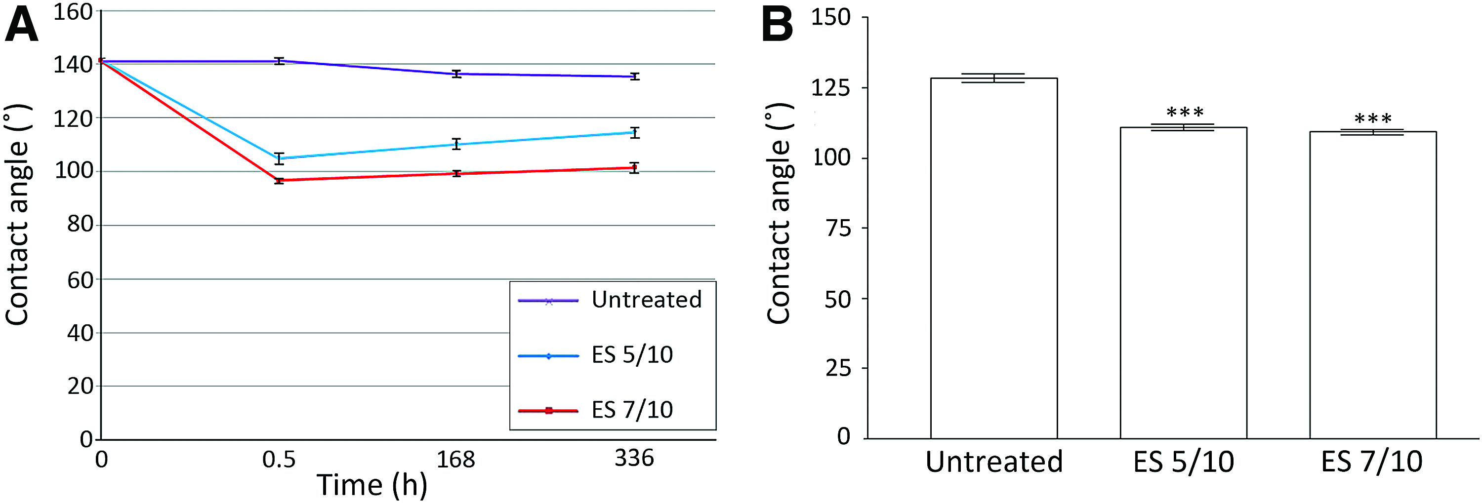
Contact angle measurements of
Surface chemistry of electrospun PLCL membranes
The atomic concentration (% at. conc.) values and O/C ratios obtained by quantification of the XPS spectral data (Fig. 3) revealed a decrease in carbon content and a significant increase (p<0.001) in oxygen content on the PLCL surfaces after DBD plasma treatment (Table 1). This increase in oxygen content was dependent on the DBD plasma dose administered. XPS scans for the Col IV-coated plasma-treated PLCL samples showed an N1s (Nitrogen) peak associated with the protein layer (Fig. 3B). From the percentage atomic concentration values it can be seen that increasing the DBD plasma dose before Col IV adsorption has a substantial impact on the amount of nitrogen detected with the Col IV-coated ES 5/10 samples displaying significantly lower nitrogen content than Col IV-coated ES 7/10 membranes (p<0.05) (Table 2). The amount of nitrogen on Col IV-coated DBD-treated membranes after both plasma doses was significantly higher compared with Col IV-coated untreated membranes (Table 2).
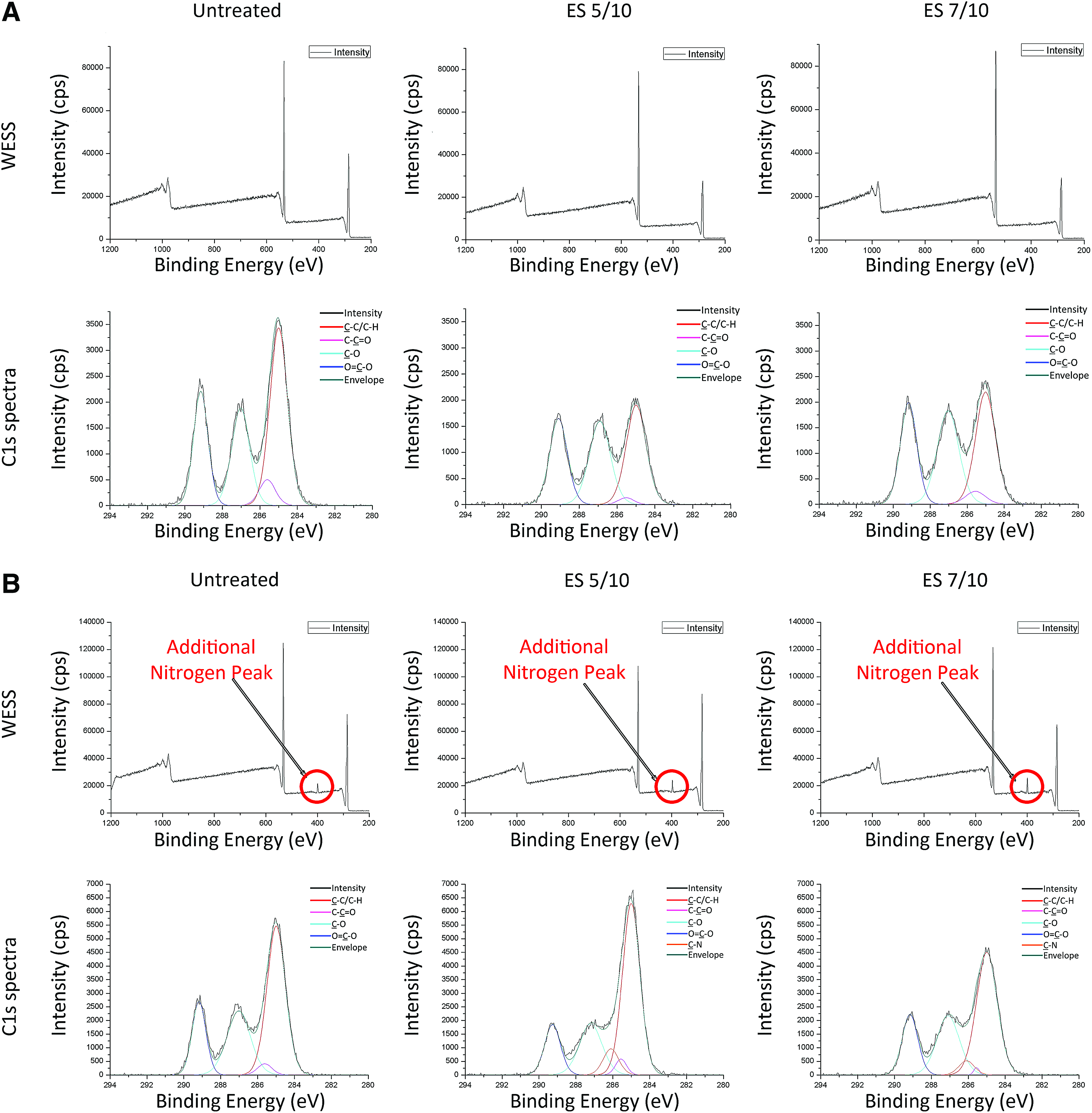
XPS wide energy survey scan (WESS) and high-resolution C1s spectra for
Distribution of collagen IV on electrospun PLCL membranes
Distribution of Col IV on porous PLCL membranes was assessed with immunofluorescence staining. Col IV adhered to the fibers of all the PLCL membranes under investigation, which was distributed in a uniform manner (Fig. 4).
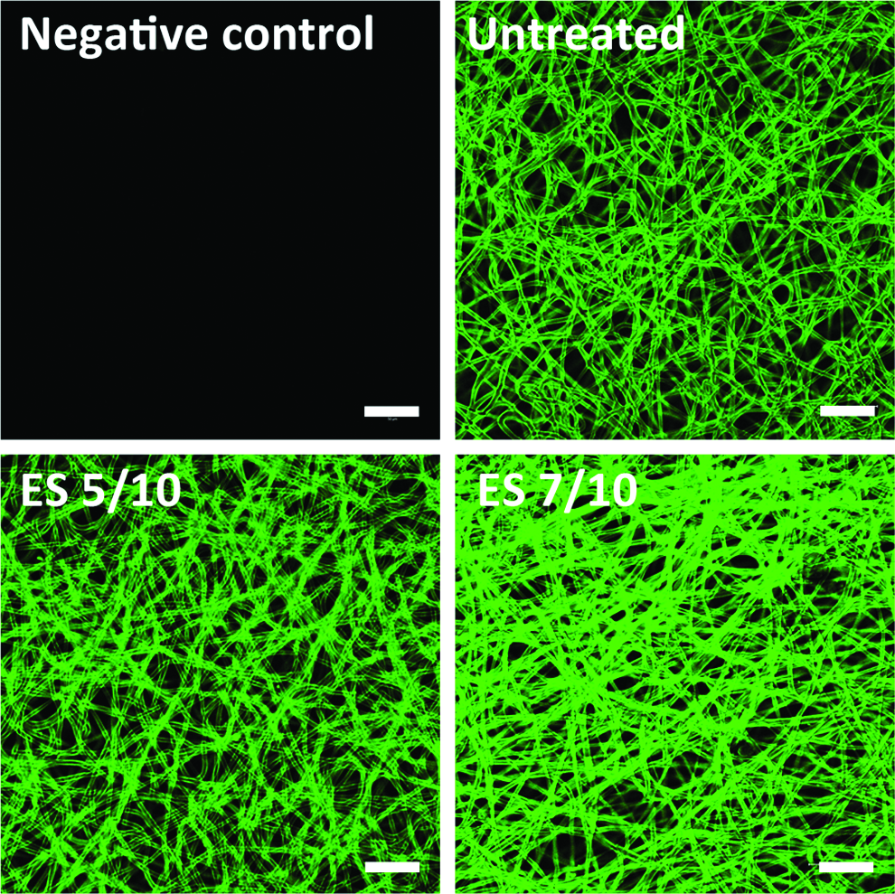
Confocal microscopy images of immunostained Col IV-coated PLCL electrospun membranes. Scale bar 50 μm. Color images available online at www.liebertpub.com/tea
Cell attachment and proliferation on biodegradable PLCL electrospun membranes
Without additional Col IV coating, hESC-RPE cells did not attach on any of the investigated PLCL membranes (data not shown). After the 7-day culture period, a significant (p<0.005) increase was detected in hESC-RPE cell attachment on the Col IV-coated DBD plasma-treated PLCL membranes compared with the number on the untreated Col IV-coated samples in both investigated cell lines (Fig. 5A). The number of Regea 08/023 hESC-RPE cells present on ES 5/10 Col IV-coated membrane was higher (p<0.05) compared with the number on ES 7/10 Col IV-coated membrane, whereas no significant difference was observed between the DBD plasma treatments with Regea 11/013 hESC-RPE cells.
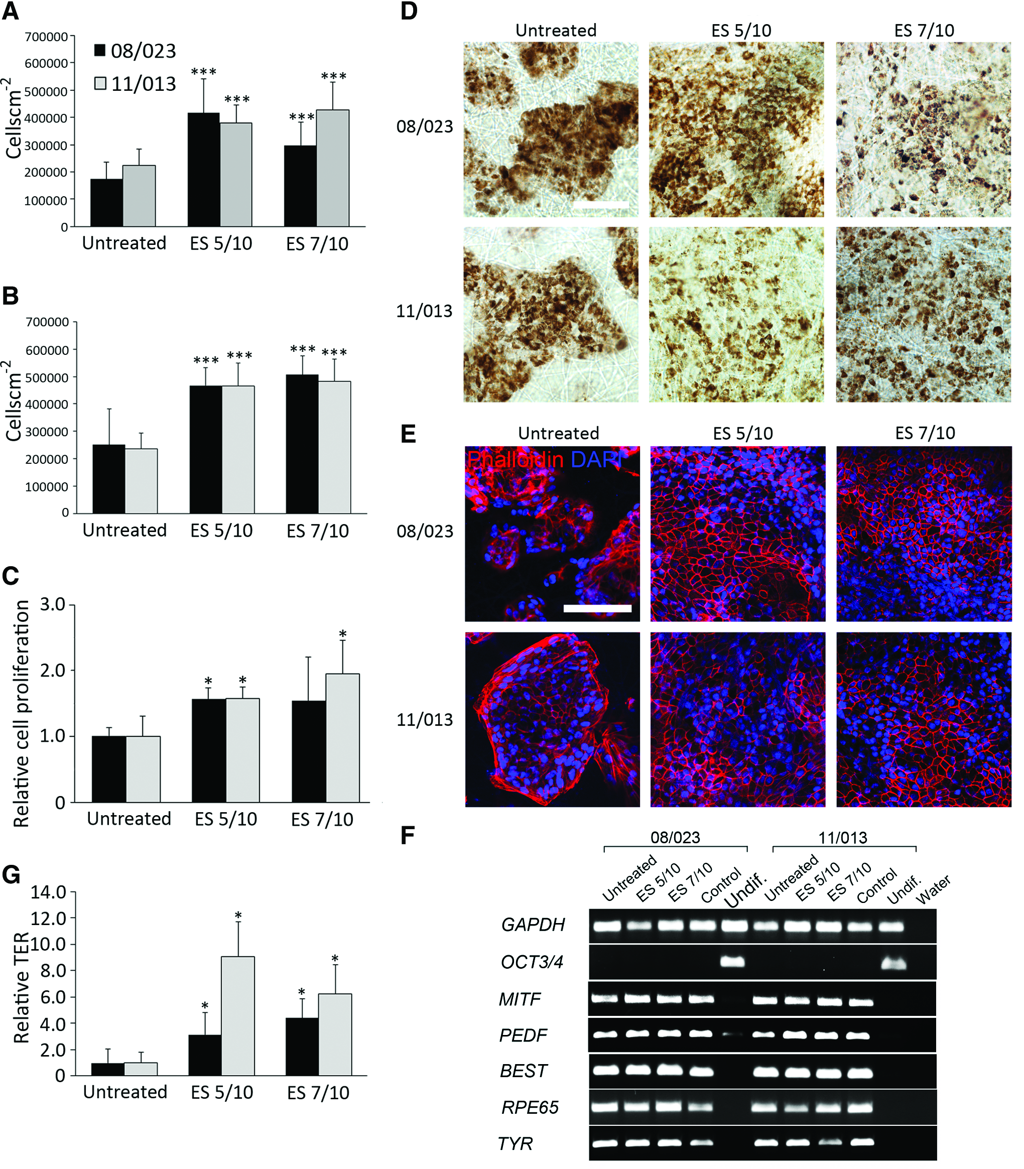
Cell counts of hESC-RPE on electrospun untreated and atmospheric plasma-treated PLCL membranes with Col IV layer after
After 42 days in culture, cell numbers were significantly higher on Col IV-coated DBD plasma-treated PLCL fiber membranes when compared with the untreated Col IV-coated sample (Fig. 5B). However, there was no difference between the numbers detected on the ES 5/10 and ES 7/10 Col IV-coated DBD plasma-treated membranes at this time point.
The results obtained from the cell proliferation analysis with alamarBlue® assay after 21 days in culture were consistent with the cell count data; higher cell proliferation was measured for both hESC-derived RPE cell lines on all of the Col IV-coated DBD plasma-treated samples compared with that on the Col IV-coated untreated membrane (Fig. 5C).
hESC-RPE cell maturation on biodegradable electrospun membranes
After 42 days in culture, the hESC-RPE cells on both the ES 5/10 and ES 7/10 Col IV-coated samples had formed a confluent and uniform RPE monolayer with abundant pigmentation and a typical hexagonal RPE cell morphology (Fig. 5D, E). By contrast, the hESC-RPE cells on Col IV-coated untreated membranes grew as raft-like heterogeneous structures and comprised spherical cell clumps such that confluent layers were not attained during the 42-day culture period (Supplementary Fig. S2A). Gene expression analysis demonstrated the expression of mature RPE markers BEST, MITF, PEDF, RPE 65, and TYR on all investigated membranes (Fig. 5F). Furthermore, there was no expression of the pluripotency marker OCT3/4 (Fig. 5F).
The hESC-RPE maturation on Col IV-coated DBD plasma-treated membranes was further examined using immunofluorescence staining.
The hESC-RPE cells on ES 5/10 and 7/10 Col IV-coated membranes showed homogenous expression of the RPE-specific markers MITF (Fig. 6A), bestrophin (Fig. 6B), CRALBP (Fig. 6C), and the tight junction marker ZO-1 (Fig. 6D). hESC-RPE cells on untreated Col IV-coated PLCL membranes expressed bestrophin, CRALBP, and MITF on protein level, however, cells failed to form homogenous monolayer (Supplementary Fig. S2A–D). The tight junction protein ZO-1 was localized on the apical membrane of hESC-RPE cells on Col IV-coated DBD plasma-treated PLCL membranes, which showed positive labeling for CRALBP, indicating that DBD plasma-treated PLCL membranes supported the growth of cells as an adherent RPE monolayer (Supplementary Fig. S3). Furthermore, Na+/K+ATPase was expressed on the apical membrane of the cells thereby demonstrating polarization of the epithelium (Fig. 6E).
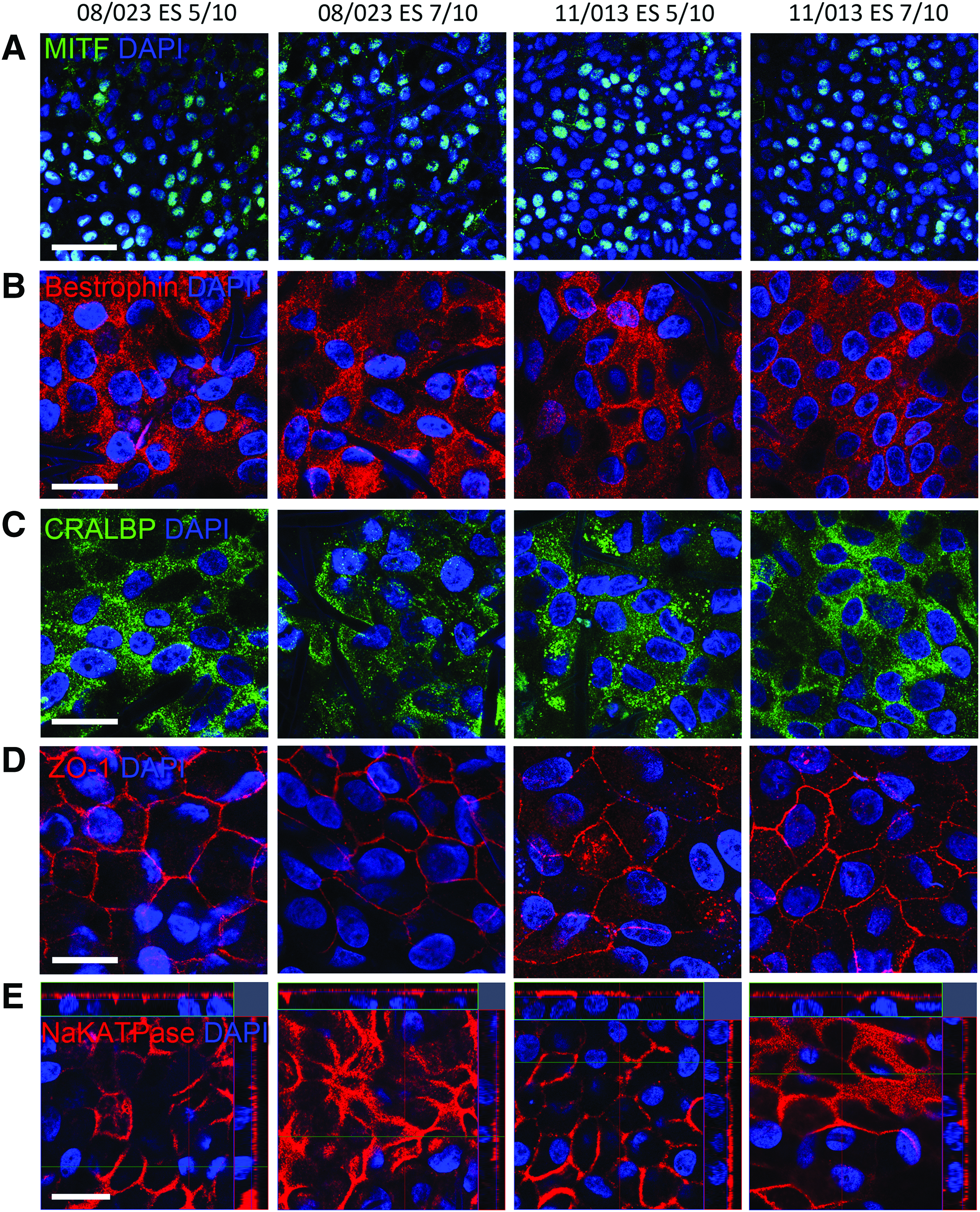
Immunofluorescent (IF) staining showing expression and localization of RPE-specific proteins:
Functionality of hESC-RPE cells on biodegradable PLCL electrospun membranes
For both hESC lines, the TER-values after 42 days in culture were significantly higher (p<0.05) on the Col IV-coated DBD plasma-treated ES 5/10 and ES 7/10 samples compared with those on the Col IV-coated untreated sample (Fig. 5G). No notable differences were found between the ES 5/10 and 7/10 Col IV-coated electrospun PLCL membranes. Functionality of the hESC-RPE cells on the ES 5/10 and ES 7/10 Col IV samples was further assessed with an in vitro phagocytosis assay after 92 days in culture. Internalized POS was seen in both hESC-RPE cell lines on each of the DBD plasma-treated membranes (Fig. 7).
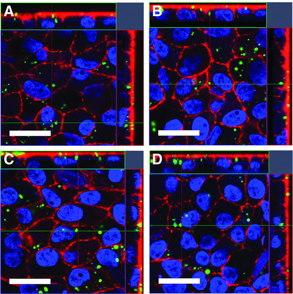
The phagocytosis of photoreceptor outer segments (POS) after 92 days in culture on atmospheric plasma-treated electrospun PLCL membranes with Col IV layer: Regea 08/023 hESC-RPE cells on
In addition, hESC-RPE cells on the DBD plasma-treated materials showed positive expression of MERTK at the apical side of the cells (Supplementary Fig. S4a). Internalized POS was not found in human foreskin fibroblasts, which were used as negative control (Supplementary Fig. S4b, c). Moreover, blocking hESC-RPE cells with anti-MERTK antibody reduced the amount of attached POS in the apical membrane and the amount of internalized POS (Supplementary Fig. S4b, c).
Discussion
Replacement of diseased RPE cells and Bruch's membrane with a healthy RPE cell layer on a thin membrane that can mimic the lost structure and function offers a viable regenerative transplantation treatment for retinal degeneration.
In this study, thin porous electrospun PLCL membranes coated with human Col IV were investigated as a carrier for the formation of a functional hESC-RPE cell monolayer. The primary aim of this work was to promote attachment, proliferation, and maturation of the hESC-RPE cells toward a functional RPE monolayer on a biodegradable thin membrane in serum-free culture conditions. Gaseous plasma-based processing is a widely utilized method for modifying a polymeric surface to enhance the cellular response thereon. 34
The DBD plasma processing technique utilized here generates a gaseous plasma discharge in air at atmospheric pressure that is capable of permanently modifying the surface properties in a cost-effective manner. In this regard, DBD has been previously shown to enhance protein absorption and cellular responses of human lens epithelial cells.34–36,42 Here, we assessed the impact of DBD treatment on the chemical and physical properties of the membranes and on the cellular response of hESC-RPE cells.
Biodegradable polymeric biomaterials provide the significant advantage of being able to be broken down or removed without surgical revision after they have served their 3D structural function. 43 Furthermore, they allow for the incorporation of therapeutic agents, such as anti-inflammatory drugs and growth factors, which can significantly improve cell survival in transplantation therapy. 44
Biodegradable scaffolds fabricated from poly(α-hydroxy esters) have been investigated as potential substrates for human RPE cells and ARPE-19 cells19,20 and Liu et al. have shown that electrospun PLCL fibers also support the growth of human fetal RPE cells18,21 However, the lack of published data implies and our own empirical tests have shown that hESC-RPE cells do not adhere well on biodegradable scaffolds in serum-free conditions. Furthermore, comparison to studies carried out with immortalized RPE cell lines should be done carefully because hESC-RPE have been shown to differ, that is, in pigmentation and gene expression. 45
hESC-RPE cells produce their own ECM including the major components of the uppermost layers of the native Bruch's membrane. 38 By using a biodegradable scaffold with a slow in vivo degradation time, the transplanted cells have a physical support during the transplantion. Thereafter, they can potentially generate their own matrix to replace the defects in the damaged Bruch's membrane while the scaffold is degrading.
Previous studies have indicated that electrospun PLCL fibrous membranes show no material degradation over 3-month cell culture period and demonstrate no detrimental effect on cellular response. 46 Our observations are in line with this study: hESC-RPE cells were cultured on DBD plasma-treated membranes up to 92 days without evidence of any degradation or harmful effects in the cells. In a previously reported in vivo study for highly porous PLCL scaffolds, the mass of the scaffold decreased to 81% after 15 weeks of subcutaneous implantation, indicating a slow degradation. 47 However, the in vivo degradation of electrospun PLCL membranes in intraocular tissue and subretinal space has not been studied and therefore in vivo experiments need to be carried out in the future to determine the suitability of these scaffolds for tissue-engineered retinal repair applications.
One important function of Bruch's membrane is allowing the reciprocal flow of small molecular substances through its layered structure. 11 Here, we showed that the prepared electrospun PLCL films had porous structure and were permeable for a small molecular weight fluorescent marker. The lowest DBD plasma dosage condition ES 5/10 did not affect the permeability of the electrospun PLCL membrane compared to the untreated membrane, whereas a slight reduction in membrane permeability was seen with the higher dosage condition ES 7/10.
In previous studies, inert PET carriers have been used as a support for electrospun poly(lactic-co-glycolic acid) (PLGA) 19 and PLCL 21 fibers in scaffolds designed for human fetal and primary RPE cells. In contrast, the manufactured electrospun films in this study were easy to handle and mechanically durable as such; no additional support was required for the scaffold to maintain its porous fibrillar structure during handling and culture. Previously, a novel shooter instrument has been shown to enable transvitreal delivery of ultrathin rigid-elastic-carriers into the subretinal space of rabbits. 48 However, the feasibility of the instrument and surgical procedure with a biodegradable electrospun PLCL scaffold-hESC-RPE complex introduced in this study needs to be addressed in future experiments.
The polymers commonly used in tissue engineering applications are highly hydrophobic, which can hinder cellular attachment and proliferation. 34 The untreated electrospun PLCL fibers exhibited a highly hydrophobic surface, whereas atmospheric pressure DBD plasma treatment significantly increased fiber wettability, that is, increased hydrophilicity.
Moreover, XPS analysis indicated that DBD plasma treatment significantly increased the O/C ratio on the surface of electrospun PLCL fiber membranes compared with that for an untreated sample. This is attributed to the fact that DBD plasma treatment produces polar oxygen groups on the surface of the electrospun PLCL fibers and can also increase surface roughness, both of which effect the surface energy. 49 These data are consistent with the previous literature findings that also observed an increase in oxygen content on the surface of electrospun polymers after plasma treatment.39,50
Analysis by SEM here revealed that there was no thinning or destruction of the fibers compared between untreated and DBD plasma-treated PLCL electrospun fiber membranes, a phenomenon previously reported with low pressure (vacuum based) plasma treatment of electrospun fibers. 39 However, AFM analysis confirmed that the DBD plasma treatment increased the surface roughness of electrospun PLCL fiber membranes. Interestingly, the lower DBD plasma dosage condition ES 5/10 resulted in higher surface roughness compared with the larger DBD plasma dosage condition ES 7/10. These data support previous research, which illustrated that higher doses of plasma treatment produces a lower fiber surface roughness compared with the lower dose treatment. 50
In this study, all the investigated membranes showed a good level of bound protein immunofluorescence staining, which uniformly dispersed across the fibers. However, XPS analysis indicated that the DBD plasma-treated electrospun PLCL membranes had a significantly higher level of nitrogen present, attributed to the introduction of the protein, 51 compared with that detected on the untreated membranes. XPS is a highly surface-sensitive quantitative spectroscopic technique that has been extensively used to determine the amount of protein on functionalized surfaces. 52 Thus, the increased nitrogen content reported in this study indicates enhanced protein binding of Col IV on DBD plasma-treated PLCL membranes compared with the untreated membranes. Furthermore, the Col IV-coated untreated electrospun PLCL membranes had an average contact angle only slightly lower than that of the untreated PLCL surface, whereas DBD plasma treatment before Col IV coating had a significant influence on the contact angle. Previously, Ai et al. observed a similar trend for plasma-treated poly(hydroxybutyrate-valerate) fibrous mats coated with collagen. 53
In this study, electrospun PLCL fibers that were DBD plasma treated before deposition of a Col IV adlayer had a major beneficial effect on hESC-RPE cell attachment, proliferation, and maturation. By comparison, Col IV-coated untreated fibers failed to support an adequate response from the same hESC-RPE cells. These results are in line with previous studies that have investigated the effect of plasma treatment on immortalized RPE cells.54,55
In this study, the beneficial effects of the DBD plasma treatment on the hESC-RPE cell attachment and maturation were significant only with Col IV coating. It is suggested that the chemical and topographical surface conditions created by DBD plasma treatment of the PLCL fibers provides a Col IV protein conformation on the fiber surface that not only promotes attachment but also influences cell division in a way that results in the formation of a monolayer with mature epithelial function. It has been established previously that a more hydrophilic surface condition can induce the adsorption of a finer network of Col IV that enhances cellular response.56,57
A definite prerequisite for a biomaterial carrier intended for the clinical use of hESC-RPE cells is that it supports the formation of homogenous, mature and functional hESC-RPE monolayer. The Col IV-coated untreated PLCL membranes did not support the formation of a homogenous monolayer, and thus failed to support proper maturation of hESC-RPE. Instead, hESC-RPE monolayers on the Col IV-coated DBD plasma-treated PLCL membranes showed expression of RPE-specific markers at both gene and protein levels, abundant pigmentation, high degree of polarity, and uniform expression of tight junction protein ZO-1. Importantly, TER measurements of the hESC-RPE cells on Col IV-coated DBD plasma-treated electrospun PLCL membranes supported the formation of a tight and polarized cell monolayer, indicative of a functional RPE.
In addition, hESC-RPE cells on DBD plasma-treated electrospun PLCL membranes exhibited phagocytic activity. All of these functions are generally considered to be hallmarks of mature RPE.4,58 Thus, this study shows that the DBD plasma-treated porous biodegradable membranes supported the growth of mature and functional hESC-RPE monolayer in serum-free culture conditions and are therefore suitable carrier candidates for retinal repair applications.
Conclusions
In this study, were produced a thin biodegradable electrospun PLCL membranes as a carrier for hESC-RPE cells for retinal cell therapy applications. The prepared membranes were modified by atmospheric pressure plasma processing and coated with Col IV to enhance cell growth and maturation. The DBD atmospheric pressure plasma treatment changed the surface charasteristics of the PLCL electrospun membranes, which led to enhanced Col IV protein binding. These DBD plasma-treated electrospun PLCL membranes had a porous structure and allowed the flow of small molecular substance through their structure. Here, we report for the first time, successful culture of mature and functional hESC-RPE cells on a porous biodegradable electrospun scaffold in serum-free culture conditions, which have potential as a tissue-engineered construct for regenerative retinal repair applications.
Ethical Issues
The National Authority for Medicolegal Affairs Finland has approved this research with human embryos (Dnro 1426/32/300/05). We also have a supportive statement from the local ethics committee of the Pirkanmaa hospital district Finland to derive and expand hESC lines from surplus embryos not used in the treatment of infertility by the donating couples, and to use these cell lines for research purposes (R05116). No new lines were derived for this study.
Footnotes
Acknowledgments
The authors wish to thank the Academy of Finland (grant numbers 218050 and 137801), TEKES (the Finnish Funding Agency for Technology and Innovation), the Finnish Cultural Foundation, Pirkanmaa Regional fund (grant number 50121465), Invest Northern Ireland (grant number RD0212954), and the Northern Ireland Department for Employment and Learning (DEL) for their support. The funders had no role in study design, data collection and analysis, decision to publish, or preparation of the article. Outi Melin, Hanna Pekkanen, and Outi Heikkilä are thanked for technical assistance. Authors one and two equally contributed to this work.
Disclosure Statement
No competing financial interests exist.
References
Supplementary Material
Please find the following supplemental material available below.
For Open Access articles published under a Creative Commons License, all supplemental material carries the same license as the article it is associated with.
For non-Open Access articles published, all supplemental material carries a non-exclusive license, and permission requests for re-use of supplemental material or any part of supplemental material shall be sent directly to the copyright owner as specified in the copyright notice associated with the article.