
Abstract
The irritancy of topical products has to be investigated to ensure the safety and compliance. Although several reconstructed human epidermal models have been adopted by the Organization for Economic Cooperation and Development (OECD) to replace in vivo animal irritation testing, these models are based on a single cell type and lack dermal components, which may be insufficient to reflect all of the components of irritation. In our study, we investigated the use of acellular porcine peritoneum extracellular matrix as a substrate to construct full-thickness human skin equivalents (HSEs) for use as irritation screening tool. The acellular peritoneum matrix (APM) exhibited excellent skin cell attachment (>80%) and proliferation for human dermal fibroblasts (HDF) and immortalized human keratinocytes (HaCaT). APM-HSEs based on coculture of HDF and HaCaT were prepared. Increased HDF seeding density up to 5 × 10⁴/cm2 resulted in APM-HSEs with a thicker and more organized epidermis. The epidermis of APM-HSEs expressed keratin 15, a keratinocyte proliferation marker, and involucrin, a differentiation marker, respectively. To assess the use of APM-HSEs for irritation testing, six proficiency chemicals, including three nonirritants (phosphate-buffered saline, polyethylene glycol 400, and isopropanol) and three irritants (1-bromohexane, heptanol, and sodium dodecyl sulfate) were applied. The APM-HSEs were able to discriminate nonirritants from irritants based on the viability. Levels of cytokines (interleukin [IL]-1α, IL-1ra, IL-6, IL-8, and granulocyte macrophage colony-stimulating factor [GM-CSF]) in these treatment groups further assisted the irritancy ranking. In conclusion, we have developed partially differentiated full-thickness APM-HSEs based on acellular porcine peritoneum matrix, and these APM-HSEs demonstrated utility as an in vitro irritation screening tool.
Introduction
S
Alternative in vitro testing methods have been developed to replace animal studies, including quantitative structure–activity relationship, computational approaches, monolayer cell cultures, and three-dimensional (3D) skin models.6,7 Among the above approaches, 3D human skin equivalents (HSEs) models are promising for in vitro testing, based on the potential to reflect cell differentiation, polarization, and cell–matrix interactions of skin. Therefore, HSEs have been used to determine drug irritation,8,9 corrosivity, 10 permeation,11,12 and genotoxicity 13 of topically applied formulations.
HSEs can be classified into three major categories: (a) epidermal models, (b) dermal models, and (c) full-thickness skin models. The epidermal models contain multiple layers (i.e., stratum corneum, stratum granulosum, stratum spinosum, and stratum basale) of primary human keratinocytes or immortalized cell lines (e.g., HaCaT, 14 NIKS 15 ) on a thin supporting porous membrane such as polycarbonate and cellulose acetate filters.16,17 The dermal models feature fibroblasts embedded in extracellular matrix (ECM) such as type I collagen hydrogel, hyaluronic acid, or on the surface of de-epidermized dermis.18,19 The full-thickness models have stratified keratinocytes over fibroblast-populated matrices. 20 Recent literature focuses on two major advances in full-thickness models: (a) higher complexity models that incorporate multiple cell types such as addition of dendritic cells, melanocytes, endothelial cells and stem cells have been reported21–25 ; (b) decellularized tissues (such as allodermis 26 ) that can be used for constructing full-thickness HSEs models. Decellularized tissues are extensively studied in skin grafting applications based on the fact that they retain high similarity in composition and structure to native ECM and could support the proliferation and differentiation of skin cells in vivo.27,28 Despite the advance in various HSEs models, there were little efforts in validating full-thickness models for in vitro irritation screening. Currently, only four reconstructed human epidermis models (Episkin™, EpiDerm™, SkinEthic™, LabCyte EPI-MODEL) have been adopted by the Organization for Economic Cooperation and Development (OECD) to provide alternative in vitro methods for classifying irritated chemicals. 29 All of these models are epidermis models that are based on keratinocyte cells and do not contain other cell populations in the dermis layer, for example, fibroblasts. Full-thickness HSEs that contain both keratinocytes and fibroblasts may better predict the irritation response since their physiology is more relevant to native skin than that for epidermal models.
The aim of this study is to construct full-thickness HSEs on porcine acellular peritoneum matrix (APM) and investigate the function of APM-HSEs as in vitro irritation screening tool. The peritoneum lines the abdominal cavity and has a basement membrane supporting a uniform layer of mesothelial cells. The APM was produced by DSM Biomedical's proprietary OPTRIX™ decellularization process, which removed cellular components while maintaining ECM composition (i.e., collagen I, collagen IV, laminin, fibronectin) and growth factors (i.e., vascular endothelial growth factor [VEGF], fibroblast growth factor [FGF], and transforming growth factor-β [TGF-β]). 30 Due to the nature of the peritoneum, the APM is a polar scaffold with a compact basement membrane side (abbreviated as the MesoBM side) and an opposing side with open fibrous architecture that faced the preperitoneal space (abbreviated as the MesoPP side). The cellular response of human skin cells on the two sides of the APM was investigated. The structure of the constructed APM-HSEs was characterized by histology and immunohistochemistry analyses. Then, the function of the APM-HSEs was studied to assess whether they can predict irritancy ranking for several selected chemicals. To the best of our knowledge, this is the first time that an APM has been used to construct a 3D full-thickness skin model and for such model to be tested for in vitro skin irritation.
Materials and Methods
Acellular peritoneum matrix
The APM was produced from porcine parietal peritoneum using DSM Biomedical's OPTRIX™ processing method. Tissue was harvested from market weight pigs. The peritoneum was dissected from the adjacent preperitoneal fat before the decellularization process. The OPTRIX™ process consists of a series of agitated washes, including an organic solvent, a detergent, salts, an enzyme, and interposed rinses. The processed tissue was freeze dried and ethylene oxide sterilized in gas permeable packaging. Experimental samples were equivalent to Meso BioMatrix® and Meso Wound Matrix™, which are commercially available.
Mechanical properties
Mechanical properties were characterized using a mechanical testing instrument (Sintech 5D MTS). APM and cadaver human skin (NY Firefighters Skin Bank) specimens were cut into rectangular shape measuring 1 × 6 cm. Hydrated APM specimens were prepared by soaking in phosphate-buffered saline (PBS; Life Technologies) for at least 1 h. The cadaver skin samples were thawed in PBS and the epithelial side was dapped with Kimwipes to remove excess fluid before testing. Thicknesses of samples were measured with a micrometer immediately before testing (thickness ∼150–260 μm for APM; 420–550 μm for skin). Samples were mounted between the two grips of Sintech 5D MTS instrument and the distance of grip-to-grip was measured without use of an extensometer. Test speed was set at 10 mm/min. Young's modulus was determined based on calculating the slope of the linear portion of the stress–strain curve. The analyses were performed in triplicate.
Cell culture
Neonatal human dermal fibroblasts (HDF) were purchased from Life Technologies. Early passages, with passage number not more than 5, were used. Spontaneously immortalized human keratinocyte (HaCaT 14 ; passage 20–30), green fluorescent protein (GFP)-expressing HaCaT and red fluorescent protein (RFP)-expressing HDF were generous gifts from the New Jersey Center for Biomaterials. GFP-HaCaT and RFP-HDF were transduced with LifeAct lentiviral vectors 31 ; the transduced cells express GFP and RFP on actin filaments of HaCaT and HDF, respectively. All cells were cultured in Dulbecco's modified Eagle's medium (DMEM) with 10% fetal bovine serum (Life Technologies) and 100 units/mL of penicillin (Life Technologies), 100 μg/mL of streptomycin (Life Technologies), and 0.25 μg/mL of amphotericin B (Life Technologies) at 37°C/5% CO2.
Cellular attachment and growth of skin cells on collagen hydrogel and APM
Collagen hydrogel was prepared by diluting 8 mL of bovine type I collagen solution (6 mg/mL, Nutragen®; Advanced BioMatrix) with 1 mL chilled 10× PBS, 1 mL of DMEM medium, and adjusted to pH = 7 with 0.1 N NaOH. The collagen solution (500 μL) was added to a 48-well tissue culture polystyrene (TCPS) plate and incubated at 37°C overnight before cell seeding. The APM specimens were cut into 12 mm diameter disks and conditioned in the 48-well TCPS plate. Monoculture of HDF or HaCaT (5 × 10⁴/cm2) was seeded onto the type I collagen gel, basement membrane side (MesoBM) of the APM, the opposite side (MesoPP) of the APM, and TCPS wells, respectively. After 5 and 24 h incubation at 37°C, cells were washed with PBS three times, and then 500 μL of cell culture medium containing 10% (v/v) alamarBlue® reagent (Life Technologies) was added to each well and incubated for another 3 h. The fluorescence intensity of the supernatant (λex = 560 nm, λem = 590 nm) was measured through a plate reader (Tecan Infinite). Attachment percentage was determined by normalizing the fluorescence intensity with that from TCPS group. Nine replicates were used in each group for the attachment experiment. To assess cellular metabolic activity at different time points, same seeding parameters and alamarBlue assay as described above were used on day 1, 4, 7, 11, and 14. Six replicates were prepared for each time point. The alamarBlue results indicate total cellular metabolic activity at different time points.
The skin cells on APM were visualized by fluorescence imaging. GFP-HaCaT and RFP-HDF (5 × 10⁴/cm2) were seeded onto MesoBM side and MesoPP side of APM, respectively. Images were taken using a fluorescence microscope (Zeiss) after 4 days postseeding.
Construction of HSEs based on APM
Disks of APM (diameter = 12 mm) were placed in 12-well Transwell® insert plates (polycarbonate 0.45 μm pore size; Corning). At the start of the experiment, 0–5 × 10⁴/cm2 of HDF were seeded to the MesoPP side of the APM. The following day, the ECM specimens were inverted and 5 × 10³–5 × 10⁵/cm2 of HaCaT were seeded on the MesoBM side of the APM. The HSEs were submerged in culture medium at 37°C/5% CO2 with medium changes every other day. After 1 week, the constructs were fed with a differential medium merely from the outer well and cultured at air–liquid interface. The differentiation medium contained 3:1 (v/v) of DMEM: EpiLife® with bovine pituitary extracts (Life Technologies) and final concentration of 25 μM palmitic acid (Sigma-Aldrich), 15 μM linoleic acid (Sigma-Aldrich), 25 μM oleic acid (Sigma-Aldrich), 7 μM arachidonic acid (Sigma-Aldrich), 100 μg/mL ascorbic acid (Sigma-Aldrich), 10 μM carnitine (Spectrum), 300 μM clofibrate (Sigma-Aldrich), 100 μM bovine serum albumin (Sigma-Aldrich), and 2 mM calcium chloride (Sigma-Aldrich). The relative humidity of the incubator was reduced to 75% to stimulate keratinocyte differentiation. The medium was changed every 2 days and the constructs were cultured for another 2 weeks before harvest. Schematic of the HSEs construction is shown in Figure 1.

Schematic representation of coculturing human dermal fibroblasts (HDF) and human immortalized human keratinocytes (HaCaT) on acellular peritoneum matrix (APM) to construct APM-human skin equivalents (HSEs). Arrow indicates the basement membrane side of the APM (MesoBM); the opposite side, which faced the preperitoneal space, has open fibrous architecture (MesoPP). Color images available online at www.liebertpub.com/tea
Hematoxylin and eosin staining and immunohistochemical staining
Human cadaver skins (NY Firefighters Skin Bank; male/left posterior leg) and APM-HSEs were fixed with 10% buffered formalin (Sigma-Aldrich) at room temperature overnight, dehydrated with ethanol and xylene following routine procedures, and embedded in paraffin blocks. The samples were cut into 8 μm thick, deparaffinized with xylene, rehydrated in a gradient of ethanol solutions, and stained for standard Hematoxylin and eosin (H&E) or immunohistochemical analyses. For immunohistochemical staining, sectioned samples were treated with 0.3% hydrogen peroxide for 10 min to block the endogenous peroxide activity. Then the samples were heat treated in 10 mM citrate buffer (Sigma-Aldrich), pH 6.0 at 95–100°C for 30 min for antigen retrieval and with protein blocking reagent (mouse-specific horseradish peroxidase/3′3′-diaminobenzidine tetrahydrochloride [HRP/DAB] detection kit; Abcam) overnight at 4°C. On the following day, the samples were incubated with 1:1500 anti-involucrin (mouse monoclonal primary antibody; Abcam) and 1:50 anti-keratin 15 (Abcam) at room temperature for 1 h. Then staining procedure was followed according to the manufacturer's protocol of mouse-specific HRP/DAB detection kit, and Hematoxylin counterstaining was performed to label the nuclei.
In vitro irritation testing
Polyethylene glycol 400 (PEG 400; EMD), isopropanol (RICCA), 1-bromohexane (Sigma-Aldrich), heptanol (Fisher), and sodium dodecyl sulfate (SDS; Sigma-Aldrich) were used as the test groups (Table 1); PBS (Life Technologies) was used as control. Freshly prepared APM-HSEs (after 3 weeks' culture; 5 × 10⁴/cm2 HaCaT and 5 × 10⁴/cm2 HDF) were used in the in vitro irritation validation testing. The medium was removed from the wells and then 25 μL of chemicals were added to the epidermis side of HSEs. The use of 25 μL was enough to uniformly cover the epidermal surface of our APM-HSEs (∼0.8 cm2, i.e., 31.3 μL/cm2) while avoiding an infinite dose. This was also based on OECD guideline TG439 that suggested applying the test articles 26–83 μL/cm2. All the groups were incubated at 37°C for 60 min and followed by washing with PBS for three times. The HSEs were then fed with serum-free DMEM medium and incubated at 37°C/5% CO2 for another 42 h before viability assay and cytokine secretion detection was performed.
Known in vivo score from albino rabbit Draize test (OECD T404).
Viability assay (n = 6)
Cellular viability of APM-HSEs after exposure to chemical groups was evaluated through alamarBlue assay. After washing with PBS three times, DMEM containing 10% (v/v) alamarBlue reagent was added to both the inner insert (0.5 mL) and outer well (0.5 mL) to be in contact with both MesoBM and MesoPP side of HSEs. After incubation at 37°C for 3 h, the contacted solutions from the inner insert and outer well were mixed and fluorescence intensity (λex = 560 nm, λem = 590 nm) of the mixture was measured. Cellular viability of APM-HSEs was normalized to that of the PBS-treated group (control).
Cytokine detection (n = 4)
The levels of extracellular cytokines from chemical-treated APM-HSEs were determined by combined solution from the inner and outer wells. Secretion of interleukin (IL)-1α in the medium was detected by ELISA (BioLegend), and secretion of IL-1ra, IL-6, IL-8, and granulocyte macrophage colony-stimulating factor (GM-CSF) was detected by Bio-Plex multiplex system (Bio-Rad Laboratories).
Statistical analyses
Results were expressed as mean ± standard deviation. Differences between various chemical treatments and control group were analyzed by ANOVA with post hoc Dunnett's test. Statistical significance analyses of p < 0.05 and p < 0.01 were performed. The cutoff point to differentiate irritant from nonirritant was based on receiver-operating characteristic analysis through Prism Software (GraphPad).
Results
Mechanical properties
Mechanical properties of dry and hydrated APM specimens and cadaver human skin were evaluated by tensile testing (Fig. 2). Dry APM specimens were stiff, and they exhibited a high modulus of 268 ± 27.9 MPa. Hydrated APM specimens showed two slopes in the stress–strain curves, with an initial modulus of 3.5 ± 0.12 MPa, and secondary modulus of 57 ± 1.63 MPa. This two-slope feature in stress–strain curves is a typical characteristic of human skin. Excised human skin specimens showed an initial modulus of 2.9 ± 1.2 MPa, and a secondary modulus of 36 ± 12 MPa.
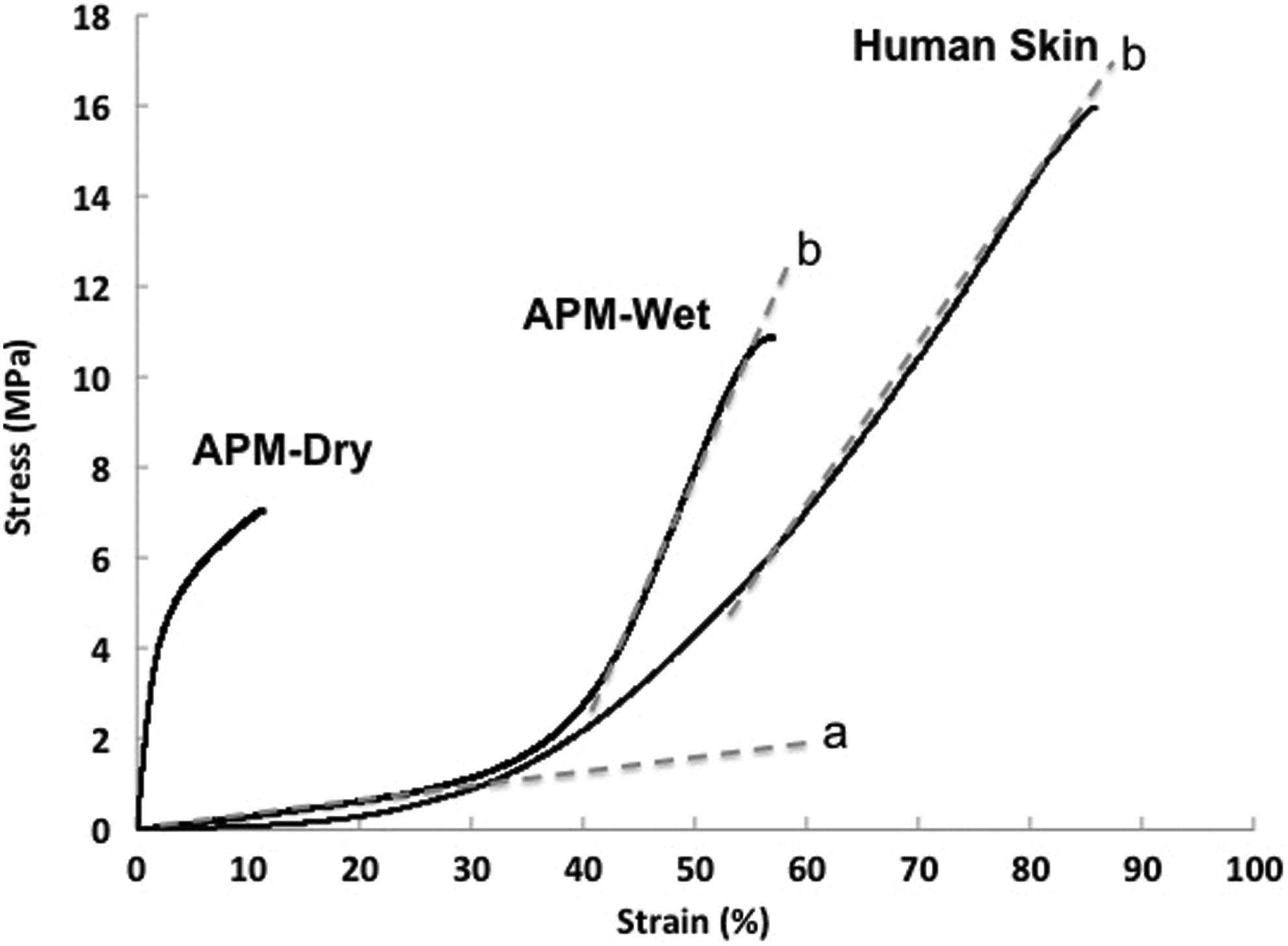
Stress–strain curves of APM specimens at dry (APM-Dry), hydrated (APM-Wet) conditions, and cadaver human skin. All the tests were performed at room temperature. a, initial slope; b, secondary slope.
APM supports fibroblasts and keratinocytes attachment and growth
Cell adhesion provides essential cues for cells to maintain normalization of tissue architecture and homeostasis in vitro and in vivo. 32 In this study, we investigated whether APM can support skin cell adhesion and subsequent proliferation, and compared the cellular adhesion and proliferation on the basement membrane and preperitoneal sides of APM. Moreover, we compared the APM with bovine type I collagen gel, which is a matrix that is widely used as a dermal substrate in many commercialized HSEs for in vivo skin grafting (e.g., Apligraf®, Orcel™) and in vitro use (i.e., EpiDerm-FT™).33–35 Human skin cells, HDF and HaCaT were seeded on the surface of bovine collagen gel and each side of the APM; TCPS was used as control. At 5 and 24 h time points, alamarBlue assay was performed to assess the cellular attachment percentage of human skin cells. For cell attachment at 24 h time point, the proliferation of HaCaT and HDF was not taken into account, due to the reported lag time for both HaCaT (24–48 h 36 ) and HDF (24 h 37 ). Our results showed that APM had significantly higher cell attachment compared to type I collagen gel with both types of cells (Fig. 3a, b), and there was no significant difference of attachment between the basement membrane (MesoBM) side and type I collagen-rich side (MesoPP) of APM.

Cellular attachment at 5 and 24 h time point of
To visualize the skin cells on APM, GFP-expressing HaCaT and RFP-expressing HDF were seeded to the MesoBM side and MesoPP side of the APM, respectively. The seeding of cells in this pattern was to mimic in vivo skin tissues where keratinocytes proliferate and differentiate over a basement membrane structure, while the fibroblasts are resident in a type I collagen-rich environment. Figure 3c shows the image of cells 4 days postseeding. The fibroblasts on MesoPP side showed a typical spindle, elongated shape of fibroblast morphology and the keratinocytes on MesoBM side presented their normal morphology in cobblestone-like and mosaic shape.
To determine the metabolic activity of the cells on matrices, HDF and HaCaT were monocultured on each side of the APM for 14 days. The metabolic activity of the cells on APM was compared to that of the cells on type I collagen gel and TCPS (Fig. 4). For HDF, Figure 4a shows that the metabolic activity on APM was similar to that on TCPS, and significantly higher than that on type I collagen gel throughout 14 days of culture. For HaCaT (Fig. 4b), interestingly, on type I collagen gel, the metabolic activity began to increase on day 7. The metabolic activity of HaCaT on APM was higher than that on TCPS (day 11 and 14), and that metabolic activity of HaCaT on TCPS was higher than that on type I collagen gel. Overall, APM promoted the attachment and viability of HDF and HaCaT in vitro as compared to collagen gel.

Metabolic activity of
Development of APM-HSEs with multiple cell layers
APM-HSEs were constructed by seeding HDF on the MesoPP side and followed by seeding HaCaT on the MesoBM side of APM to simulate the cellular distribution of in vivo environment. The cocultured constructs were then submerged in culture medium for 1 week and transferred to an air–liquid interface for another 2 weeks to induce keratinocyte differentiation. 38 The APM-HSEs were then harvested on day 21 and fixed for histology analysis. Histological H&E analysis of cross-sections showed that HaCaT and HDF were able to grow into multiple layers on MesoBM side and MesoPP side of APM, respectively (Fig. 5a). Migration of cells to the inner layers of ECM was not observed. In HaCaT layers, flattening of nuclei and formation of stratum spinosum-like structures were observed, demonstrating differentiation of the HaCaT. The epithelial thickness and structure formation from HaCaT were highly influenced by the seeding density of HDF in vitro (Fig. 5b). Increased HDF seeding density to 5 × 10⁴/cm2 on APM resulted in the formation of thickest and most organized epidermis (Fig. 5b, d), whereas increasing the seeding density of HaCaT did not improve the thickness or organization of the epidermal or HDF layer (Fig. 5c, e). Overall, our H&E images indicated that APM supported the growth of HDF, proliferation, and differentiation of HaCaT to form a partially differentiated full-thickness HSEs, and the thickness of epidermal layers was affected by the seeding density of HDF.
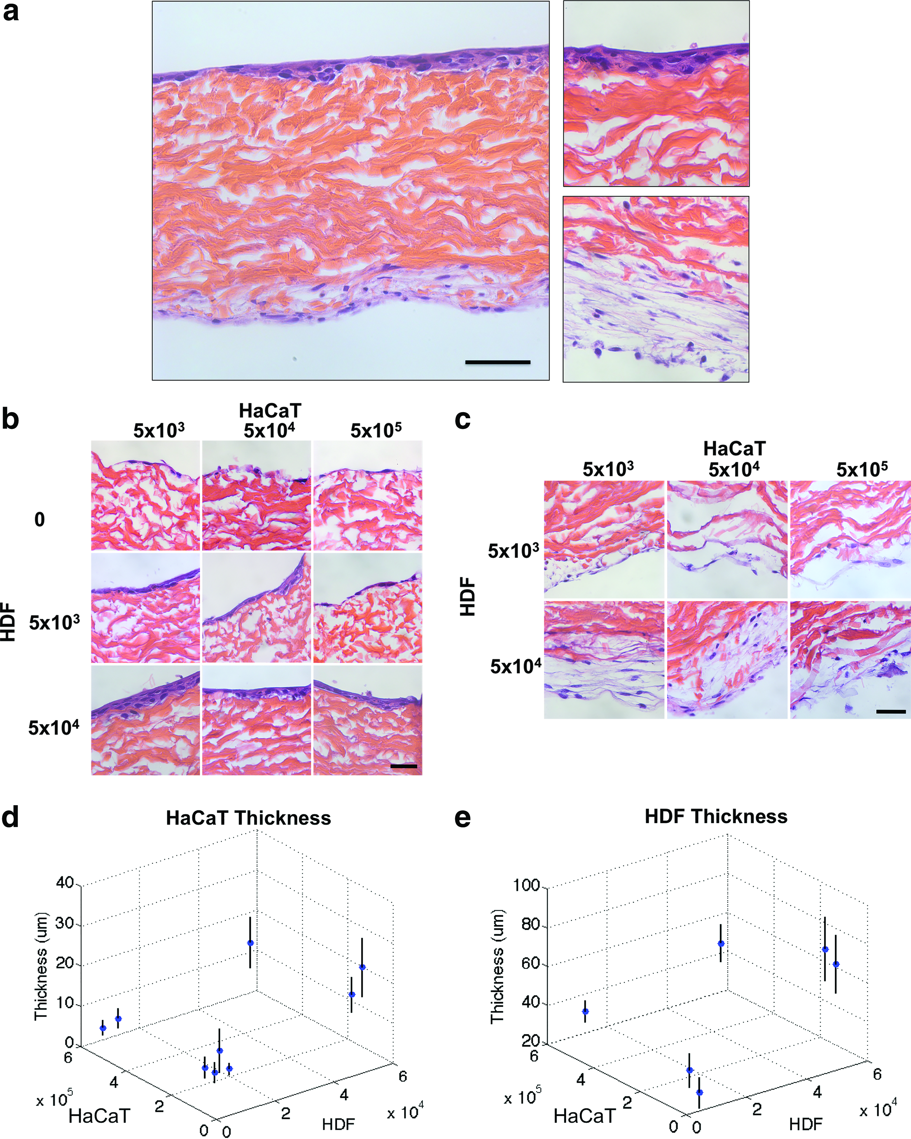
Histological Hematoxylin and eosin analysis and cell layer thickness of constructed APM-HSEs at day 21.
Expression of keratinocyte proliferation and differentiation markers on APM-HSEs
The keratinocyte proliferation and differentiation on APM-HSEs were further examined by performing immunohistochemistry staining of keratin 15 and involucrin markers, respectively (Fig. 6). Cadaver human skin specimens were used as reference material. Keratin 15 is a keratinocyte proliferation marker and is found to mark putative stem and progenitor cells located in stratum basale. 39 Involucrin, a keratinocyte differentiation marker, is a cornified envelope protein marker that is highly expressed in the supra stratum granulosum layer of the epidermis in native human skin. 40 Immunohistochemistry staining of cadaver human skin specimens indicated well-stratified viable epidermis layers, that is, basal layer expressing keratin 15 (Fig. 6a) and supra stratum granulosum layers expressing involucrin (Fig. 6c). For APM-HSEs specimens, there was very low level of keratin 15 detected (Fig. 6b), and the expression pattern of keratin 15 was irregular compared to cadaver human skin (staining on basal layer is evident) (Fig. 6a). The expression of involucrin was evident in the outermost layer of APM-HSEs, which was similar to human skin (Fig. 6c, d). While in normal native skin, proliferation and differentiation of keratinocytes are well balanced and hence the stratum corneum is formed; our immnohistochemistry images suggested the epidermal layers of the APM-HSEs contained differentiated and viable cell layers, but had no stratum corneum and were less organized than native human skin.

Immunohistochemistry labeling of keratinocyte proliferation marker, keratin 15
In vitro irritation testing on APM-HSEs
To examine the potential of APM-HSEs models for irritancy screening, six proficiency chemicals (Table 1) were selected based on OECD T439 guidelines for in vitro skin irritation. 29 The choice of irritants and their concentrations is based on either OECD TG439 guidelines (i.e., PBS, 1-bromohexane and heptanol), or published concentrations from literature 41 (i.e., 20% SDS, PEG 400). The selected chemicals were applied to the freshly prepared APM-HSEs (at week 3 time point of HaCaT/HDF coculture) for 60 min at 37°C, and end point analyses were performed 42 h posttreatment. End point analyses included cellular viability and quantification of the extracellular release of five cytokines into the medium (Fig. 7). APM-HSEs treated with PBS were considered as control group based on the OECD T439 guidelines.

Cellular viability
Viability data showed that APM-HSEs responded to irritant ranking in a consistent manner. Cellular viability decreased with increasing ranking of irritation of the chemicals (Fig. 7a). When comparing the viability to PBS control group without any chemicals (set as 100%), the cellular viabilities of treated group can be used to discriminate between nonirritants and irritants (Table 2). The cutoff point to differentiate irritant from nonirritant was 71.7% viability based on the receiver-operating characteristic analysis (Supplementary Fig. S1 and Supplementary Table S1; Supplementary Data are available online at www.liebertpub.com/tea). All chemicals excepting heptanol and SDS (these two groups resulted in viability <5%) were further analyzed for cytokine secretions to confirm ranking order of the irritants.
APM, acellular peritoneum matrix; HSE, human skin equivalent; I, irritating; NI, nonirritating; SD, standard deviation.
During skin inflammatory process in vivo, proinflammatory cytokines, including IL-1α, IL-6, IL-8, and GM-CSF, are secreted together by keratinocytes, fibroblasts, Langerhans, monocytes, and T cells. Upon irritant stimuli, keratinocytes secrete IL-1α, a primary cytokine that initiate inflammatory cascade. The activation of IL-1α stimulates further production of secondary proinflammatory cytokines (e.g., IL-8, IL-6, GM-CSF) by surrounding epidermal and dermal cells. 42 In our study, both primary and secondary proinflammatory cytokines were detected. The concentrations of secreted IL-1α were detected in isopropanol (12.37 ± 3 pg/mL) and 1-bromohexane (49.5 ± 16.7 pg/mL) treatments, and it was under detection limit for PBS and PEG 400 treatments (Fig. 7b). IL-1ra, a natural antagonist of IL-1α that prevents damages of exceeded IL-1α significantly increased from 1-bromohexane treatment compared with PBS and PEG 400 groups (Fig. 7c). IL-6 and IL-8 are secondary proinflammatory cytokines and chemotactic for neutrophils. Secretion of secondary cytokines IL-6 and IL-8 followed a similar trend as mentioned above. Importantly, increase in the in vivo irritation scores resulted in significantly increased cytokine productions (Fig. 7d, e). Also the secretion of GM-CSF, a cytokine that enhances effector function of monocytes and neutrophils was significantly increased for those treated with 1-bromohexane (Fig. 7f).
Discussion
Predictive in vitro assays that can identify skin irritation are in demand to reduce and replace the use of animal testing. Ideal 3D skin models for in vitro assays should possess (a) high accuracy, sensitivity, specificity, (b) represent native skin composition and physiology, (c) resemble native skin barrier properties, (d) be highly available, (e) provide high-throughput screening, (f) be consistent in batch-to-batch culture, and (g) have long shelf life. However, the ideal 3D skin models that fulfilled the above criteria are still yet to be developed.
Over the past decade, type I collagen hydrogel has been the dominant material used to construct full-thickness HSEs, based on the high biocompatibility and the nature of collagen representing ECM properties. However, the extraction process of collagen utilizes harsh treatments of acid and proteolytic solutions, which alter its molecular structures by cleaving terminal telopeptides. This results in decreased tropocollagen self-assembled fibrils and partially denatures the collagen. 43 The collagen gels lack the mature collagen fiber hierarchical structure and biological diversity of ECM proteins present in decellularized tissues. The partially denatured collagen affects the mechanical properties of the constructed HSEs and results in variable surface area, shape, and thickness of HSEs models. The extent of contraction is also affected by collagen concentration, number of fibroblasts seeded in the matrix, and HSEs culture time. On the other hand, decellularization processes remove cells and antigenic compounds, while preserving the native ECM structure, tissue strength, and distribution of cell attachment proteins and growth factors.
The feasibility of using APM as a substrate to develop full-thickness skin tissue was evaluated. The hydrated APM specimens exhibited nonlinear tensile properties that represent mechanical properties of human skin; both the hydrated APM and excised skin specimens showed a two-slope stress–strain feature. The modulus values of hydrated APM (initial slope = 3.5 ± 0.12 MPa; secondary slope = 57 ± 1.63 MPa, Fig. 2) were similar to those of the excised human skin (initial slope = 2.9 ± 1.2 MPa, secondary slope = 36 ± 12 MPa). This phenomenon is due to the degree of alignment from the collagen and elastic fibers. 44 In comparison, the modulus of collagen gels has been reported in the range of 1.5–24.3 kPa. 45 The intact mature ECM structure of APM enables the development of APM-HSEs with relatively controlled dimensions and excellent handling properties. The APM supported the attachment and growth of HDF and HaCaT, significantly better than the type I collagen gel (Figs. 3 and 4). This could be explained by mechanical cues that result from the increased modulus of APM versus collagen gels.
Further, we attempted to construct a full-thickness HSE based on coculturing human skin cells on the decellularized ECM. Primary keratinocytes, although easy to stratify into stratum corneum structures, have limited life span, rapidly die after induced differentiation, and are difficult to expand into large quantity. HaCaT, a spontaneously immortalized human keratinocyte cell line was used in this study, as it exhibits basal cell properties, is highly available, and easier to expand compared to primary keratinocytes, which might be more suitable for in vitro assays. Immortalized cell lines have shown utility in the construction of HSEs, for example, normal immortal keratinocytes (NIKS) mutated from the BC-1 EP strain of normal human neonatal foreskin provide indefinitely culture, near-diploid karyotype in monoculture and established fully stratified epidermis, and less batch-to-batch variability in HSEs. 15 However, for commercialization, there will be licensing issues associated with the use of HaCaT.
In our study, although excellent support of attachment and proliferation of fibroblasts and HaCaT on APM was observed, the 3D coculture of HDF and HaCaT resulted in HSEs with partially differentiated epidermis layer (Figs. 5a and 6). This partial differentiation may be due to the natural difference of HaCaT from primary keratinocytes, where deficiency in epithelial–stromal interactions and decreased secretion of paracrine IL-1α were found in the HaCaT. 46 Ideally, stratum corneum should be present to provide proper barrier to reflect the permeation of irritation compounds to viable epidermal and dermal layers. However, commercially marketed skin models with fully stratified stratum corneum still face the challenge of exhibiting inferior barrier properties compared with those of human skin.47,48 Despite the differences of HaCaT from primary keratinocytes, supplementing additional growth factors, such as TGF-α, keratinocyte growth factor, and GM-CSF during in vitro culture, can rescue the normal differentiation. 46 Interestingly, we found that increasing seeding density of HDF improved the thickness and organized morphology of HSEs epidermis layer in vitro (Fig. 5b). This finding is in agreement with several studies that suggested paracrine effect from stromal cells could also affect epidermal homeostasis.20,46 Therefore, by further optimizing in vitro culture conditions, we may obtain fully stratified epidermis from HaCaT.
Skin irritation is a complex process that involves penetration of chemicals through skin layers, disruption of skin barrier, and induction of proinflammatory cytokine cascades through keratinocytes, fibroblasts, Langerhans cells, and T cells. Ideally, full-thickness HSEs should be applied to study skin irritation as they resemble skin more than epidermal models due to their higher complexity in cell types and skin strata. However, all the current OECD-approved HSE models for irritation tests are epidermis models. 29 Few reports have used full-thickness models that contain keratinocytes on fibroblasts-populated collagen matrices to predict the secretion of secondary proinflammatory cytokines (e.g., IL-6, IL-10) that are not detected in the epidermal model.49,50 Mallampati et al. used a full-thickness model, EpiDerm-FT (MatTek) to study the toxic effect and structural activity relationship from saturated aliphatic hydrocarbons. Endpoint evaluations such as cellular viability and proinflammatory cytokines (IL-1α, IL-6, IL-8) demonstrated that it is possible to rank the toxicity effect of various hydrocarbon chain lengths and the in vitro results were correlated with in vivo data. 49 Similarly, in another study, Black et al. has demonstrated the use of EpiDerm-FT to identify the molecular mechanism of skin inflammation caused by sulfur mustard. 51 It should be noted that EpiDerm-FT is based on collagen hydrogels. The contraction of collagen gel by the skin cells often results in unpredictable variations in specimen thickness and surface area. Collagen gels are fragile with undesirable mechanical strength and are often difficult to handle. Decellularized tissues, such as APM, are attractive biomaterials to construct full-thickness HSEs for in vitro testing, as they highly retain the 3D structure, mechanical properties, and biochemical composition of ECM. 30
In our study, APM-HSEs containing both the viable epidermis and dermis components were used for skin irritation test. Three nonirritants (i.e., PBS, PEG 400, and isopropanol) and three irritants (i.e., 1-bromohexane, hepanol, and SDS) were applied on the epithelial surface of APM-HSEs model and used to evaluate the in vitro prediction of irritancy through viability and cytokine secretion. In the OECD TG439 guideline, the viability cutoff point is 50%. The guideline is established for reconstructed human epidermis models; therefore, the viability is simply based on viable keratinocytes. Comparison between APM-HSEs and established models are shown in Table S2. In our study using APM-HSEs, our cutoff point is 71.7%. This is due to the feature of our APM-HSEs model, comprising not only viable keratinocytes, but also fibroblasts, which will also contribute to the overall viability. In addition, the test compounds would have to diffuse through APM-HSEs to reach the underneath fibroblast. While viability itself can distinguish irritants from nonirritants, primary and secondary cytokine production can further differentiate the irritation potency of various compounds. However, to evaluate the true predictive power of APM-HSEs in skin irritation testing, accuracy, sensitivity, and specificity of the test needs to be validated with an expanded panel, with variants from concentrations and types of chemicals and topical formulations.
Conclusions
In this study, we demonstrated that APM supports attachment and proliferation of fibroblasts and HaCaT, and coculture procedures resulted in partial epidermal differentiation. The APM can be used as a substrate to aid the development of advanced full-thickness skin models with coculture of multiple cell types. Further, the cultured APM-HSEs models have the potential to function as in vitro irritation screening tool, based on the ability to classify topically applied nonirritants from irritant chemicals and to differentiate their irritation rankings through the viability of epidermis and dermis cells and the secretion levels of multiple cytokines. In conclusion, we have demonstrated the use of APM to support the development of HSEs and envision them to be valuable tools for future in vitro screening of topically applied drugs.
Footnotes
Acknowledgments
The authors thank DSM Biomedical Corporation for providing APM matrices as well as antibodies and reagents for immunohistochemistry assay. They also thank Kathleen Roberts for her assistance in histology, Dr. Joachim Kohn from the New Jersey Center for Biomaterials for RFP and GFP-expressing skin cells, Dr. Yong Mao for discussions, and Meng-Chen Lo for MATLAB code. This work was financially supported by the Center for Dermal Research and New Jersey Center for Biomaterials.
Disclosure Statement
No competing financial interests exist.
References
Supplementary Material
Please find the following supplemental material available below.
For Open Access articles published under a Creative Commons License, all supplemental material carries the same license as the article it is associated with.
For non-Open Access articles published, all supplemental material carries a non-exclusive license, and permission requests for re-use of supplemental material or any part of supplemental material shall be sent directly to the copyright owner as specified in the copyright notice associated with the article.