
Abstract
Cartilage has a poor healing response, and few viable options exist for repair of extensive damage. Hyaluronic acid (HA) hydrogels seeded with mesenchymal stem cells (MSCs) polymerized through UV crosslinking can generate functional tissue, but this crosslinking is not compatible with indirect rapid prototyping utilizing opaque anatomic molds. Methacrylate-modified polymers can also be chemically crosslinked in a cytocompatible manner using ammonium persulfate (APS) and N,N,N′,N′-tetramethylethylenediamine (TEMED). The objectives of this study were to (1) compare APS/TEMED crosslinking with UV crosslinking in terms of functional maturation of MSC-seeded HA hydrogels; (2) generate an anatomic mold of a complex joint surface through rapid prototyping; and (3) grow anatomic MSC-seeded HA hydrogel constructs using this alternative crosslinking method. Juvenile bovine MSCs were suspended in methacrylated HA (MeHA) and crosslinked either through UV polymerization or chemically with APS/TEMED to generate cylindrical constructs. Minipig porcine femoral heads were imaged using microCT, and anatomic negative molds were generated by three-dimensional printing using fused deposition modeling. Molded HA constructs were produced using the APS/TEMED method. All constructs were cultured for up to 12 weeks in a chemically defined medium supplemented with TGF-β3 and characterized by mechanical testing, biochemical assays, and histologic analysis. Both UV- and APS/TEMED-polymerized constructs showed increasing mechanical properties and robust proteoglycan and collagen deposition over time. At 12 weeks, APS/TEMED-polymerized constructs had higher equilibrium and dynamic moduli than UV-polymerized constructs, with no differences in proteoglycan or collagen content. Molded HA constructs retained their hemispherical shape in culture and demonstrated increasing mechanical properties and proteoglycan and collagen deposition, especially at the edges compared to the center of these larger constructs. Immunohistochemistry showed abundant collagen type II staining and little collagen type I staining. APS/TEMED crosslinking can be used to produce MSC-seeded HA-based neocartilage and can be used in combination with rapid prototyping techniques to generate anatomic MSC-seeded HA constructs for use in filling large and anatomically complex chondral defects or for biologic joint replacement.
Introduction
C
Anatomic, 3D tissue engineering has made significant advances over the past two decades, made possible by the incorporation of rapid prototyping techniques in scaffold fabrication. 11 Rapid prototyping relies on information garnered through advanced imaging modalities, such as magnetic resonance imaging (MRI) and microcomputed tomography (μCT), and in silico models generated through computer-assisted drawing (CAD) programs, which render 3D shapes into a series of two-dimensional cross sections. 12 Rapid prototyping techniques have gained popularity because they allow for precise control of pore size and distribution13,14 and provide the ability to fabricate a scaffold in any desired shape. For these reasons, image-guided tissue engineering using rapid prototyping techniques is ideal for cartilage engineering, as it allows for precise size matching for each patient.
Early work in this area for the engineering of image-guided anatomic cartilage scaffolds has shown promising results.15–17 For example, Chang et al. developed replica molds for facial implants that were filled by injection molding with cell-seeded alginate hydrogels. 18 This work has recently been extended to fashion anatomic meniscus implants. 19 Hung et al. applied these techniques to articular cartilage, fashioning stainless steel molds to produce replicas of the retropatellar articular cartilage based on chondrocyte-seeded agarose hydrogels. 15 Still more recently, Hollister and others have developed direct fabrication methods (3D printing) of solid porous materials with controlled geometry and pore spacing. These and similar methods have been implemented by Mao and colleagues to develop fully anatomic replacements for the humeral head in a rabbit model. 16
In addition to matching an individual patient's anatomy, the use of patient-specific cells to form the regenerated tissue would eliminate the risk of disease transmission that must be considered when using any allograft tissue. For almost two decades, autologous cell therapies have been available in the form of ACI, first commercialized in the United States as Carticel® (Genzyme). Furthermore, ex vivo expansion and culture of chondrocytes in 3D hydrogels have produced cartilage constructs with mechanical properties and proteoglycan content that meets or exceeds native tissue values.20,21 However, the use of chondrocytes is limited by the amount of healthy tissue in patients with widespread joint damage. 22 As an alternative cell source, mesenchymal stem cells (MSCs) are a precursor cell population present in patient bone marrow that can be isolated and expanded in vitro without losing their ability to differentiate into chondrocytes. 23 When coupled with various hydrogels and cultured in chondrogenic media, these cells can take on a chondrocyte-like phenotype, deposit cartilage-like extracellular matrix (ECM), and produce constructs with native-like mechanical properties.24,25
With respect to materials to support anatomic tissue engineering, the choice largely depends on whether the intent is to form an anatomic scaffold that will later be seeded with cells or invaded by endogenous cells when implanted, or whether the intent is to generate a cell-seeded anatomic replica that is matured in vitro. For the latter approach, the general scheme is to form a negative mold of the desired anatomic structure and then use injection molding to create a cell-seeded construct. Various hydrogels have been used for such applications, including agarose (which sets when the temperature falls below a certain level) 15 and alginates (which are combined with a crosslinking agent just before injection into the mold). 18
Our group has optimized the use of hydrogels based on hyaluronic acid (HA), a polysaccharide consisting of alternating molecules of D-glucoronic acid and N-acetyl-D-glucosamine, naturally found in hyaline cartilage.26,27 HA can be chemically modified through the addition of methacrylate groups to enable free radical-mediated crosslinking between HA molecules.28,29 Using these photopolymerizable HA networks (formed through ultraviolet (UV)-induced free radical generation), chondrocytes and MSCs can be encapsulated to generate neocartilage with biochemical and mechanical properties approaching that of native tissue.24,29–32 However, this UV-mediated crosslinking method is generally not compatible with rapid prototyping techniques in which cells suspended in liquid monomer are injected into opaque anatomic 3D molds made from metal or plastic. Recently, Temenoff et al. developed a thermal radical initiation system that was used to encapsulate rat marrow stromal cells. They reported that the addition of 25 mM ammonium persulfate (APS) and 25 mM N,N,N′,N′-tetramethylethylenediamine (TEMED) and heating to 37°C for 8 min induced free radical formation and crosslinking of an oligo(poly(ethylene glycol) fumarate) (OPF) hydrogel. Importantly, marrow stromal cells encapsulated in this thermally crosslinked OPF hydrogel survived the crosslinking process and produced mineralized bone matrix after 28 days in culture. 33
Based on the above, we carried out the following studies. First, we implemented the APS/TEMED crosslinking method and compared functional and biochemical outcomes to our traditional UV-mediated crosslinking method for MSC-seeded HA networks. After establishing equivalence between these two gelation methods, we next developed an anatomic cartilage molding process based on contrast-based μCT imaging of the Yucatan minipig femoral head articular cartilage. Using these molds, we fabricated anatomic 3D MSC-seeded HA hydrogel constructs and evaluated their functional maturation over 12 weeks of in vitro culture. Our findings advance the field of anatomic cartilage tissue engineering and define new challenges that must be overcome if such methods are to be realized in clinical practice for the restoration of large, degenerated cartilage surfaces.
Materials and Methods
Fabrication of anatomic 3D femoral head molds
Juvenile (8 month old) Yucatan minipig femoral heads were obtained as remnant tissue from unrelated studies that were performed with approval by our institute's Institutional Animal Care and Use Committee. These femoral heads were imaged using μCT (VivaCT 45; Scanco Medical) with the following settings: energy = 45 kV, intensity = 177 μA, and resolution = 76 μm between image slices. Femoral heads were then soaked in an iodinated contrast agent, Lugol's solution (Sigma), for 48 h and reimaged using the same settings. MicroCT images were input into ITK-SNAP, an open source software. The autosegmentation tool in ITK-SNAP was used initially to perform slice-by-slice segmentation of the cartilage layer from the underlying subchondral bone 34 ; the segmentation was then refined manually. An STL shape file was exported from ITK-SNAP with the cartilage surface rendered as 2,196,340 interconnected triangular faces. This STL shape file was processed and refined using MeshLab to smooth the 3D model and reduce the number of triangular faces to 26,000, allowing import of the file into SolidWorks, a CAD program. SolidWorks was used to generate anatomic negative molds with a cavity between the male and female pieces of the mold in the shape of the cartilage layer. Molds were printed using fused deposition modeling (Objet 30 Desktop 3D Printer; Stratasys) in thermoresponsive plastic (VeroBlack) with a 28-μm layer thickness.
HA hydrogel synthesis
Methacrylated HA (MeHA) was synthesized through reaction with methacrylic anhydride (Sigma) and 74 kDa HA (Lifecore) as previously described. 24 Using 1 H NMR characterization, the final product was determined to be 25–30% methacrylated. Lyophilized MeHA was sterilized by exposure to a biocidal UV lamp for 1 h and dissolved to 1% (w/v) in either 0.05% photoinitiator Irgacure-2959 (2-methyl-1-[4-(hydroxyethoxy)phenyl]-2-methyl-1-propanone; Ciba-Geigy) in sterile phosphate-buffered saline (PBS) or in 25 mM APS in sterile PBS.
MSC isolation, expansion, and 3D culture
MSCs were harvested from 3 to 5 juvenile bovine femurs or tibias, 35 combined, expanded to passage 2–4, and suspended in either 1% MeHA in 0.05% I-2959/PBS or 25 mM APS/PBS at 60 million cells/mL. Cell/MeHA suspensions were pipetted between glass plates separated by 2.25 mm spacers and polymerized through UV exposure for 10 min or via the addition of 25 mM TEMED just before casting, followed by heating of the casting apparatus to 37°C for 10 min, as described by Temenoff et al. 33 After polymerization, dermal biopsy punches (4 mm diameter) were used to create MSC-seeded hydrogel cylinders.
To generate molded constructs using the anatomic molds, MSCs were first suspended at 60 million cells/mL in 1.2 mL of 1% MeHA in 25 mm APS/PBS. TEMED was then added to a final concentration of 25 mM, and the cell-MeHA suspension was pipetted into the mold. A 2.25 mm spacer was used to separate the male and female segments of the mold to generate thicker constructs that could be more readily handled. The mold was then heated to 37°C for 10 min, and the polymerized construct was transferred into culture media.
All constructs were cultured in a chemically defined chondrogenic medium containing high-glucose DMEM with 1× PSF, 0.1 μM dexamethasone, 50 μg/mL ascorbate 2-phosphate, 40 μg/mL L-proline, 100 μg/mL sodium pyruvate, and ITS (6.25 μg/mL insulin, 6.25 μg/mL transferrin, 6.25 ng/mL selenous acid, 1.25 mg/mL bovine serum albumin, and 5.35 mg/mL linoleic acid) and supplemented with TGF-β3 (10 ng/mL; R&D Systems). Constructs were maintained at 21% oxygen tension, and media were changed thrice weekly with media volume scaled to the volume of the construct (i.e., 0.5 mL per 4 mm diameter construct, 8 mL per molded anatomic construct). Molded constructs were cultured in six-well flat bottom plates (35-mm well diameter, 18-mm height) with their concave side facing up (Fig. 1).
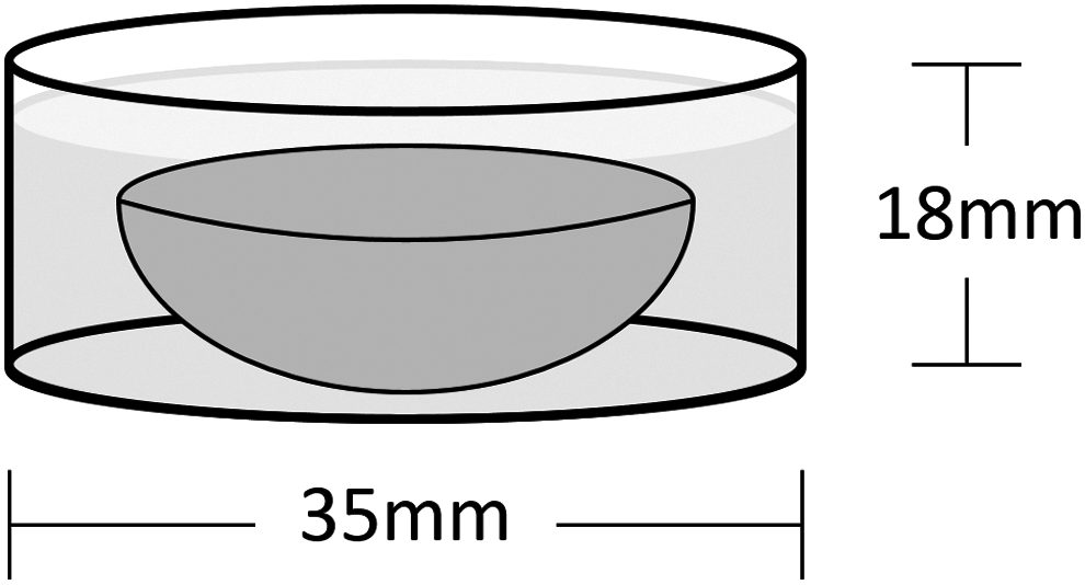
Schematic showing culture geometry for anatomic molded hyaluronic acid (HA) constructs.
Mechanical and biochemical analyses
At specific time points (2, 4, 6, 8, and 12 weeks for APS/TEMED versus UV comparison studies, 6 and 12 weeks for molded HA constructs), mechanical properties and biochemical content were determined. Before testing, each construct was weighed to determine wet weight. Constructs were then trimmed with a freezing stage microtome to create a flat surface for mechanical testing. Samples were placed in a custom-built mechanical testing apparatus, and a 2N creep tare load was applied to ensure consistent contact between surfaces before the initiation of the stress–relaxation test. The unconfined equilibrium compressive modulus was derived from a stress–relaxation test (10% strain, 1000 s relaxation). 36 After reaching equilibrium, the dynamic modulus was determined from five sinusoidal cycles of compression at 1 Hz (1% strain amplitude). 37
After testing, each construct was digested in papain and then analyzed for sulfated glycosaminoglycan (sGAG) and collagen content. 31 Glycosaminoglycan content was assessed using the 1,9-dimethylmethylene blue (DMMB) dye binding assay. 38 Orthohydroxyproline (OHP) content of digested samples was determined through reaction with chloramine T and diaminobenzaldehyde, 39 and collagen content was extrapolated from a 1:7.14 ratio of OHP:collagen. 40
Mechanical and biochemical analyses were performed on molded constructs by first using a 4 mm dermal biopsy punch to remove cylinders from four sites in the middle and four sites at the edge of the constructs. The cylinders were removed from the flattest surfaces possible and planed with a freezing stage microtome to ensure completely flat surfaces. These cylinders then underwent unconfined compression, papain digest, and biochemical analyses for sGAG and collagen as for the smaller cylindrical constructs.
Cell viability and histologic analysis
Cell viability was assessed by cutting cylindrical constructs in half and staining with calcein AM and ethidium homodimer (Live/Dead Kit; Invitrogen). Cylindrical and anatomic molded constructs were also fixed in 4% paraformaldehyde, paraffin embedded, and sectioned (8 μm). Sections were stained with Alcian Blue (proteoglycan) and Picrosirius Red (collagen) before imaging at 2×, 4×, 10×, and 20× magnification. Immunohistochemistry was performed to identify the presence of collagen (col) type I (MAB 3391 at 10 μg/mL; Millipore) and type II (II-II6B3 at 10 μg/mL; Developmental Studies Hybridoma Bank) as previously described. 25
Statistical analyses
Data are reported as mean and standard error of the mean (SEM). Two-way ANOVA with Bonferroni post-test analysis was performed with time in culture and polymerization method (for UV versus APS/TEMED comparison studies) or location (middle versus edge for anatomic molded constructs) as independent variables, with significance set at p < 0.05. All statistical analyses were performed using GraphPad Prism.
Results
Comparison of APS/TEMED-polymerized versus UV-polymerized HA constructs
MSCs were suspended in 1% HA, which then underwent either UV polymerization or thermal/chemical polymerization with APS/TEMED. MSC-seeded cylindrical HA constructs were cultured in vitro in chemically defined media supplemented with TGF-β3 for up to 12 weeks. At 2, 4, 6, 8, and 12 weeks, constructs were assayed for cell viability, mechanical properties, biochemistry, and histology. After 12 weeks in culture, both UV- and APS/TEMED-polymerized constructs had a large proportion of viable cells and demonstrated similar levels of proteoglycan and collagen deposition (Fig. 2).
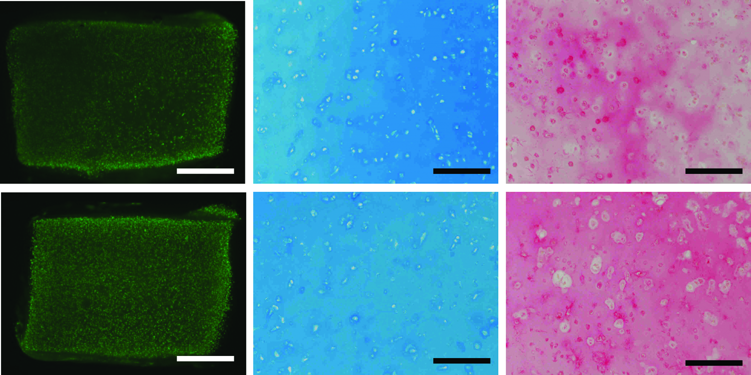
Comparison of UV-polymerized (top) and APS/TEMED- polymerized (bottom) cylindrical HA constructs after 12 weeks in culture. Both constructs maintained cell viability (calcein staining, left) and demonstrated equivalent proteoglycan deposition (Alcian Blue staining, middle) and collagen deposition (Picrosirius Red staining, right). Calcein stain magnification, 2×; scale bar, 1 mm. Alcian Blue and Picrosirius Red stain magnification, 10×; scale bar, 0.2 mm. Color images available online at www.liebertpub.com/tea
When assayed for mechanical properties, both types of constructs were very weak at early time points as expected. However, over time, both types of constructs showed increasing equilibrium and dynamic moduli (p < 0.05, Fig. 3A, B). At 12 weeks, the equilibrium moduli of the UV- and APS/TEMED-polymerized constructs reached 185 kPa (±22.0) and 286 kPa (±25.2), respectively. At 12 weeks, the dynamic moduli of the UV- and APS/TEMED-polymerized constructs were 1461 kPa (±148.6) and 2518 kPa (±202.2), respectively. There were no significant differences in equilibrium moduli between either type of construct at early time points (p > 0.05). However, by 12 weeks, constructs formed using the APS/TEMED method had significantly higher equilibrium and dynamic moduli than their UV-polymerized counterparts (p < 0.001).
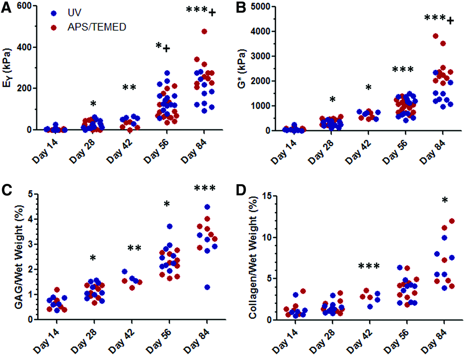
Mechanical testing and biochemical assays comparing UV- and APS/TEMED-polymerized HA constructs over a 12-week culture period.
Over time, both types of constructs also accumulated increasing amounts of proteoglycan and collagen (p < 0.05, Fig. 3C, D). At 12 weeks, GAG/wet weight for UV- and APS/TEMED-polymerized constructs were 3.01% (±0.43) and 3.48% (±0.17), respectively. At 12 weeks, collagen/wet weight for UV- and APS/TEMED-polymerized constructs were 6.76% (±0.89) and 7.37% (±1.42), respectively. There were no significant differences in the proteoglycan and collagen content between both types of constructs at any time point (p > 0.05).
Fabrication, culture, and characterization of anatomic molded HA constructs
Juvenile porcine femoral heads were imaged using μCT, stained with Lugol's solution, and then reimaged using the same settings. Images were input into ITK-SNAP to allow for segmentation of the cartilage layer from the underlying subchondral bone. The 3D cartilage model was further refined in MeshLab, and the final STL shape file was input into SolidWorks to generate an anatomic negative mold of the cartilage layer. The mold consisted of male and female pieces that formed a cavity and was 3D printed through fused deposition modeling from thermoresponsive plastic (Fig. 4).

Process for generating anatomic molds of porcine femoral heads. The femoral head was imaged with microcomputed tomography (μCT), stained with Lugol's solution, and reimaged. μCT images were imported into ITK-SNAP to allow slice-by-slice segmentation of cartilage from subchondral bone. The three-dimensional (3D) cartilage model was first simplified and refined in MeshLab and imported into SolidWorks to generate anatomic negative 3D molds, which were then 3D printed through fused deposition modeling. Color images available online at www.liebertpub.com/tea
MSCs were suspended in 1% HA in 25 mM APS/PBS, TEMED (25 mM) was added, and the suspension was pipetted into the cavity of the anatomic femoral head mold with a 2.25 mm spacer block separating the male and female pieces. The mold containing the MeHA-MSC suspension was placed in the incubator at 37°C for 10 min to crosslink the hydrogel. Polymerized cell-laden HA constructs were then removed from the molds and cultured. Initially, HA constructs were extremely weak and lucent (Fig. 5). When cultured for up to 12 weeks in a chemically defined prochondrogenic media, constructs retained their hemispherical shape in culture and became stiffer and opacified over time.
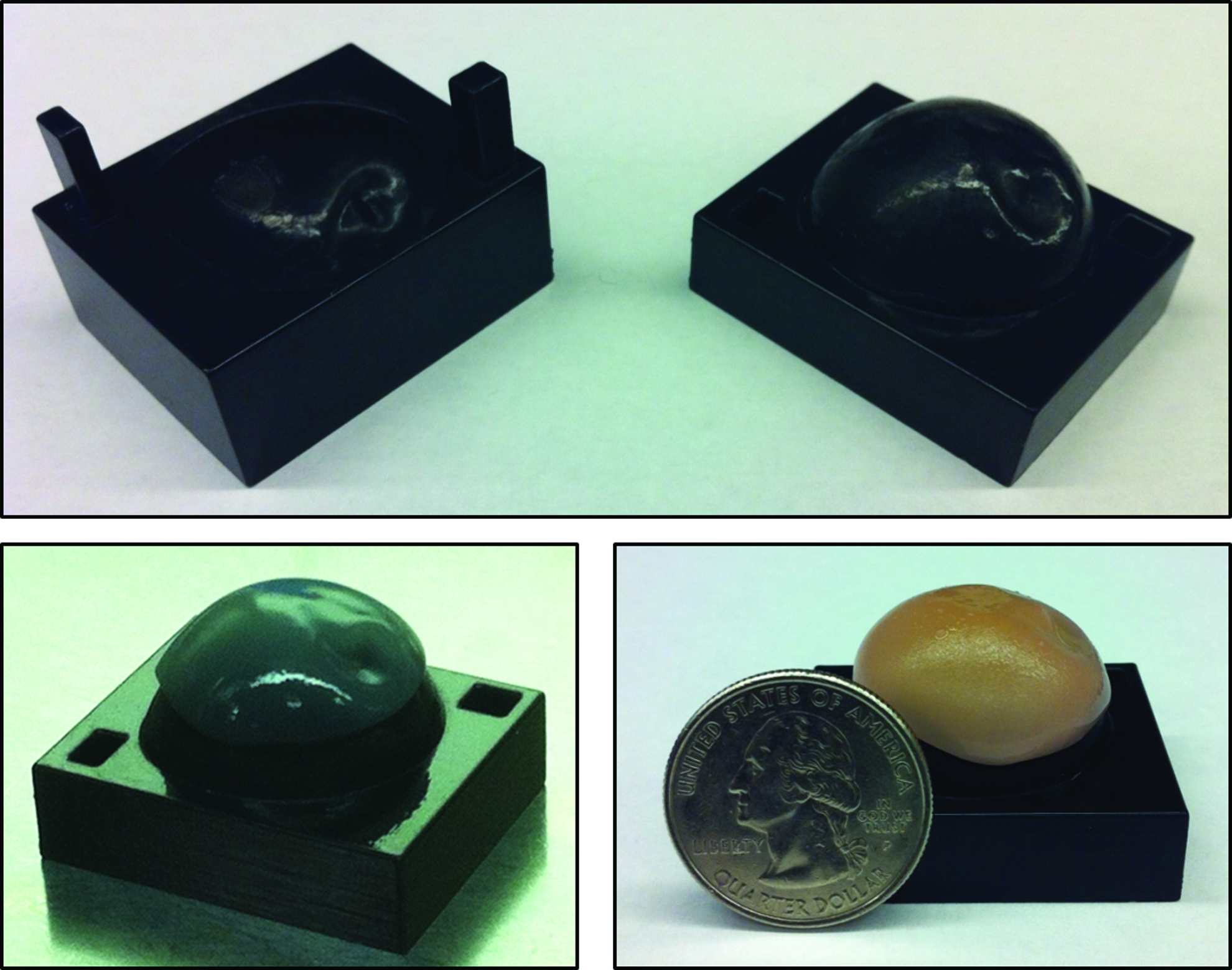
Anatomic porcine femoral head molds (top). After polymerization at Day 0, HA constructs were weak and lucent (bottom left). However, over time in culture, constructs became stiffer and opaque (bottom right, sample at 12 weeks). Color images available online at www.liebertpub.com/tea
These anatomic constructs were assayed for cell viability, mechanical properties, biochemistry, and histology at 6 and 12 weeks. After 6 weeks, constructs remained extremely weak throughout with low equilibrium and dynamic moduli. MSCs remained viable with no gross differences in viability between the middle and edge of the constructs at this time point (not shown). After 12 weeks, equilibrium moduli and dynamic moduli throughout the constructs increased (p < 0.05 except for equilibrium moduli in the middle, Fig. 6A, B), although moduli of test samples removed from the middle of the construct remained relatively low. Equilibrium and dynamic moduli at the edges increased to 48 kPa (±11.4) and 680 kPa (±71.8), respectively, and were significantly greater than moduli of samples taken from the middle of constructs (p < 0.001). At this 12-week time point, cell viability was noticeably lower in the middle of constructs compared to at the edge (Fig. 7E–H).
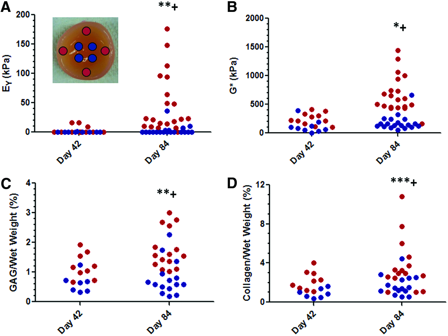
Mechanical testing and biochemical assays for molded HA constructs over a 12-week culture period.

Stitched images of histologic analysis of molded HA constructs. Alcian Blue staining
Analysis of proteoglycan and collagen content was consistent with cell viability and mechanical testing results. The proteoglycan and collagen content increased with time in culture (p < 0.05 for proteoglycan at the edge and collagen in the middle at 12 weeks compared to 6 weeks, Fig. 6C, D). After 12 weeks, GAG/wet weight at the middle and edge reached 0.79% (±0.15) and 1.77% (±0.17), respectively. Collagen/wet weight at the middle and edge were 1.73% (±0.27) and 3.76% (±0.69), respectively. The edges of constructs had significantly higher proteoglycan and collagen content than the middle (p < 0.01). Histologic analysis at 12 weeks supported these findings, demonstrating greater deposition of proteoglycans and collagen at the edge of these larger constructs compared to in the middle (Fig. 7A, B), consistent with mechanical testing and biochemical analysis. Immunohistochemical analysis showed predominantly collagen II deposition with little collagen I apparent, consistent with previous findings using this hydrogel and cell culture system (Fig. 7C, D).
Discussion
The objectives of this study were to compare an alternative APS/TEMED chemical polymerization method for use with crosslinkable methacrylated HA networks and to use this polymerization method to fabricate anatomic molded MSC-seeded HA constructs in the shape of a porcine femoral head through rapid prototyping. Cylindrical HA constructs polymerized using APS/TEMED had excellent cell viability and showed evidence of proteoglycan and collagen deposition after 12 weeks of culture. In addition, APS/TEMED-polymerized constructs increased in mechanical properties and produced similar proteoglycan and collagen content after 12 weeks of in vitro culture in comparison to constructs polymerized through UV crosslinking. These findings validate the APS/TEMED polymerization method for use with methacrylated HA hydrogels and offer excellent promise in the generation of functional neocartilage using MSCs.
Having established this polymerization method with our hydrogel, we next fabricated anatomic MSC-seeded HA constructs in the shape of a porcine femoral head using chemical polymerization with APS/TEMED. Molds were able to reproduce the anatomy of the pig femoral cartilage, and cell-seeded constructs could be readily cast. These molded constructs were cultured for up to 12 weeks. Constructs were relatively weak at 6 weeks with low equilibrium and dynamic moduli in the middle and edges. After 12 weeks, constructs showed increased equilibrium and dynamic moduli throughout, especially at the edges. However, the central region remained relatively soft. This finding was supported by biochemical and histological analysis, which showed less proteoglycan and collagen staining in the center of the constructs, along with a decrease in viability in this region.
This work illustrates the potential for using rapid prototyping technologies to fabricate anatomic 3D hydrogel constructs for cartilage engineering. However, the lower compressive moduli and decreased proteoglycan and collagen content of molded constructs compared to hyaline cartilage and the smaller cylindrical HA constructs, especially in the central region, after 12 weeks of in vitro culture represent a significant area for improvement. The inferior mechanical properties and ECM deposition at the center of the constructs may be due to reduced cell viability that arises from poor diffusion of nutrients, as evidenced by higher moduli in smaller MSC-seeded HA constructs.
This result is not surprising given the relatively large size of the molded constructs. Indeed, our work and that of others show that even small hydrogel constructs develop significant mechanical heterogeneities over relatively small thicknesses.41–43 Thus, the in vitro culture conditions for molded constructs will need to be optimized to produce improved cell viability, better ECM deposition, and higher mechanical stiffness at the center. This may be accomplished by dynamic loading, 20 culture in hydrodynamic environments that encourage nutrient exchange,24,44 or even the ordered introduction of channels within the construct to facilitate more consistent nutrient exchange to the central regions of these large constructs.45,46
A limitation in this study is the use of juvenile bovine MSCs to develop engineered cartilage constructs that clinically will be applied to elderly patients. Our group has previously shown that increased age decreases the chondrogenic potential of bovine MSCs. 47 Increased age has also been shown to reduce the chondrogenic potential of human MSCs. 48 Recent studies have shown that allogeneic MSCs can be used in humans without increased complication rates, and some companies have marketed allogeneic juvenile chondrocytes for cartilage repair (DeNovo®; Zimmer, Inc.). The ultimate application of this technology may involve the use of allogeneic MSCs.
Although the large molded constructs grossly maintained their hemispherical shape in culture, another limitation in our study was that we did not assay construct geometry over time. Our prior work using this HA formulation and MSC seeding density has shown that the geometry of small cylindrical constructs is relatively stable over time. In future studies, we plan to monitor changes in width, depth, thickness, and curvature of these large engineered cartilage constructs as they develop.
While the formed constructs were able to replicate the macroscopic shape of the cartilage lining a complex diarthrodial joint, this study did not consider the major challenge of integration of in vitro engineered constructs with the underlying subchondral bone. Several prior research groups have circumvented this problem by using rapid prototyping to engineer whole osteochondral units with stems that allow press-fitting of these constructs into medullary canals.16,17 Several materials exist for potentially improved integration of anatomic HA constructs with subchondral bone, including devitalized subchondral bone itself. 49 Other materials, such as porous poly(epsilon-caprolactone) (PCL) composite sponges, have been shown to enable cellular infiltration, attachment, and proliferation.50–52 Another alternative is coupling the molded HA constructs with porous tantalum, which has been used in prosthetic joint replacement and allows bony ingrowth into the implant.53–55 Thus, a molded MSC-seeded HA construct backed by a porous PCL sponge or porous tantalum may provide improved integration with subchondral bone.
In conclusion, this work provides a proof of concept that rapid prototyping technology can be used to create anatomic MSC-seeded HA constructs for potential use in either filling large, irregular chondral defects or as a biologic total joint replacement. Although this study used the porcine femoral head to establish proof of principle, theoretically, any articular geometry could be used. Molded HA constructs retained their gross shape in in vitro culture, matured, and demonstrated increased ECM deposition and mechanical properties over time. With further optimization of this in vitro growth and integration, these constructs may provide for an autologous means by which to achieve functional, biologic total joint replacement.
Footnotes
Acknowledgments
This work was funded by the National Institutes of Health (R01 EB008722), the Department of Veterans Affairs (I01 RX000700), and the Penn Center for Musculoskeletal Disorders (P30 AR050950).
Disclosure Statement
No competing financial interests exist.