
Abstract
Incorporating therapeutic genes into three-dimensional biomaterials is a promising strategy for enhancing tissue regeneration. Alginate hydrogels have been extensively investigated for cartilage and bone tissue engineering, including as carriers of transfected cells to sites of injury, making them an ideal gene delivery platform for cartilage and osteochondral tissue engineering. The objective of this study was to develop gene-activated alginate hydrogels capable of supporting nanohydroxyapatite (nHA)-mediated nonviral gene transfer to control the phenotype of mesenchymal stem cells (MSCs) for either cartilage or endochondral bone tissue engineering. To produce these gene-activated constructs, MSCs and nHA complexed with plasmid DNA (pDNA) encoding for transforming growth factor-beta 3 (pTGF-β3), bone morphogenetic protein 2 (pBMP2), or a combination of both (pTGF-β3–pBMP2) were encapsulated into alginate hydrogels. Initial analysis using reporter genes showed effective gene delivery and sustained overexpression of the transgenes were achieved. Confocal microscopy demonstrated that complexing the plasmid with nHA before hydrogel encapsulation led to transport of the plasmid into the nucleus of MSCs, which did not happen with naked pDNA. Gene delivery of TGF-β3 and BMP2 and subsequent cell-mediated expression of these therapeutic genes resulted in a significant increase in sulfated glycosaminoglycan and collagen production, particularly in the pTGF-β3–pBMP2 codelivery group in comparison to the delivery of either pTGF-β3 or pBMP2 in isolation. In addition, stronger staining for collagen type II deposition was observed in the pTGF-β3–pBMP2 codelivery group. In contrast, greater levels of calcium deposition were observed in the pTGF-β3- and pBMP2-only groups compared to codelivery, with a strong staining for collagen type X deposition, suggesting these constructs were supporting MSC hypertrophy and progression along an endochondral pathway. Together, these results suggest that the developed gene-activated alginate hydrogels were able to support transfection of encapsulated MSCs and directed their phenotype toward either a chondrogenic or an osteogenic phenotype depending on whether TGF-β3 and BMP2 were delivered in combination or isolation.
Introduction
A
Recombinant growth factor administration has been widely investigated for tissue engineering; however, the delivery of such factors in vivo for therapeutic effect is hampered by issues, such as their short half-life, transient action, and side effects associated with the need for delivery of high concentrations of protein to elicit a therapeutic outcome. 11 Nonviral gene therapy may provide a more physiological, durable, and cost-effective alternative.12,13 Expression of the gene product also guarantees authentic post-translational modifications reducing possible immunogenicity and increasing biological activity in comparison to presynthesized recombinant proteins. 14 Additionally, gene therapy allows for a simpler way of simultaneous and sequential delivery of cell-mediated growth and transcription factors that could enhance the multifactorial process of articular cartilage regeneration.
In tissue engineering applications, gene therapeutics can be combined with biomaterials for a prolonged, sustained, and localized in situ delivery of a protein of interest. This approach may overcome the limitations associated with two-dimensional (2D) transfection15,16 and direct injection, which are not ideal for targeting a specific tissue or cell type. 17
Hydrogels are extensively used in tissue engineering to provide cells with a three-dimensional (3D) environment similar to native extracellular matrix (ECM). Hydrogels offer many advantages over prefabricated porous scaffolds; they are typically more compatible with minimally invasive delivery strategies, permit efficient oxygen and nutrient transport, facilitate homogeneous cell distribution, and can be used in the treatment of irregular-shaped defects. However, to date, only a small number of studies have explored the use of hydrogels as gene delivery platforms or gene-activated matrices for articular cartilage tissue engineering.18,19
Alginate is a naturally occurring anionic polymer that is ideally suited to cartilage and bone tissue engineering due to its biocompatibility, tailorable degradation kinetics, and low toxicity. 20 Encapsulation of MSCs in alginate hydrogels has demonstrated potential for both cartilage and bone regeneration in vitro and in vivo.21–25 Alginate hydrogels have also been used as a 3D gene delivery platform for bone tissue engineering applications26–29 and to support transfected chondrocytes for articular cartilage regeneration.18,19,30 This hydrogel, therefore, has broad potential applications in orthopedic medicine if appropriate strategies can be developed to control therapeutic gene delivery and hence cell fate within such constructs.
The overall objectives of this study were thus to develop and characterize a novel nanohydroxyapatite (nHA)-mediated nonviral gene-activated alginate hydrogel capable of directing MSC fate by delivering the therapeutic genes transforming growth factor-beta 3 (TGF-β3) and/or bone morphogenetic protein 2 (BMP2). It was hypothesized that chondrogenesis and the progression toward hypertrophy and endochondral ossification within such an alginate construct can be either suppressed or enhanced by the delivery of specific genes to encapsulated MSCs.
Materials and Methods
Experimental design
To achieve the objectives of this study, a series of different experiments were performed. First, nHA transfection efficiency and its effects on cell viability were assessed in 2D using polyethylenimine (PEI) as a control. Second, nHA gene-activated alginate hydrogels were produced, and reporter genes encoding for green fluorescent protein (pGFP) and luciferase (pLuc) were used to determine the capacity of these gels to transfect MSCs over time and explore their potential cytotoxicity. Additionally, tracking of nHA–pGFP complex uptake in the gene-activated alginate hydrogels was performed through plasmid DNA (pDNA) labeling and cell fluorescent staining using confocal microscopy. Third, the effects of the delivery of the therapeutic genes TGF-β3 and BMP2 in isolation or combination on MSCs encapsulated within these hydrogels were assessed. Specific protein expression was confirmed using enzyme-linked immunosorbent assay (ELISA), and differentiation of the MSCs was assessed through biochemical, histological, and immunohistochemical analysis of the secreted ECM components. Mechanical testing of the gene-activated hydrogels was also conducted.
Isolation and expansion of bone marrow-derived MSCs
Bone marrow-derived mesenchymal stem cells (BMSCs) were isolated from the femora of porcine donors (3–4 months, >50 kg) within 3 h of sacrifice according to a modified method developed for human MSCs.
31
Mononuclear cells were plated at a seeding density of 5 × 10³ cells/cm2 in standard culture media containing high-glucose Dulbecco's modified Eagle's medium (4.5 mg/mL
Plasmid propagation
Four different plasmids were used in the current study: two plasmids encoding for the reporter genes luciferase (pGaussia Luciferase; New England Biolabs) and green fluorescent protein (pGFP) (Amaxa; Lonza) and another two encoding for the therapeutic genes BMP2 (the BMP2 plasmid was a donation from Prof. Kazihusa Bessho, Kyoto University, Japan) and TGF-β3 (InvivoGen). Plasmid amplification was performed by transforming One Shot® TOP10 chemically competent Escherichia coli bacterial cells (Biosciences) according to the manufacturer's protocol.
The transformed cells were cultured on LB plates with 50 mg/L kanamycin (Sigma-Aldrich) as the selective antibiotic for pGFP and 100 mg/L ampicillin (Sigma-Aldrich) as the selective antibiotic for pLuc-, pTGF-β3-, and pBMP2-competent bacteria. Bacterial colonies were harvested and inoculated in LB broth (Sigma-Aldrich) overnight for further amplification. The harvested bacterial cells were then lysed, and the respective pDNA samples were collected using a MaxiPrep Kit (Qiagen). Nucleic acid content was analyzed using NanoDrop 1000 spectroscopy, taking the 260:280 ratio and 230 nm measurement to determine the ng/μL measurement. Plasmids in this study were used at a concentration of 0.5 μg plasmid in 1 μL Tris–EDTA (TE) buffer.
nHA–pDNA complex formation and monolayer transfection
The synthesis of the nHA nanoparticles was performed as previously described. 32 Briefly, a solution of 12 mM sodium phosphate, containing 0.017% DARVAN 821A (RT Vanderbilt), was added to an equal volume of a 20 mM calcium chloride solution and filtered through a 0.2-μm filter. The nHA–pDNA transfection mix consisted of 150 μL (monolayer transfections) or 50 μL (gene-activated alginate hydrogels) of the nHA solution added to the pDNA (specific amounts detailed below) combined with 0.25 M CaCl2. Monolayer nHA–pGFP and nHA–pLuc transfections were performed adding the 2 μg nHA–pGFP or nHA–pLuc complexes suspended in 500 μL standard culture media to BMSCs seeded on six-well plates at a cell density of 5 × 10⁴ cells/well. Residual complexes were removed after 4 h of incubation and replaced with fresh culture media.
PEI–pDNA complex formation and monolayer transfection
PEI was used as a positive control for the determination of the transfection efficiency of the MSCs in monolayer. Branched 25-kDa PEI (Sigma-Aldrich) was condensed with pDNA encoding for GFP in an N:P ratio of 7. PEI–pGFP monolayer transfections were performed adding 2 μg of the PEI–pGFP complexes suspended in 500 μL Opti-MEM Reduced Serum Media (Life Technologies) to BMSCs seeded on six-well plates at a cell density of 5 × 10⁴ cells/well. PEI–pGFP complexes were removed after 4 h of incubation, and new standard culture media were added.
Assessment of MSC monolayer transfection efficiency
Analysis of monolayer transfection efficiency was conducted by determining the percentage of green fluorescent cells at 3 and 7 days posttransfection in relation to the whole population through cell sorting using a BD Accuri™ C6 Flow Cytometer.
Assessment of metabolic activity and cell viability
Cell metabolic activity was evaluated using alamarBlue™ (Biosciences), a nonendpoint nontoxic assay, after 3 and 7 days of nHA- and PEI-mediated gene delivery in monolayer and after 1 and 7 days in the 3D gene-activated alginate hydrogel system. 10% alamarBlue in 1 mL standard culture media was used for the assay. All samples were incubated for 4 h at 37°C. After the incubation time, 200 μL of the supernatant was plated in triplicate into a 96-well plate; absorbance was read at 570 and 600 nm, and its reduction was translated to cell activity relative to the untransfected control.
For the nHA–pLuc-transfected groups, cell viability was assessed using a LIVE/DEAD® Viability/Cytotoxicity Assay Kit (Invitrogen, Biosciences). Images were taken by confocal microscopy using an Olympus FV-1000 Point-Scanning Confocal Microscope at 488- and 543-nm channels and analyzed using FV10-ASW 2.0 Viewer software, and cell viability was calculated using ImageJ software.
DNA analysis was performed by digesting the samples with papain (125 μg/mL, pH 6.5) in 0.1 M sodium acetate, 5 nM
Production of the control and gene-activated alginate hydrogels
Expanded BMSCs were trypsinized and counted using trypan blue exclusion staining. For the untransfected control groups, they were encapsulated in 2% alginate, yielding a final concentration of 1 × 10⁷ cells/mL of alginate (5 × 10⁵ cells per hydrogel). For the gene-activated alginate hydrogels, the trypsinized BMSCs were incubated with different concentrations of pDNA complexed with nHA nanoparticles for 60 min before alginate encapsulation. In the case of pGFP gene-activated hydrogels, 2 μg pDNA per hydrogel was used. For pLUC gene-activated hydrogels, 2, 5, or 10 μg per hydrogel were used. For therapeutic gene delivery, 2 μg pBMP2 (pBMP2 group), 2 μg pTGF-β3 (pTGF-β3 group), and a combination of 2 μg pBMP2 and 2 μg pTGF-β3 (pTGF-β3–pBMP2 group) per hydrogel were used. After incubation, the BMSCs and the pDNA–nHA complexes were encapsulated in 2% alginate for a final concentration of 1 × 10⁷ cells/mL of alginate.
Alginate/cell suspensions were pipetted into 3% agarose/100 mM CaCl2 cylindrical molds, and the gels were allowed to ionically cross-link within these molds at room temperature for 15 min to form cylindrical constructs (Ø5 × H3 mm).
Characterization of nHA–pDNA complex uptake and internalization in the gene-activated alginate hydrogels
Cellular uptake and internalization of labeled pDNA on its own and complexed with nHA nanoparticles were assessed through confocal microscopy using an Olympus FV-1000 Point-Scanning Confocal Microscope. Actin cellular cytoskeleton staining with Alexa 488 Phalloidin (Invitrogen) and nuclear staining with diamidino-2-phenylindole (DAPI; Invitrogen) and cyanine 3 (Cy3; Life Technologies) labeling of pDNA encoding for luciferase were performed. Constructs were fixed in 4% paraformaldehyde at 4, 24, and 72 h posttransfection and subsequently imaged using confocal microscopy.
Hydrogel culture
For assessment of reporter gene expression, alginate hydrogels were cultured in standard culture medium in 24-well plates with 1 gel per well in a humidified atmosphere at 37°C, 5% CO2, and 20% O2. Each construct was maintained in 1.5 mL medium, with complete medium changes performed twice weekly.
For differentiation of MSCs, after 1 day in standard culture media, the alginate hydrogels were cultured in a chemically defined medium consisting of DMEM GlutaMAX™ supplemented with penicillin (100 U/mL)–streptomycin (100 μg/mL) (both from Gibco, Biosciences), 100 μg/mL sodium pyruvate, 40 μg/mL
ELISA for BMP2 and TGF-β3 quantification posttransfection
The levels of BMP2 and TGF-β3 in the culture medium expressed by transfected MSCs encapsulated in the gene-activated alginate hydrogels (n = 4) were quantified using ELISAs (Koma Biotech). The cell culture supernatant was collected and analyzed at the following time points: days 1, 3, 7, 10, and 14. Assays were carried out according to the manufacturer's instructions, and the absorbance of each sample was read at 450 nm using a plate reader, whereby the quantity of either BMP2 or TGF-β3 protein present was deduced by calculating against a standard curve.
Quantitative biochemical analysis
Samples were digested with papain (125 μg/mL, pH 6.5) in 0.1 M sodium acetate, 5 nM
Histological and immunohistochemical analysis
Constructs were fixed in 4% paraformaldehyde, dehydrated in a graded series of ethanol baths, embedded in paraffin wax, sectioned at 8 μm, and affixed to microscope slides. The sections were stained with 1% alizarin red (Sigma-Aldrich) to assess calcium accumulation, aldehyde fuchsin/alcian blue (Sigma-Aldrich) to assess sGAG content, and picrosirius red (Sigma-Aldrich) to assess collagen production after 28 days of in vitro culture. Collagen types II and X were evaluated using a standard immunohistochemical technique as described previously. 9 Negative and positive controls of porcine ligament, cartilage, and growth plate were included for each immunohistochemical analysis.
Mechanical testing
Constructs were mechanically tested (n = 3) in unconfined compression using a standard material testing machine with a 5 N load cell (Zwick Roell Z005). Briefly, constructs were kept hydrated through immersion in DMEM (Gibco, Biosciences) bath maintained at room temperature. A preload of 0.01 N was applied to ensure that the construct surface was in direct contact with the impermeable loading platens. Stress relaxation tests were performed consisting of a ramp displacement of 1 m/s up to 10% strain, which was maintained until equilibrium was reached (∼30 min).
Statistical analyses
Statistical analyses were performed using GraphPad Prism (version 5) software with three to four samples analyzed for each experimental group. Pairwise comparisons between means of different groups were performed using a Student's t-test. Two-way ANOVA was used for analysis of variance with Tukey's post hoc test to compare between groups. Numerical and graphical results are displayed as mean ± standard deviation. Significance was accepted at a level of p < 0.05.
Results
nHA can be used to effectively transfect MSCs
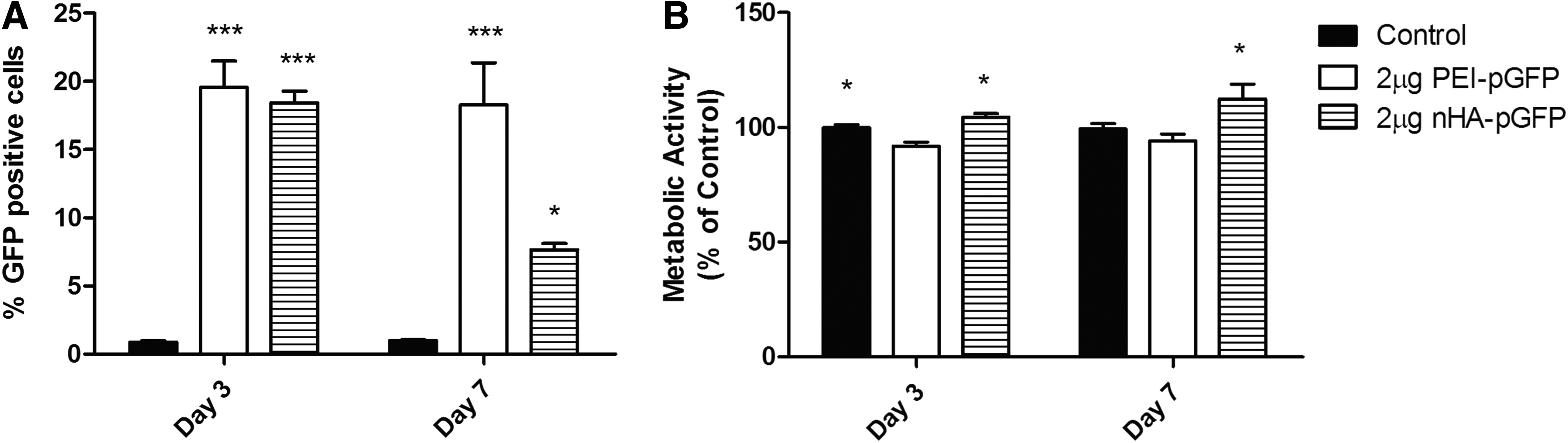
PEI and nHA demonstrated a comparable capacity to transfect BMSCs in monolayer culture, with ∼20% of cells GFP positive at day 3 in both groups. At day 7, the percentage of GFP-positive cells significantly decreased in the nHA–pGFP group but stayed constant in the PEI group (Fig. 1A). Assessment of cell viability using alamarBlue showed significantly higher cell metabolic activity in the untransfected control and the nHA–pGFP group in comparison to the PEI-transfected cells at day 3 (Fig. 1B), suggesting that PEI has a negative effect on the metabolic activity of MSCs. Increased metabolic activity observed in the nHA–pGFP group showed at day 7 compared to the control and PEI–pGFP constructs (Fig. 1B).
Alginate hydrogels are able to support nHA-mediated gene delivery and sustained expression of the transgene over time without negatively impacting cell viability
Quantification of luciferase expression confirmed effective gene delivery to MSCs in the gene-activated alginate hydrogels containing nHA complexed with different concentrations of pLuc, with sustained expression of the transgene over 14 days (Fig. 2A). The luciferase expression peaked at day 7 in all gene-activated groups (Fig. 2A), in comparison to 2D transfection in which the luciferase expression peaked at day 3, and was not significantly different from the untransfected control by day 7 (Supplementary Fig. S1; Supplementary Data are available online at www.liebertpub.com/tea). The use of 2 μg pLuc per gel appeared to be the optimal concentration of pDNA, as evidenced by significantly higher levels of luciferase expression in comparison to 5 and 10 μg pLuc at specific time points (Fig. 2A). Importantly, luciferase expression was also sustained over 14 days within the 2 μg pLuc per hydrogel group.

Fluorescent inverted microscope images of the gene-activated alginate hydrogels containing MSCs and nHA–pGFP complexes revealed the presence of green fluorescent cells inside the hydrogels, indicating effective transfection and GFP production over 23 days (Fig. 2B). Fluorescent staining of the cellular actin cytoskeleton and the nucleus of the encapsulated MSCs, and pDNA encoding for luciferase, showed naked pLuc and nHA–pLuc uptake over 72 h (Fig. 2C). pDNA could be observed inside the cell cytoplasm and nucleus of the MSCs in the nHA–pLuc group at 4, 24, and 72 h after encapsulation in alginate. In contrast, the pDNA was completely degraded after 72 h in the naked pLuc group, and it could not be observed inside the cellular compartment or around the cell membrane (Fig. 2C). Additionally, imaging of the nHA–pLuc group suggested the presence of stable nHA–pDNA complexes inside and outside the cell over 72 h posttransfection (Fig. 2C).
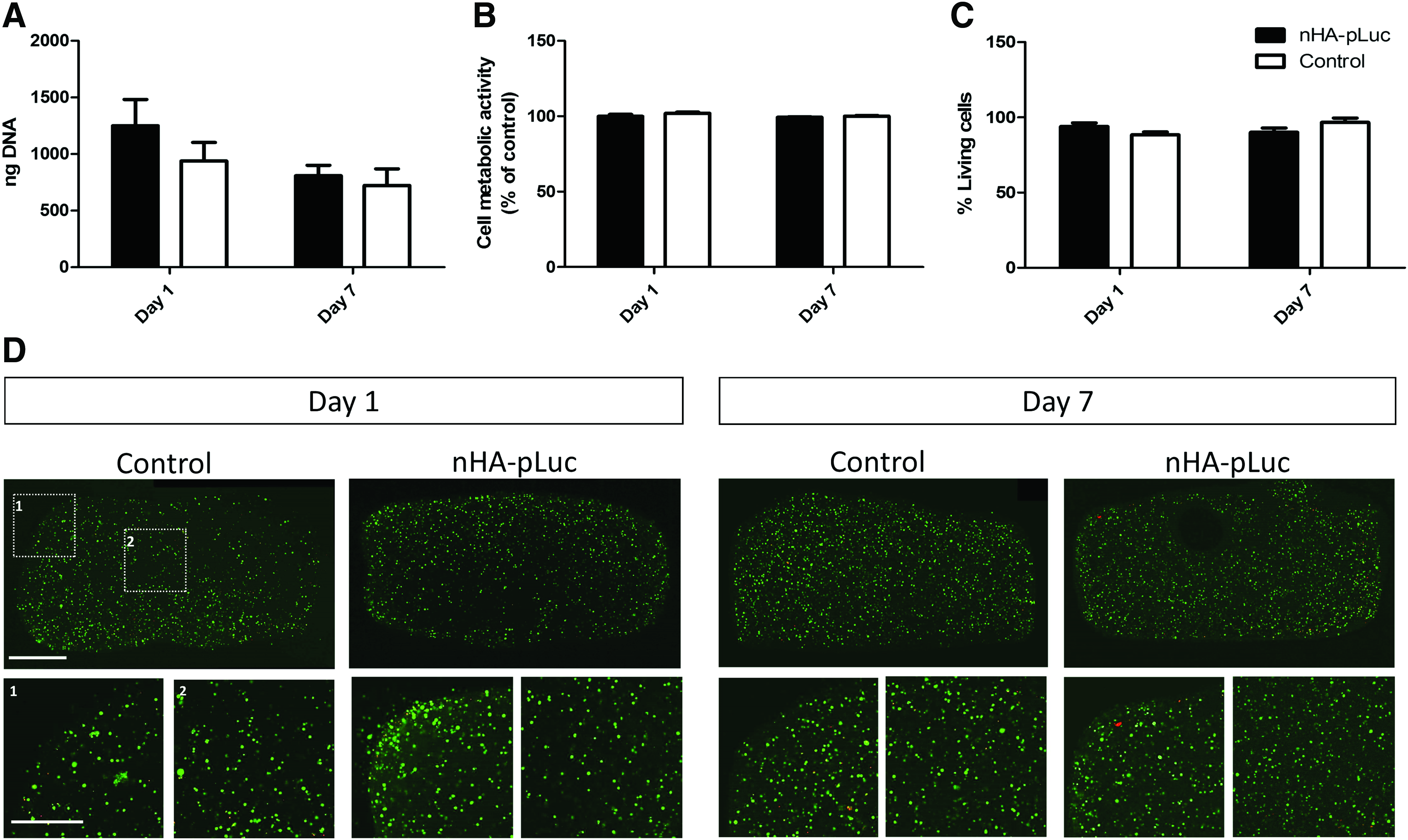
No significant differences were observed between the DNA content and the cell metabolic activity of the nHA–pLuc gene-activated hydrogels and the untransfected controls at days 1 and 7 (Fig. 3A, B), suggesting that cell viability was unaffected by the incorporation of the complexes into the alginate hydrogels. Confocal imaging of live and dead cells revealed a homogeneous distribution of the cells throughout each gel, with no obvious differences in cell viability between the groups (Fig. 3C, D).
Addition of therapeutic genes into 3D alginate hydrogels results in effective delivery and sustained expression of the transgene
Successful expression of therapeutic proteins was achieved using nHA-mediated transfection of MSCs within the alginate hydrogels. This resulted in increased levels of TGF-β3 (Fig. 4A) and BMP2 (Fig. 4B) in the culture media of the pTGF-β3, pBMP2, and pTGF-β3–pBMP2 codelivery groups (Fig. 4). The peak of expression was observed at day 7 for TGF-β3 and day 3 for BMP2, and a high reduction in the expression of the gene products was observed at day 14. These results verify effective MSC transfection with pTGF-β3 and pBMP2 within the gene-activated hydrogels and demonstrate sustained expression of the genes of interest. A similar trend and level of expression were observed for each protein, independent of whether the pDNA had been delivered in isolation or combination with another pDNA, implying that codelivery of two plasmids did not hinder the expression of either gene.

Codelivery of TGF-β3 and BMP2 genes enhances chondrogenesis and suppresses hypertrophy and calcification compared to delivery of either gene in isolation
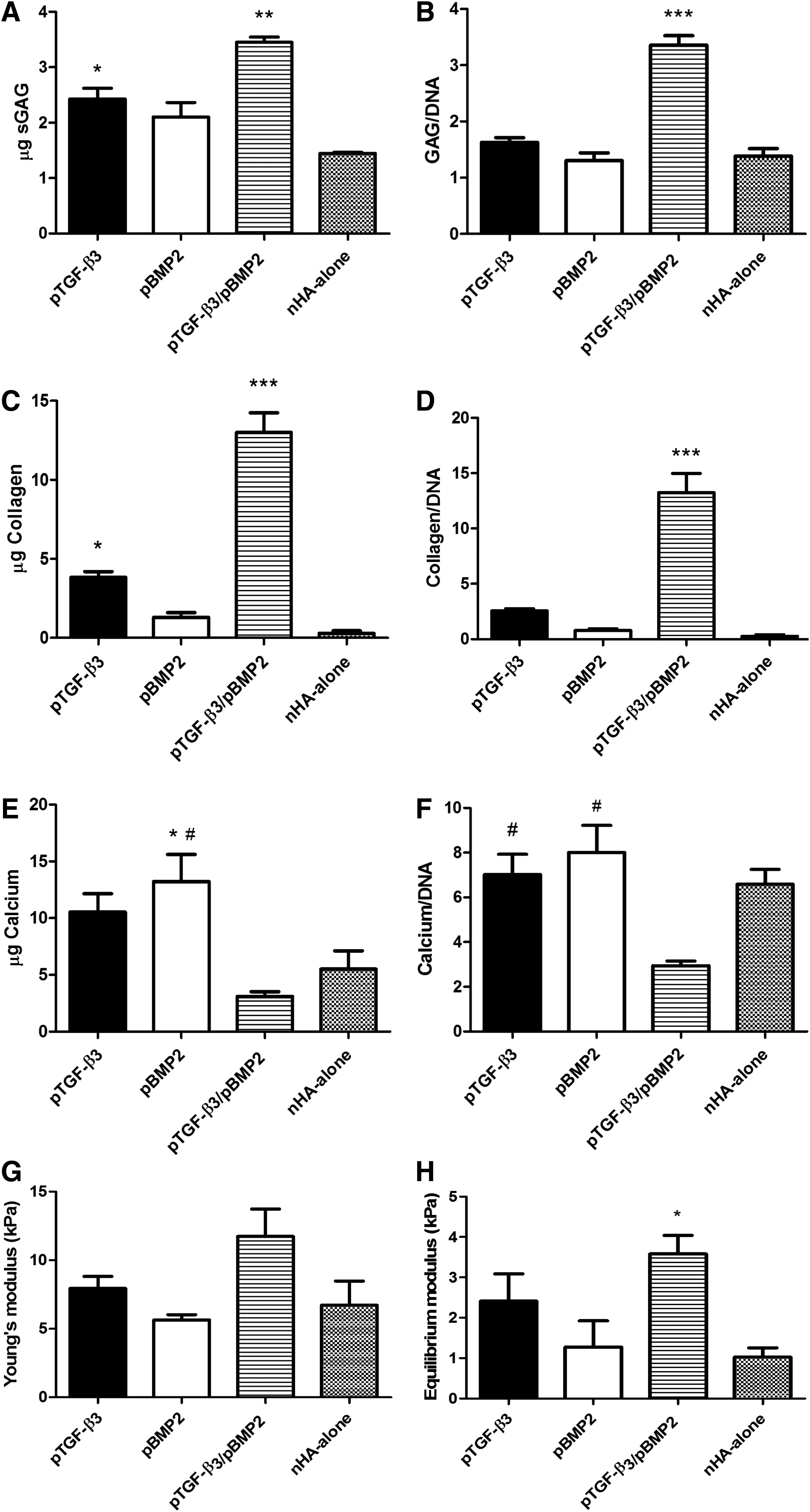
pTGF-β3–pBMP2 codelivery led to significantly higher levels of sGAG and collagen accumulation compared to all other groups after 28 days of culture (Fig. 5A–C). Collagen accumulation in the pTGF-β3 group was significantly higher than that in the pBMP2- and nHA-alone groups (Fig. 5C). Similar trends were found when the sGAG and collagen content were normalized to the DNA levels (Fig. 5B, D). In contrast, significantly higher levels of calcium deposition were observed in both the pTGF-β3- and pBMP2-only groups in comparison to the codelivery of both genes (Fig. 5E, F). At the levels of nHA used within this study, no evidence of osteoinductivity or calcification was observed in an nHA-alone control group (Fig. 5E, F).
Biochemical and mechanical analysis of the constructs after 28 days of in vitro culture.
The equilibrium modulus (Fig. 5H) of the pTGF-β3–pBMP2 codelivery group was significantly higher than that of the nHA-only control after 28 days of in vitro culture, with a similar trend observed for the Young's modulus (Fig. 5G).
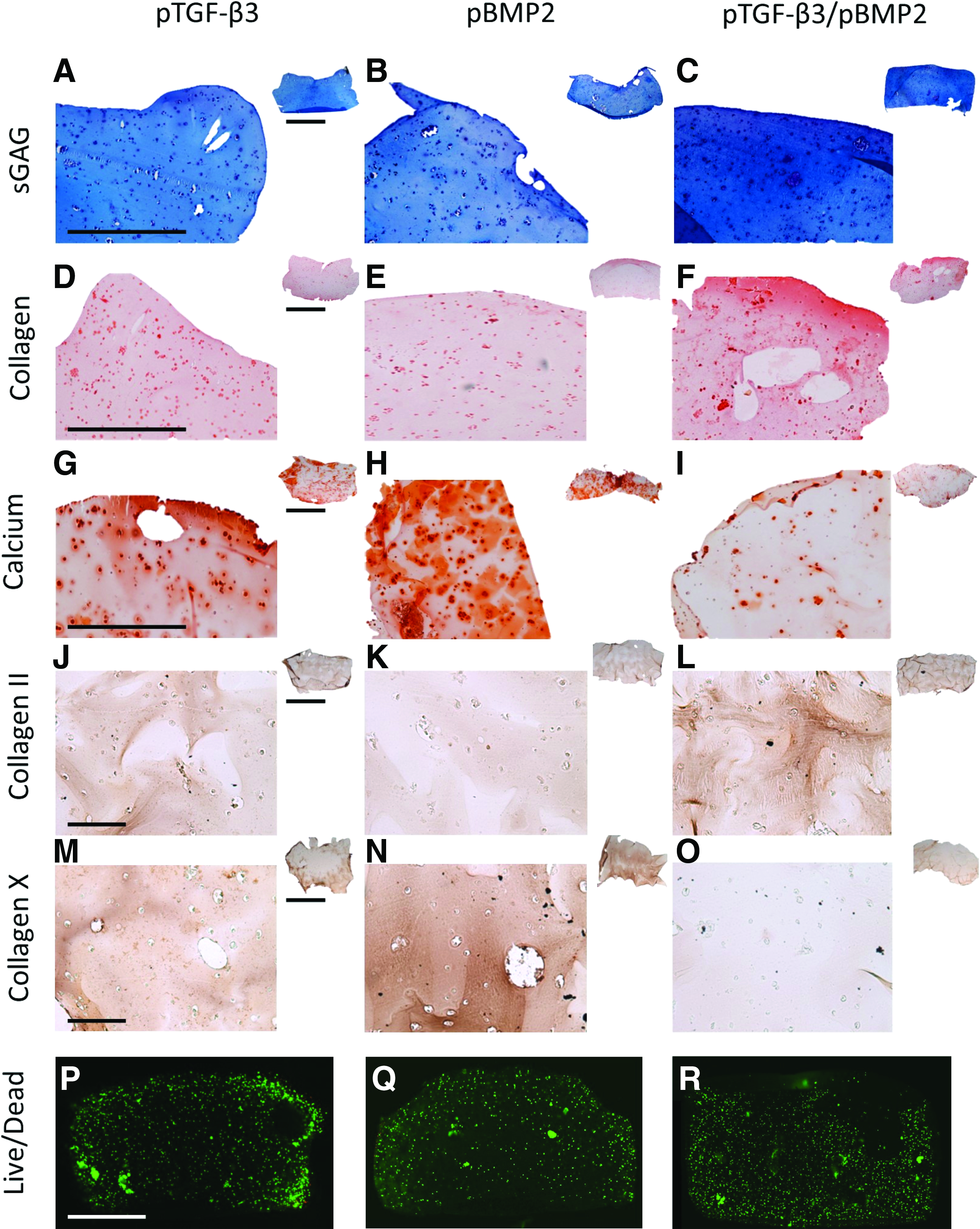
Histological evaluation of the hydrogels after 28 days in culture demonstrated higher pericellular sGAG and collagen type II deposition in the pTGF-β3–pBMP2 codelivery constructs compared to all other groups (Fig. 6). Greater calcification (Fig. 6G, H) was observed in the pBMP2- and pTGF-β3 delivery-only groups. Aldehyde fuchsin/alcian blue and picrosirius red staining revealed higher sGAG (Fig. 6A–C) and collagen accumulation (Fig. 6D–F) in the pTGF-β3–pBMP2 codelivery group, confirming the results of the biochemical analysis. Minimal immunostaining for collagen type X, a marker of hypertrophy, was seen in the pTGF-β3–pBMP2 codelivery group compared to the pTGF-β3- and pBMP2-only constructs (Fig. 6M–O), with the most intense staining in the pBMP2 group. These results suggest enhancement of chondrogenesis in the codelivery group and the promotion of hypertrophy and calcification when TGF-β3 and BMP2 genes are delivered in isolation using nHA as a delivery vector. Confocal imaging of live and dead cells confirmed the presence of living cells in all the groups after 28 days of in vitro culture (Fig. 6P–R).
Histological examination of the gene-activated hydrogels and the controls after 28 days of in vitro culture.
Discussion
The overall goal of the present study was to develop a novel nHA-mediated gene-activated alginate hydrogel capable of supporting sustained delivery of pDNA encoding for therapeutically relevant factors to MSCs to control their differentiation for either articular cartilage or endochondral bone tissue engineering. Luciferase expression analysis, fluorescent imaging, and cell viability assessment confirmed effective gene delivery to MSCs encapsulated within the gene-activated hydrogels without any toxic effects. Therapeutic gene delivery of TGF-β3 and BMP2 and subsequent expression of the transgenes promoted sGAG and collagen type II production when the two genes were delivered in combination or calcification and collagen type X deposition when these genes were delivered in isolation. Taken together, these results indicate that the nHA gene-activated hydrogels are able to efficiently sustain nonviral transfection and expression of the genes of interest over a temporary time frame, providing relevant concentrations of proteins that modulated the phenotype of MSCs either toward a cartilage or down an osteogenic/endochondral pathway depending on whether TGF-β3 and BMP2 were delivered in combination or isolation.
In-house synthesized hydroxyapatite (HA) nanoparticles showed transfection efficiencies similar to PEI but without the cytotoxicity associated with this cationic polymer. nHA nanoparticles have been investigated as delivery vectors for diverse biomedical applications, including growth factors, 35 anticancer drugs, 36 enzymes and antibodies, 37 and nucleic acids, such as pDNA38,39 and small interfering RNA (siRNA). 40 In terms of gene delivery, nHA nanoparticles have a high binding affinity for pDNA due to the interactions between the calcium ions in the apatite and the negatively charged phosphate groups of DNA. 41 Previous studies have reported MSC transfection efficiency to be higher using nHA than using commercial calcium phosphate (CaP) kits and to offer better cell viability than Lipofectamine 2000, 39 the most referenced transfection reagent. Additionally, nHA-mediated gene delivery within collagen scaffolds has shown greater therapeutic benefits for bone regeneration in vivo in comparison to PEI, which is often considered the gold standard for cationic gene delivery. 42 Although relatively low levels of nHA-mediated transfection efficiency were reported in this study in comparison to viral vectors, such as recombinant adeno-associated viruses (rAAV),43,44 or physical cell stimulation methods, such as electroporation, 45 the use of nonviral particles for gene delivery is a promising option for their application in tissue engineering approaches due to their low immunogenicity, transient effect, and the ease of incorporation to 3D matrices for a sustained and localized delivery of the gene of interest.
In this study, these nHA nanoparticles were incorporated into alginate hydrogels to facilitate and sustain gene delivery to MSCs over time. This resulted in effective transfection with pDNA encoding for Luc and GFP and subsequent expression of the transgenes over 23 days as confirmed by fluorescent imaging and Luc analysis. Luc expression peaked at day 7 in the gene-activated hydrogels, suggesting a sustained uptake of the nHA–pLuc complexes over time, in comparison to the transfection with pLuc or pGFP in 2D monolayer culture, where expression peaked at day 3 and showed a significant reduction by day 7. Different pLuc concentrations (2 μg, 5 μg, and 10 μg/gel) were also assessed. Two micrograms per gel was found to be the optimal in terms of gene expression, with significantly higher Luc expression at every time point, which may be due to the formation of larger complexes with increasing concentrations of pDNA preventing cellular uptake. 46 Cytotoxicity analysis of the system showed similar levels of DNA content, metabolic activity, and percentage of living cells across the gene-activated groups and the untransfected controls, confirming the monolayer results and previous studies that highlighted the nontoxic effects of these nanoparticles, 39 making them ideal for many tissue engineering applications.
To track the pDNA uptake in the gene-activated hydrogels and better understand the efficacy of the nHA nanoparticles in the system, the internalization of pDNA was monitored through fluorescent staining at 4, 24, and 72 h after alginate encapsulation of the nHA–pDNA complexes and MSCs. Confocal imaging of the fluorescent-labeled pDNA, cellular cytoskeleton, and cellular nucleus showed that the combination of nHA nanoparticles and pDNA resulted in formation of pDNA complexes, sustained cellular internalization, and protection of the internalized pDNA overtime inside the alginate hydrogels. In contrast, in the absence of the nHA nanoparticles, the naked pDNA was not able to be internalized by the MSCs, and it was not been observed in the cells or their periphery after 72 h, suggesting degradation of the plasmid. HA particles have been previously reported to protect pDNA from degradation driven by serum and nucleases, such as DNase I,47,48 and these results further demonstrate this protective action. The presence of nHA also resulted in effective gene transfection, confirming that these nanoparticles acted as a successful gene delivery vector in the developed system.
nHA nanoparticles were able to effectively deliver pDNA encoding for TGF-β3 or BMP2 in the 3D alginate hydrogels and sustain their expression over 14 days. Medium supplementation with growth factors from the TGF family, such as transforming growth factor-beta 1 (TGF-β1) and beta 3 (TGF-β3) or BMP2, has previously been used to direct differentiation of MSCs encapsulated in alginate hydrogels toward either a chondrogenic or an osteogenic phenotype.9,49,50 Although relatively low levels of the transgene expression were quantified in the media (compared to studies that directly supplement the media with the gene product), it is likely that a significant proportion of the TGF-β3 and BMP2 proteins being produced is retained within the alginate construct, particularly within the pericellular matrix deposited by the encapsulated cells. Such matrix components have previously been shown to bind strongly with such growth factors. 51 In comparison to recombinant growth factor medium supplementation, this approach offers a continuous production of proteins of interest, which overcomes the limitations related to the short half-life and quick degradation of proteins in vivo and the adverse effects associated with the administration of supraphysiological amounts of growth factors. 11
Gene delivery of either TGF-β3 or BMP2 in isolation showed only moderate sGAG and collagen synthesis and significantly increased calcification in comparison to the pTGF-β3/BMP2 codelivery group. Furthermore, the delivery of these genes in isolation enhanced the production of collagen type X, a marker for chondrocyte hypertrophy and endochondral ossification. While it is perhaps unsurprising that the delivery of BMP2 appeared to support a more osteogenic phenotype,29,52 the finding that pTGF-β3 delivery supported a similar phenotype was less expected. It is likely that the combination of the osteogenic stimulus provided by the nHA, with the overexpression of pTGF-β3, led to promotion of hypertrophic/osteogenic differentiation in the MSCs. 53 In contrast, nHA-mediated codelivery of pTGF-β3 and pBMP2 resulted in suppressed calcification and collagen type X deposition and promoted a more stable chondrogenic phenotype characterized by increased GAG and collagen type II production.
Combined medium supplementation of either TGF-β3 or TGF-β1 with BMP2 has been shown previously to synergistically enhance chondrogenic differentiation of MSCs encapsulated in alginate beads.49,54 This synergistic effect on MSC chondrogenesis may be produced through modulation of the Smad and MAPK signaling pathways55,56 and suppression of Runx2 expression, 57 resulting in the promotion of collagen type II, cartilage oligomeric matrix protein (COMP), and aggrecan (ACAN) gene expression and sGAG synthesis and, at the same time, decreasing the expression of bone-specific alkaline phosphatase.49,54 While the present study shows that the codelivery of TGF-β3 and BMP2 produced more stable chondrogenesis and suppressed hypertrophy, Simmons et al. showed that the combination of TGF-β3 and BMP2 growth factors in peptide-modified alginate laden with MSCs led to higher bone formation compared to when either growth factor was delivered in isolation. 24 This could be due to the presence of peptide modification and/or the reduced molecular weight of the alginate used in that study. 58 Other factors to be considered are the relatively low concentrations of TGF-β3 and BMP2 produced by the MSCs in the gene-activated hydrogels and the hypoxic conditions of the in vitro culture, which have previously shown to suppress the hypertrophic phenotype of MSCs.59,60 The exact molecular mechanism by which combined delivery of TGF-β3 and BMP2 suppressed calcification in vitro and promoted chondrogenesis remains unclear. Its elucidation could help understand and modulate hypertrophy and endochondral ossification of MSCs and will be addressed in future studies.
In conclusion, the nHA gene-activated alginate hydrogels developed in this study were capable of sustaining gene transfection of MSCs, leading to transgene expression over at least 14 days of culture. The expression of the transgenes was capable of modulating stem cell fate toward either a chondrogenic or an osteogenic/endochondral phenotype depending on whether pTGF-β3 and pBMP2 were delivered in isolation or combination. This is the first study to show that nHA-mediated gene delivery is capable of inducing chondrogenic differentiation of MSCs and that alginate hydrogels can be used as gene delivery platform for both cartilage and endochondral bone tissue engineering. Altogether, these results may be of clinical importance for the treatment of osteochondral defects as this system offers control of MSC fate toward either a chondrogenic or an endochondral phenotype while avoiding the risks and drawbacks associated with recombinant protein administration.
Footnotes
Acknowledgments
Funding from Science Foundation Ireland through the Advanced Materials and Bioengineering Research (AMBER) center and an Investigator Programme grant (12/IA/1554), as well as through the European Research Council (StemRepair–E12406).
Disclosure Statement
No competing financial interests exist.
References
Supplementary Material
Please find the following supplemental material available below.
For Open Access articles published under a Creative Commons License, all supplemental material carries the same license as the article it is associated with.
For non-Open Access articles published, all supplemental material carries a non-exclusive license, and permission requests for re-use of supplemental material or any part of supplemental material shall be sent directly to the copyright owner as specified in the copyright notice associated with the article.