
Abstract
Stem cell differentiation is guided by contact with the physical microenvironment, influence by both topography and mechanical properties of the matrix. In this study, the combined effect of substratum nano-topography and mechanical stiffness in directing mesenchymal stem cell (MSC) chondrogenesis was investigated. Three polyesters of varying stiffness were thermally imprinted to create nano-grating or pillar patterns of the same dimension. The surface of the nano-patterned substrate was coated with chondroitin sulfate (CS) to provide an even surface chemistry, with cell-adhesive and chondro-inductive properties, across all polymeric substrates. The surface characteristic, mechanical modulus, and degradation of the CS-coated patterned polymeric substrates were analyzed. The cell morphology adopted on the nano-topographic surfaces were accounted by F-actin distribution, and correlated to the cell proliferation and chondrogenic differentiation outcomes. Results show that substratum stiffness and topographical cues affected MSC morphology and aggregation, and influenced the phenotypic development at the earlier stage of chondrogenic differentiation. Hyaline-like cartilage with middle/deep zone cartilage characteristics was generated on softer pillar surface, while on stiffer nano-pillar material MSCs showed potential to generate constituents of hyaline/fibro/hypertrophic cartilage. Fibro/superficial zone-like cartilage could be derived from nano-grating of softer stiffness, while stiffer nano-grating resulted in insignificant chondrogenesis. This study demonstrates the possibility of refining the phenotype of cartilage generated from MSCs by manipulating surface topography and material stiffness.
Introduction
M
To be more directed in generating cartilage of specific properties, extensive studies have been performed to investigate how stem cells respond to individual types of microenvironmental cues, especially at the level of cell–substratum interaction.16–18 MSC chondrogenesis can be influenced by the biochemical cues provided by the ECM composition,19–23 and also by the biophysical cues, which include mechanical stiffness24–26 and topographical features27–31 of the matrix. However, it is important to investigate how the complex interplay among these niche cues collectively influence stem cell fate and function, and to elucidate how stem cells respond to interactive niche signals.
Our previous study on nano-topographical influence has found MSC to differentiate to chondrocytes with specific phenotype depending on the nano-topographical patterning; with nano-grating inducing formation of fibro/superficial zone-like cartilage, while nano-pillar promoting hyaline-like cartilage. 31 The purpose of this study is to investigate the combined effect of substratum topography and mechanical stiffness in directing MSC chondrogenesis. To vary the mechanical property of the substratum, different polyesters, polycaprolactone (PCL), polylactide (PLA), and polyglycolide (PGA) constituting varying mechanical stiffness (PCL<PLA<PGA) were used. Thermal imprinting technology was employed to create precise nano-topographical patterns, with the same dimension of either grating or pillar patterns, on the three polyester films. The surface of the nano-patterned substrate was coated with chondroitin sulfate (CS) to provide an even surface chemistry, with cell-adhesive and chondro-inductive properties.19,31 The mechanical modulus and surface characteristic of the chondroitin sulfate-coated patterned surfaces were analyzed. The cell morphology adopted on the nano-topographic surfaces were accounted by F-actin distribution, and correlated to the cell proliferation and chondrogenic differentiation outcomes.
Materials and Methods
Preparation of nano-imprinted polymeric films
PCL, PLA, and PGA (PURAC Biomaterials, The Netherlands) solutions (10% w/v) were casted on the glass surface to form films of 150 ± 20 μm. Thermal nano-imprinter (Obducat AB Nanoimprinter), with silicon molds of inverse pattern of nano-grating (250 nm line, 250 nm space, and 150 nm height; Institute of Microelectronics) or nano-hole (225 nm diameter, 400 nm pitch and 300 nm depth; NIL Technology) were placed in direct contact with the polymeric films at an imprinting temperature of 80°C for PCL, and 120°C for PLA and PGA, at a pressure of 60 bar for a duration of 5 min. Films were demolded after system temperature was cooled to 25°C.
Chondroitin sulfate conjugation of the polymeric surfaces
The polymeric films were aminolyzed with 20% 1,6-diaminohexane/ethanol solution for 15 min, washed by phosphate-buffered saline (PBS), and treated with a carbodiimide solution (48 mM EDC and 6 mM NHS in 50 mM MES buffer, pH = 5.5) and 10% CS MES solutions (CS concentration at 10 mg/mL; Sigma-Aldrich) for 24 h at 37.5°C. The CS coupling reaction was allowed for 24 h to ensure saturation of all surfaces with CS according to our previous publication. 31 The amount of CS immobilized on the polymeric films was measured using Blyscan sulfated glycosaminoglycan assay kit (Biocolor Ltd, Newtownabbey, Ireland) following the manufacturer's protocol.
Scanning electron microscope of the nano-patterned polymeric films
The morphology of the CS-coated nano-patterned polymeric films was examined by using a scanning electron microscope (JEOL 5600).
Atomic force microscopy measurement of substrate compressive stiffness
A Nanoscope IV multimode atomic force microscopy (AFM) with a picoforce scanner (Digital Instruments, Inc.) and silicon nitride AFM cantilever (NovaScan) with spring constant of 0.01 N/m and a 4.5 μm diameter polystyrene bead tip was used for indentation. The apparent elastic modulus, E, was obtained using the formula: H × k = [4E(RD 3 )1/2]/[3(1 − v2)], where E is the apparent elastic modulus to be determined, v is the Poisson's ratio, R is the radius of the spherical bead, D is the indentation depth, H is the cantilever deflection, and k is the spring constant of the cantilever.
Surface wettability characterization
The hydrophilicity of the nano-topographic surfaces was determined by water contact angle measurement by a sessile drop method using a Water Contact Angle Locator (SL-200B; Solon information technology Co., Ltd.). A 5 μL droplet of water was placed on the surface of the films at room temperature, and the sample was measured at four corners and four center locations.
Attenuated total reflectance-Fourier transform infrared spectroscopy
The degree of CS coating to the different polymeric surfaces was qualified by attenuated total reflectance-Fourier transform infrared (ATR-FTIR) spectroscopy (ATR, Spectrum One; FTIR, PerkinElmer). The scanning was set from 500 to 4000 cm−1, light source set at 1 cm−1 and each sample received 16 scans. The data were exported and analyzed by machine software. The spectrum was graphically presented to show the peaks of different surface chemical properties.
Molecular weight determination
The polymeric films was incubated in PBS at 37°C in 5% CO2 atmosphere, with medium changed every 3 days, for up to 28 days. The average molecule weight of the polymeric films between day 0 (un-incubated) and day 28 was measured by Gel Permeation Chromatography (GPC) (Agilent 1260 GPC/SEC system, column: PLgel MIXED LS, detector: viscometry). Dried films were dissolved in chloroform: methanol (5:1) at concentration 10 mg/mL and the weight-averaged molecular weights (Mw) were determined, and degradation percentage and dispersity, based on the difference in molecular weight between day 0 and day 28 samples, were calculated by Agilent software.
MSC culture and chondrogenic differentiation
MSCs were generated from bone marrow aspirates of consented human donors (Institutional Review Board-NUS reference code: 08–097). Bone marrow cells were cultured in Dulbecco's modified Eagle's medium (DMEM; Invitrogen) supplemented with 10% fetal bovine serum (Invitrogen). After removal of nonadherent cells, adherent MSCs were further expanded and used between passage 3 and 5.
For chondrogenic differentiation studies, 25 μL MSCs suspended in expansion medium at 106 cell/mL were seeded onto 5 mm diameter of nano-topographic surface and allowed for attachment at 37°C for 2 h. Attached cells on the nano-topographic surfaces were incubated in expansion medium for 24 h before subjecting to chondrogenic differentiation condition following our published protocol. 31
Cell proliferation analysis
Cell proliferation was performed from day 1 to 7 as our previous publication has shown that with the seeding condition, proliferation proceed only within the first week of culture. 31 Cell proliferation was determined by DNA quantification using Hoechst Dye 33258 solution (Sigma) following manufacturer's protocol. Fluorescence was determined using a FLUOstar Optima fluorescent plate reader (BMG Labtech, Offenburg, Germany) at 350 nm excitation and 445 nm emission. DNA concentration was extrapolated from standard curve generated using calf DNA.
F-actin staining
Chondrogenic induced MSCs on polymeric films at day 3 were fixed in 4% paraformaldehyde, incubated in 0.1% Triton X-100, then TRITC-conjugated Phalloidin (diluted 1:500 in PBS; Invitrogen). Nucleus were counterstained with 50 ng/mL of 4′,6-diamidino-2-phenylindole (DAPI). The images were taken by laser confocal microscopy (Olympus FV-1000). The F-actin length was quantitated using ImageJ. Day 3 was chosen to analyze cell morphology as cells at this early time point remained mostly as nonaggregated cells, which allow fluorescence analysis of F-actin fiber length per cell.
Type I and II collagen quantification
The levels of type I and type II collagen were analyzed by ELISA following the manufacturer's protocols (Chondrex, Redmond, WA).
Real-time polymerase chain reaction analysis
Real-time polymerase chain reaction (PCR) analysis was according to our published protocol. 31 Real-time PCRs for GAPDH, aggrecan, COMP, collagen I (Col 1), collagen II (Col 2), Col 9, and collagen X (Col 10) were conducted using the SYBR green system (primer sequences as previous publication). 32 PRG4 and cartilage intermediate layer protein (CILP) were determined with the customized TaqMan probe-based gene expression system (Applied Biosystems). Expression of the target gene were normalized to GAPDH, and presented as fold changes with reference to the undifferentiated MSC.
Statistical analysis
All assays were repeated a minimum of three times. Data were analyzed using SPSS 10.0 software. Standard deviation was calculated by excel software. Statistically significant values were defined as p < 0.05 based on one-way analysis of variance (ANOVA and T-test).
Results
Fabrication of nano-pattern of differing stiffness
Films of different polyesters, PGA, PLA, and PCL, were imprinted with nano-pillar or nano-grating topographical patterns and surface-treated with CS. Scanning electron microscope (SEM) images showed that the dimensionality of the pillar and grating was kept the same across the three polymers (Fig. 1).
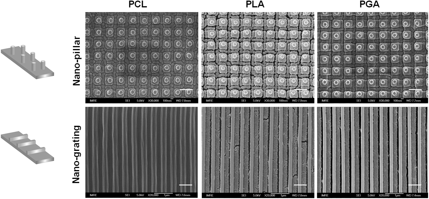
SEM images showing nano-patterned topographies on different polymeric films after chondroitin sulfate conjugation. The scale bars in the images are 500 nm. SEM, scanning electron microscope.
The compression modulus of the nano-patterned surfaces was determined in comparison to the non-patterned surface by AFM indentation. The compression modulus of nonpatterned PGA, PLA, and PCL films were ∼204, 128, and 62 MPa, respectively. Figure 2 shows that nano-patterning lowered the surface compressive modulus of the polymeric films. Although slightly higher compression modulus was detected on nano-grating than nano-pillar of the same material across the three polymeric materials, the compression range across the three different polymeric materials was maintained.

Measurement of compressive stiffness of nano-patterned and non-patterned polymeric films by AFM indentation. Average compressive moduli are listed in the table. AFM, atomic force microscopy.
Characterization of the nano-patterned surfaces
ATR-FTIR spectroscopy analysis before CS conjugation shows similar FTIR peaks across the three polymeric surfaces, with a peak around 1735 cm−1 representing the C = O stretch of polyester (Fig. 3A). After conjugation to CS, the surface chemical properties were similarly changed across the three polymeric surfaces, with the formation of a peak around 1615 cm−1, representing the N–H bend of CS molecules, while the polyester C = O stretch was not detectable, indicating saturation of all three polymeric surfaces with CS treatment.

The wettability of a surface represents the hydrophilicity of the material surface. The wettability of the nano-patterned surfaces, after CS treatment, was determined by contact angle measurement (Fig. 3B). Nano-patterning, in general, increased the wettability of the polymeric surfaces, as indicated by the decrease in contact angle. Slight difference in wettability between the two patterns of the same polymeric surface was detected (P PCL = 0.042; P PLA = 0.035; P PGA = 0.045), however, no significant difference was detected across the same nano-topography of different materials.
Given that the surface characteristic of the three polymeric nano-patterns was similar after CS-conjugation, the PGA, PLA, and PCL material will be referred as Stiff, Medium, and Soft material, respectively.
Degradation of the polymeric films
The degradation rate of the three polymeric films was determined by the change in molecular weight of the films after incubation in PBS at 37°C for a period of 28 days. Table 1 shows that after 28 days of incubation, the molecular weight of PGA has reduced much faster than PLA and PCL, with PCLs reduction the slowest. The morphology of the nano-patterns after 28 days of incubation was examined to assess the change in the pattern morphology. The SEM images (Fig. 4) reveal more pronounced alteration in the PGA nano-grating morphology, with localized portions of the gratings fused, constituting ∼5% of the surface. PLA and PCL grating surfaces remained largely intact. Nano-pillars on all three polymers remained intact, with no obvious difference across the three materials, even though PGA and PLA films became more brittle during the preparation of SEM process.

SEM images showing nano-patterned topographies on different polymeric films after 28 days of incubation.
PCL, polycaprolactone; PLA, polylactide; PGA, polyglycolide.
The characteristic of the CS-coated polymeric surfaces was analyzed after incubating the films in chondrogenic media at 37°C for a period of 6 weeks. Wettability of the three polymeric surfaces remained unchanged over the 6 week period compared to the day 0 samples (Fig. 5A). Amount of immobilized CS on the surfaces was intact at week 1, with detectable decreased at week 4, and significant decrease at week 6 (Fig. 5B). Amount of CS across all three polymeric surface remained similar at all measured time points. ATR-FTIR spectroscopy analysis at week 4 shows changes in surface chemistry on the three polymeric surfaces compared to the newly coated surfaces, with the detection of FTIR peaks at around 1609–1629 and 1735 cm−1 (Fig. 5C). Appearance of the 1735 cm−1 peak, representing C = O stretch of polyester, indicates the erosion of CS coating.

Proliferation of MSC on nano-patterned surfaces
MSC proliferation profile on different nano-patterned polymeric surfaces under chondrogenic differentiation conditions was determined by DNA quantification. Figure 6 shows that cells on the same nano-patterned surfaces have similar growth rates regardless of the stiffness of the material. MSCs on nano-grating surfaces have significantly higher growth rate than cells on nano-pillar surfaces. After 7 day culture in chondrogenic medium, DNA amount on nano-grating surface increased by ∼2-folds, while those on nano-pillar by ∼1.5-folds, compared to day 1.
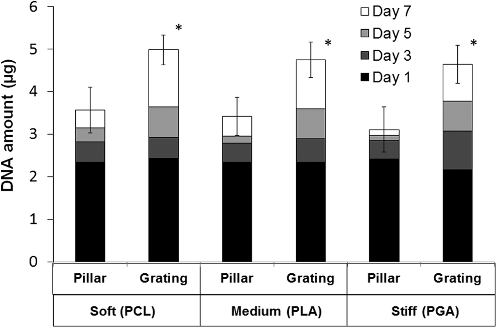
Cell proliferation profile on nano-patterned surfaces over a period of 7 days in chondrogenic differentiation conditions. *p < 0.05, significant different in DNA amount between nano-pillar and nano-grating of the same polymer.
Cell morphology adopted on nano-patterned surfaces
To study the influence of nano-pattern and material stiffness on cell morphology, F-actin distribution of MSCs was investigated after 3 days culture in chondrogenic medium. Phalloidin-stained images of single MSC show formation of fibroblastic cell morphology with F-actin stress fibers on all three nano-grating surfaces (Fig. 7A). MSCs on the Soft and Medium nano-pillar adopted round morphology with their F-actin organized at the cortical, while MSCs on the Stiff nano-pillar formed polygonal morphology with fibrous F-actin. Multiple cell analysis showed apparent cell aggregation and condensation by day 3 on the Soft and Medium nano-pillars, but not on the Stiff nano-pillar, whereas aligned fibroblastic cells on nano-gratings were observed (Fig. 7B). Quantification of the F-actin shows significant increase in fiber length in cells on nano-grating relative to nano pillar of the same polymeric surface (Fig. 7C). Cells on Stiff nano-pillars acquired less than twofolds longer F-actin fibers than cells on the two softer nano-pillars. Significant increase in fiber length was also detected in cells on Stiff nano-grating, compared to cells on the two softer nano-gratings.

Cell morphology analysis by F-actin phalloidin staining on different nano-patterned surfaces after 3 days of culture under chondrogenic condition. Representative images of single cell
Quantification of type I and II collagen expression
The amount of collagen generated by MSC-derived chondrogenic cells on different polymeric nano-patterns was measured by RNA analysis and ELISA quantification. With the same polymeric material, differentiated MSCs on nano-pillar surfaces consistently expressed higher amount of Col 2 compared to cells on the nano-grating surfaces. Real-time PCR RNA analysis (Fig. 8A, at week 2 and 4) and protein quantification (Fig. 8B, at week 4 and 6) indicate that the expression of Col 2 on nano-pillar significantly decreased on the stiff material. Differentiated MSCs on nano-gratings expressed significantly higher amount of Col 1 genes (Fig. 8A) and protein (Fig. 8B) across all three polymeric surfaces, and Col 1 formation was further enhanced on the stiffer PGA material (Fig. 8B). Notably, Col 1 expression remained low on softer nano-pillars but increased by two- to three-folds on the Stiff nano-pillars.

Quantification of type I and II collagen expression.
PCR analysis of cartilaginous genes
Expression levels of cartilaginous markers were analyzed by real-time PCR with cells differentiated for 4 and 6 weeks. Figure 9 shows that the Soft and Medium nano-pillars induced similarly higher levels of aggrecan when compared to levels expressed on nano-grating of the same materials. The differences between nano-pillar and nano-grating on the same polymeric materials were significant. However, the aggrecan expression on the Stiff nano-pillar and grating was at level five- to six-folds lower than those on the nano-patterns of the two softer materials. No significant difference in aggrecan expression was detected between the Stiff nano-grating and pillar pattern (P PGA = 0.67). Expression of middle/deep zone cartilage markers, CILP, COMP, and Col 9, were upregulated on Soft and Medium nano-pillars, but at significantly reduced levels on the Stiff nano-pillar (in both week 4 and 6 samples), in which expression levels was similar to those on all the three nano-grating surfaces. Analysis of the specific superficial zone cartilage marker, PRG4, showed that PRG4 mRNA was highly upregulated on Soft and Medium nano-gratings at week 6, while the Stiff nano-grating induced much lower levels (fourfolds lower). No significant PRG4 induction was detected on all nano-pillar surfaces. Quantification of hypertrophy marker, Col 10, shows expression enhanced significantly on the Stiff nano-pillar, at week 6.

Expression of cartilaginous genes on nano-patterned surfaces. Real-time PCR analysis of mRNA expression levels of cartilaginous genes after 6 weeks of chondrogenic differentiation. Data shown are mean ± SD, n = 3. *p < 0.05, significant different in Stiff samples compared to Soft or Medium samples of the same pattern. White bars = Soft (PCL); gray bars = Medium (PLA); and black bars = Stiff (PGA).
Discussion
The effects of either mechanical24–26 or topographical cues27–31 on cell fate and chondrogenic differentiation have been studied. How these cues collectively influence stem cell chondrogenic differentiation remains unclear. The contribution of material stiffness with nano-topography on MSC chondrogenesis was reported in randomly oriented nanofibers. 33 However, variation in fiber stiffness was achieved by electrospinning, forming fibers of different thickness. The variation in fiber dimension would invariably, by itself, influence cell adhesion, morphology, proliferation, and differentiation,30,33–35 leaving uncertainty as to the relevant contribution of these cues on the observed cellular response.
In this study, specific nano-topographical patterning (pillars vs. gratings) of differing stiffness was formed on three polyester polymers of variable mechanical properties by using thermal imprinting technology. Saturated CS-coating of the nano-patterned surfaces minimized the difference in surface properties of the three polymeric materials, as indicated by ATR-FTIR analysis (Fig. 3A) and wettability measurement (Fig. 3B). Characterization of the CS-coated polymeric surfaces over 6-week culture period indicates the maintenance of the CS immobilization up to at least 1 week, while the surface energy, as indicated by the contact angle measurement, remained unchanged throughout this period (Fig. 5). Erosion of CS coating (Fig. 5) and significant difference in molecular weight change of the polymeric materials (Table 1) were detected at week 4. The exposure of variable pendent groups from the three polyester surfaces, and the possible alteration in spacing and concentration of CS molecules on the surfaces could thus likely contribute to cellular response at the later stage of differentiation, including the presentation of absorbed proteins from the media, interaction with the adherent cells, and would contribute to diverse differentiation outcome. In addition, the different degradation rate of the three polymers would likely change the size scale and mechanical properties of the nano-topography. However, the similarity of the surface chemistry, and integrity of the nano-pattern, at the initial 1–2 weeks of differentiation would have allowed us to investigate the influence of material stiffness alone, on the topographical-directed proliferation and chondrogenesis, at the early stage of MSC differentiation, which was highly dependent on cell morphological changes and aggregation during this period.32,33
As early as 3 days after seeding, MSCs on nano-grating surfaces adopted fibroblastic morphology with extensive stress fibers, regardless of substratum stiffness, with the formation of aligned cells (Fig. 7). Morphology of MSCs on nano-pillars, however, were more sensitive to the substratum stiffness, adopting round morphology on the softer materials and subsequent cell aggregation, while forming polygonal morphology on the Stiff material, with increased stress fiber length. The adoption of distinct cellular morphology and aggregation likely primed the MSCs for subsequent cellular condensation, leading to development of unique tissue phenotypes. Cellular condensation on the softer nano-pillars predisposed MSCs to form more mature hyaline cartilage, indicated by the high expression of Col 2, accompanied by low levels of Col 1 (Figs. 8 and 9). Upregulation of aggrecan, CILP, COMP, and Col 9 characterized the phenotype as middle/deep zone-like cartilage (Fig. 9).6,36 This is in agreement with reports that implicated the importance of cellular condensation in the earlier phase of MSC chondrogenic differentiation.32,37 The effect of nano-pillar induced differentiation outcomes was, however, offset with the stiffest PGA material. Formation of a more elongated polygonal cell morphology on Stiff nano-pillar correlated to a curtailment in Col 2 expression, and drastic reduced expression of aggrecan and middle/deep zone markers. In contrast, expression levels of Col 1 and Col 10 increased significantly on the Stiff nano-pillar, suggestive of the induction of a transitional osteochondral cartilage, of mixed phenotype with fibro, hyaline, and hypertrophy characteristic. Formation of elongated MSCs on the softer nano-gratings potentiated MSCs to form fibro/superficial-like cartilage, with promotion of high PRG4 and Col 1 expression, lower aggrecan levels, while supressing Col 2 and middle/deep zone cartilage markers. On the other hand, insignificant aggrecan and PRG4 expression indicated the formation of noncartilaginous tissue on the stiffest nano-grating topography. The exposure of variable pendent groups from the three polyester surfaces and the alteration of patterns and mechanical properties with degradation of the polymeric substrates could have contributed to the differentiation outcome and heightened phenotypic characteristic at the later stage (week 4–6). However, given that the development of phenotypic characteristic between the two patterns and across the three stiffness, as indicated by Col 2/Col 1 levels, was apparent at week 2 and persisted to week 4, it is thus conceivable that stiffness have played a significant role in determining this development.
Differentiation outcomes on substratum of high stiffness, with fibro and hypertrophic cartilage characteristics promoted on the stiffest nano-pillar, while fibrous tissue resulted from the stiffest nano-grating, were similar to reports of MSC chondrogenic differentiation in hydrogel of tunable mechanical properties. A decrease in matrix output and induction of tissue calcification, 26 and the formation of fibrocartilage or fibrous tissues 25 was reported with increasing hydrogel stiffness. A determinant effect of substrate stiffness was also report by Wang et al., in a study to elucidate the interactive biochemical and mechanical signaling on chondrogenesis. 38 Effect of ECM composition on chondrogenesis is dependent on the matrix stiffness of hydrogels; with soft hydrogels augmenting chondrogenesis in a dose-dependent manner, while the trend was reversed with higher stiffness.
MSCs under chondrogenic medium showed significantly higher growth rate on nano-grating surfaces than those on nano-pillars at the early stage of differentiation (day 1–7). However, unlike differentiation, proliferation rate remained similar across all three polymers of the same topography. This is contrary to studies that showed that MSCs on soft substrates have less spreading, fewer stress fibers, 24 and lower proliferation rates than MSCs on stiff substrates.39–41 Li et al. study on the interactive effect of substrate stiffness, topography, and dimension suggested that both stiffness and dimension, but not topography, affect MSC proliferation. 42 Studies on the effect of stiffness to stem cell proliferation employed stiffness at the range of kPa to less than 5 MPa,39–41 much lower than the stiffness range used in this study. Our results indicate that the adaptation of distinct cell morphology on specific nano-pattern was probably the deciding factor in regulating proliferation.
Matrix stiffness regulates MSC differentiation toward bone, muscle, or neuronal lineages when grown on substrates that are akin to the native stiffness of their respective tissues.40,43 The equilibrium compression modulus of an adult cartilage is at ∼1 MPa, with dynamic compressive stiffness at ∼10 MPa.44,45 The compressive modulus of the PCL and PLA nano-topographies was in the 20 and 80 MPa ranges, well above that of the native articular cartilage. The compression stiffness of the materials in our study was determined by AFM indentation using a 4.5 μm microspherical tip, applied vertically over several patterns on the topographic surface. However, horizontal traction force would be exerted on individual pattern by cells upon adhesion, and during migration, on the patterned surface. Such traction force would be governed by the horizontal bending rigidity of the materials, which could differ drastically from the compression modulus obtained by vertical exertion, especially so for micro/nano-patterning surface. The study of micrometer and submicrometer (2–0.5 μm) pillars showed that the axial compression stiffness is much greater (100-folds different) than pillar bending rigidity. 46 On the other hand, the difference of axial compression stiffness and bending rigidity of grating is not as great, with the bending rigidity at 10% that of axial compression. 47 It is likely that the bending rigidity of nano-pillars for both PCL and PLA materials was in a softer range than that of compressive modulus measured. We speculate that softer PCL and PLA pillars could be bent by MSCs allowing the cells to adopt the round morphology akin to the study using elastomeric microposts, 48 while rigid PGA pillars would be less bendable, causing the attached MSCs to adopt a spread polygonal shape, modulating subsequent cytoskeletal restructuring, intracellular signaling, and differentiation outcomes that resulted in different cartilage phenotype. On the same note, although no significant difference in morphology was detected on cells across all three nano-grating surfaces, bending rigidity between PGA to that of PCL and PLA might have influenced the traction force experienced by the adhered MSCs and the resulting tissue generated.
In summary, we have shown that MSC morphology and aggregation was highly sensitive to both the substratum stiffness and topographical cues, and has the potential to influence the early differentiation direction of MSCs toward the specific cartilage phenotype. Such knowledge will be crucial in designing scaffolds for stem cell-based cartilage tissue engineering, or the derivation of phenotypically defined chondrocytes from MSC, for the generation of hierarchically organized articular cartilage.
Footnotes
Acknowledgments
We thank Prof. C.T. Lim from Mechanobiology Institute, National University of Singapore for the use of the AFM machine, and Dr. Feng Wen from School of Materials Science and Engineering, Nanyang Technological University, for assistance in ATR-FTIR microscopy analysis. This study was funded by the National Medical Research Council of Singapore (NMRC/CIRG/1403/2014) and University of Malaya HIR-MoE Grant (Reference No. UM.C/625/1/HIR/MOHE/MED/32 Account No. H20001-E000071).
Disclosure Statement
No competing financial interests exist.