
Abstract
Retracting hypertrophic scars resulting from healed burn wounds heavily impact on the patients' life quality. Biomaterial scaffolds guiding burned-out skin regeneration could suppress or lessen scar retraction. Here we report a novel silk noil-based three-dimensional (3D) nonwoven scaffold produced by carding and needling with no formic acid exposure, which might improve burn healing. Once wetted, it displays human skin-like physical features and a high biocompatibility. Human keratinocyte-like cervical carcinoma C4-I cells seeded onto the carded-needled nonwovens in vitro quickly adhered to them, grew, and actively metabolized glutamine releasing lactate. As on plastic, they released no proinflammatory IL-1β, although secreting tumor necrosis factor-alpha, an inducer of the autocrine mitogen amphiregulin in such cells. Once grafted into interscapular subcutaneous tissue of mice, carded-needled nonwovens guided the afresh assembly of a connective tissue enveloping the fibroin microfibers and filling the interposed voids within 3 months. Fibroblasts and a few poly- or mononucleated macrophages populated the engineered tissue. Besides, its extracellular matrix contained thin sparse collagen fibrils and a newly formed vascular network whose endothelin-1-expressing endothelial cells grew first on the fibroin microfibrils and later expanded into the intervening matrix. Remarkably, no infiltrates of inflammatory leukocytes and no packed collagen fibers bundles among fibroin microfibers, no fibrous capsules at the grafts periphery, and hence no foreign body response was obtained at the end of 3 months of observation. Therefore, we posit that silk noil-based 3D carded-needled nonwoven scaffolds are tools for translational medicine studies as they could guide connective tissue regeneration at deep burn wounds averting scar retraction with good functional results.
Introduction
S
Regeneration of lost skin, for example, following deep and extended burn wounds, involves complex events that result in scars, which by becoming hypertrophic and retracting compromise the working of the affected sites. To counter this, biomaterial scaffolds should cover the wounds to guide the proper engineering of the novel tissue with reduced or no scar hypertrophy and retraction. Such improved functional and esthetic upshots would positively impact on the patients' body image, coping capacities, mental conditions, social interactions, and working possibilities and significantly lessen healthcare costs.4–6
Ideally, biomaterial scaffolds for human tissue engineering should have a microfiber- or nanofiber-based organization according to the mechanical needs of the site considered. Such makeups should have optimized biophysical features (surface properties, gas and vapor permeability, mechanical strength, and pliability) and be biocompatible enough to avert immunological reactions and foreign body responses (FBRs). They might also release growth factor(s) improving tissue regeneration and/or drug(s) to suppress infection. Finally, a timely biodegradability would give a further benefit. Amid the candidate biomaterials, scaffolds based on microfibers or nanofibers of silk fibroin (SF) have stirred a growing interest because their properties match most of the just mentioned needs.7–13
SFs are natural proteins produced by wild and domesticated silkworms, wasps, honeybees, spiders, and ants.10,14 They share poly(Ala) or poly (Gly-Ala) repetitive sequences arranged in antiparallel β-sheets.11,14,15 SFs are biodegradable in vitro and in vivo,16–19
show good biocompatibility, and are only weakly inflammogenic.7,8,20,21 Genetically engineered SF variants exhibit improved properties.
22
Moreover, SF can be electrospun into pure nanofibers9,13,23–26 or blended or coated with other biomaterials, for example, hydroxybutyl chitosan, poly(
SF scaffolds are apt for engineering and regenerating soft tissues, such as nerves, skin wounds, and vascular grafts,47–58 and hard tissues, such as tendons, ligaments, cartilage, and bone.59–75 In these studies, mesenchymal stem cells from the bone marrow were often seeded onto the scaffolds to assess their differentiation into various connective tissue cell types or Schwann cells.12,53,75–78 Besides, SF scaffolds functionalized with bioactive peptides (e.g., RGD and BMP) improved cell adhesion, spreading, and proliferation.50,57,79
We have been focusing on SF scaffolds with nonwoven conformations aimed at skin connective tissue regeneration.7,8 The European Disposables and Nonwovens Association defines “nonwovens” as “a manufactured sheet, web or batt of directionally or randomly oriented fibers, bonded by friction, and/or cohesion and/or adhesion, excluding paper or products which are woven, knitted, tufted or stitch-bonded, incorporating binding yarns or filaments, or felted by wet-milling, whether or not additionally needled.” 80
A nonwoven fabric lacks yarns. Therefore, when a nonwoven fabric is torn or cut, no raveling occurs. In knitted or woven fabrics, yarns are likely to slip out at the edges. Under this respect, a nonwoven fabric is like leather or paper. At variance with conventional textiles, nonwovens have a 3D makeup of varying thickness. The fabric's geometry varies according to the unidirectional, bidirectional, or randomly multidirectional fiber orientation. Compared with usual textiles, nonwovens present several benefits such as lower material density, greater liquid permeability and/or retention, superior gas exchanges, and improved tear strength.
Nonwoven manufacturing needs three main steps: (1) preparation of a fibers-based web, (2) connection of the ingredient fibers, and (3) after-treatment(s) of the bonded fibers. At step (2) various methods may strengthen the web, such as exposure to chemical binders, carding, needle punching, hydroentanglement (spunlace), and stitch bonding, each of which produces a distinct nonwoven fabric. 81
Previously, we studied the physical features and biocompatibility of 3D SF-based formic acid (FA)-bonded nonwovens.7,8 Once grafted in mice, such nonwoven guided the de novo formation of a vascularized reticular connective tissue, and moreover interacted favorably with human keratinocytes and fibroblasts in vitro.7,8 However, looking for scaffolds with improved biophysical features, we explored the feasibility and properties of silk-based nonwovens made by the carding-needling method, thus avoiding the FA- or other chemical-mediated cross-linking of the SF microfibers. Here, we report the favorable features and connective tissue engineering capabilities of such novel nonwovens.
Materials and Methods
Production
A nonwoven fabric is made of randomly oriented individual fibers, with a texture characterized by the absence of any organized geometrical structure (like that of regularly interlaced warp and weft yarns in a woven fabric). Silk nonwovens were manufactured by using a laboratory scale drylaid system, with carding as a first processing step to form the web. The web was then mechanically consolidated by needling, which entangled the fibers by exploiting their frictional bonding strength. Short length silk fibers (silk noils) were used as starting material. These come from the so called spun silk processing cycle, which exploits all the silk materials (defective cocoons, reeling wastes, etc.) that cannot be processed as continuous filaments. 23
After scouring and cleaning, spun silk fibers, with a length from 10 mm to about 120 mm, are processed to combed sliver by drying, beating, opening, and combing. Silk noils, which represent the shortest fraction of spun silk fibers, with an average fiber length of 10–40 mm, were selected to manufacture several batches of lightweight, soft 3D carded-needled nonwovens whose pliability resembles that of skin. 82 Extraction with methanol cleaned the silk noils before use, followed by extensive washing with warm and cold water and drying at room temperature. Table 1 lists fiber length, thickness, and mass per unit area of this nonwoven product.
The tensile properties of both materials were tested under wet conditions.
FA-crosslinked nonwovens of the previous work 7 are stronger (value of stress is double), less elastic (value of strain is less than half), and much stiffer (value of initial modulus is fourfold greater) than the newly produced carded-needled silk nonwovens. Stiffness, that is, the low tendency to deform under a small applied load, makes the previous FA-crosslinked nonwovens less compliant with the mechanical behavior of native skin. 82
FA, formic acid; SF, silk fibroin.
Amino acid analysis
Amino acid composition of silk fibers was determined after acid hydrolysis with 6 N HCl, at 105°C for 24 h, under vacuum. Free amino acids were analyzed by HPLC (AccQ-Tag Method; Waters) at a 1.0 mL min−1 flow rate. Eluate was detected at 254 nm. The analysis was run in duplicate. External standard calibration (Amino Acid Standard H; Pierce) allowed assessment of quantitative amino acid composition.
Scanning electron microscopy
The morphology of 3D carded-needled nonwovens was examined in a Stereoscan 440 microscope (LEO Electronic Microscopy Ltd.). Samples were observed at 10 kV acceleration voltage after gold coating under reduced argon atmosphere with a Med 020 Coating System (BAL-TEC).
Physical properties and tensile measurements
Thickness and mass per unit area of 3D silk-based carded-needled nonwovens were measured according to UNI EN ISO 5084:1998 and UNI EN 12127:1999 standard methods, respectively.
Mechanical properties of 3D nonwovens were measured in the tensile mode on stripes of 100 × 10 mm (0.13 mm thick) by an Instron tensile testing machine mod. 4501 at 50 mm gauge length and 50 mm min−1 crossbar rate. Measurements were performed under standard conditions of temperature (20°C) and relative humidity (65%) on wet samples, soaked into distilled water for 24 h at 20°C before measurement. Stress, strain, and Young's modulus were calculated from the load–elongation curves as the average of 20 measurements.
Fourier transform-infrared spectroscopy
Measurements were performed on a NEXUS Thermo Nicolet Fourier transform-infrared (FTIR) spectrometer employing an attenuated total reflectance (ATR) accessory mod. Smart Performer with a SeZn crystal cell as internal reflection element. Spectra were recorded in the 4000–700 cm−1 wavenumber range by collecting 64 scans at a resolution of 4 cm−1.
Differential scanning calorimetry
Measurements were performed with a differential scanning calorimetry (DSC)-30 instrument (Mettler Toledo), from 18°C to 500°C, at a heating rate of 10°C min−1, on 3 mg samples. The open Al pan was swept with N2 during the analysis.
Nonwovens sterilization
Nonwovens were cut into square pieces (10 × 10 × 0.13 mm) that were sealed in sterilization pouches and sterilized by exposing them for 3 h at 55°C to a mixture of ethylene oxide/CO2 (10/90 v/v) under a pressure of 42 psi. Next, the specimens underwent a 24-h aeration at room temperature and a further 8-h degassing at 50°C in a vacuum oven. Sterilized nonwovens were systematically tested before use to assess the absence of any morphological change as well as the preservation of the mechanical properties. This is in keeping with the notion that native silk fibers with oriented β-sheet molecular conformation display a remarkably higher stability to current sterilization techniques than regenerated SF materials such as films, gels, porous scaffolds, and so on.
Cell cultures, seeding efficiency, and metabolic studies
Keratinocyte-like human-papillomavirus 18-transformed human cervical carcinoma C4-I cells 83 from our own Bio-Bank were used due to a momentary shortage of normal human keratinocytes. C4-I cells were plated in 175-cm2 plastic flasks (Sarstedt S.r.l.) and incubated at 37°C in 95% air/5% CO2 in complete medium consisting of 95% (v/v) Dulbecco's modified Eagle's minimum essential medium (DMEM; Sigma-Aldrich), 5% (v/v) heat-inactivated (56°C for 30 min) fetal bovine serum (Lonza AG), and gentamycin (0.1 mg mL−1; Lonza). Before reaching confluence, the cultures were split at a ratio of 1:6 by incubating them at 18°C ± 2°C with 0.025% (w/v) trypsin (Sigma-Aldrich). 84
Experiments were started by very slowly seeding 1.0 × 10⁶ cells suspended in 300 μL medium on sterilized square pieces (10 × 10 × 0.13 mm) of 3D carded-needled nonwovens placed in 13 mm diameter culture wells. 8 After 4 h, the nonwovens with the attached cells were transferred into new wells, each filled with 2.0 mL medium. The cell-bearing nonwovens underwent periodic checking under phase contrast or fluorescence optics. Every 2–3 days, changes of fresh growth medium substituted the cell/conditioned medium during the 15-day observation period. Cell-conditioned medium samples were stored at −80°C for subsequent biochemical analysis.
To assess cell viability through epifluorescence microscopy, before seeding, cells were intravitally stained with 1,1′-dioctadecyl-3,3,3,3′-tetramethylindocarbocyanine perchlorate (Dil-C18; Molecular Probes, Inc.), an intravital membrane phospholipid-binding probe (λex, 549 nm, λem, 565 nm), giving a bright orange-red fluorescence. 8 Dil-C18 did stain neither the cells' nuclear contents nor the nonwovens' silk microfibers.
To evaluate cell adhesion 4 h after seeding, the number of C4-I cells adhering to the nonwovens was indirectly assessed by an Alamar blue assay (Serotec). Standard absorbance curves were constructed using cultures on plastic plates, the number of cells of which were evaluated by phase contrast microscopy. 85
Lactate content of cell-conditioned growth medium samples was assessed through a lactate oxidase assay (Sigma).
ELISA of proinflammatory cytokines
Commercial ELISA kits (CLB) served to test human IL-1β and of tumor necrosis factor-alpha (TNF-α) in the cell-conditioned media. The sensitivity of both assays was around 0.8 pg mL−1.
In vivo surgical grafting
Four-week-old C57BL/6 mice (Charles River Italia, S.p.A.) were individually raised at 18–20°C in plastic cages on standard chow and water ad libitum with 12 h/12 h light/dark cycles. Before use, mice were bred for 1 week in the University's animal house. The experimental protocols were approved by the Committee for Animal Care and Use of Verona Medical School and carried out in keeping with its guidelines. Mice were anesthetized by intraperitoneally injecting 5.0 × 10⁻⁵ g per gram body weight of pentobarbital sodium (Euthatal; Merial Italia S.p.A.) as 1% (w/v) solution into the lower right or left quadrant of the abdomen. Ten minutes later mice were ready for surgery. Thirty mice were grafted with silk-based 3D carded-needled nonwovens by adapting the techniques applied to rats by Hafemann et al. 86
In short, the rostral portion of the interscapular skin was incised to excavate a 10 × 10 mm pocket in the subcutaneous tissue. Next, a 3D nonwoven sample of an alike size was implanted into the pocket and the wound stitched. Another 30 mice served as sham-operated controls: they underwent the same surgical procedure save for the nonwoven grafting step. A further 10 mice served as untreated controls. All the animals survived surgery and their postoperative course was problem free. Biopsy samples of the whole skin were taken from the operated sites of animals belonging to both groups and from untreated controls, half at 1 month and half at 3 months after the onset of the experiments.
Histology and immunohistochemistry
Tissue biopsies were instantly frozen using precooled (−80°C) isopentane in liquid N. 87 Frozen sections (5–10 for each of the 60 operated and 10 untreated mice) were cut at 10 μm thickness, picked up on aminopropyltriethoxysilane-coated slides, fixed for 20 min in acetone at −20°C, and dried. 88 Two methods served to stain sections. Mayer's hematoxylin and eosin with acid differentiation (1% HCl in 70% v/v ethanol) dyed nuclei (blue), collagen fibers (pink), and fibrous proteins (e.g., SF; red). 85 Gomori's rapid one-step trichrome method dyed nuclei (gray-blue), collagen fibers (green), and fibrous proteins (e.g., SF; magenta).7,88 All staining reagents were from Bio-Optica Milano S.p.A.
The immunohistochemistry (IHC) procedures were those of Jackson and Blythe. 87 After washing twice with phosphate-buffered saline–bovine serum albumin (PBS-BSA) (1.0% w/v)-NaN3 (0.1% w/v), primary antibodies (at 1.0 μg mL−1) applied for 60 min at 18–20°C tissue sections served to reveal the antigens CD4 or CD8 (T4 or T8 lymphocytes), CD22 (B lymphocytes), CD11b (macrophages), immunoglobulins (plasmacells), vimentin (fibroblasts and endothelial cells), collagen type-I (fibroblasts), or endothelin-1 (ET-1; endothelial cells) (all from Santa Cruz Biotechnology). Next, after washing thrice with PBS-BSA solution, specific secondary antibodies conjugated to alkaline phosphatase (at 1.0 μg mL−1; all from Santa Cruz Biotechnology) covered the tissue sections for a further 60 min at room temperature.
Specific IHC colors were developed with Fast Red AS-MX (Sigma-Aldrich) or diaminobenzidine (Dako Italia) tablet sets. Specimens were examined under an Olympus BX-60™ microscope and two to five pictures/section were taken with a QIClick CCD Colordigital camera (QImaging). Control tissue sections were always run in parallel with no primary antibody added.
Deconvolution of histological images from mouse skin tissue sections was performed using Huygens Professional X11 software (Scientific Volume Imaging) according to the seller's guide.
Statistical analysis
Data were mean values ± SEMs. One-way ANOVA followed by Holm–Sidak's post hoc test served to assess the statistical level significance among group means taking as significant values of p < 0.05.
Results and Discussion
Chemical and physical characterization of silk noil
Silk noils are a secondary product of the spun silk processing cycle, which uses silk materials that cannot be reeled as a continuous filament. 23 Specifically, silk noils are a short-length (mean <40 mm) fiber fraction recovered during combing spun silk slivers, from which the well-known “shappe” silk yarns are manufactured by carding, combing, and spinning technologies inspired to the cotton processing cycle. The silk noils sample used in this study had a mean fiber length of 25 mm (Table 1). Based on preliminary silk nonwoven manufacturing trials, this range of fiber length was the best compromise between processability of the silk noils by carding and needling technology, and the properties and performance of the resulting nonwovens (i.e., lightness, softness, strength, and pliability).
After the preliminary cleaning procedures, amino acid analysis characterized the chemical composition of the silk noils. The results listed in Table 2 show that pure SF makes the material, as shown by the high amounts of the three amino acids Gly, Ala, and Ser, which account for about 85 mol% of the protein composition. 24
nd, not determined.
The physical properties of silk noils were further investigated by FTIR-ATR spectroscopy and DSC analysis. Figure 1A shows the FTIR spectrum of silk noil fibers in the 1900–700 cm−1 wavenumber range. This spectral range is a fingerprint of SF because it contains various absorption bands strongly sensitive to the chemical and physical makeup of SF.89,90 The conformational sensitive bands at 1618/1695 cm−1 (amide I), 1510 cm−1 (amide II), and 1227/1261 cm−1 (amide III) arise from different vibrational modes of the peptide bond. Amide I is chiefly CO stretching plus CN stretching; amide II is mainly NH bending plus CN stretching; amide III contains NH bending and CN stretching vibrations.

Biophysical properties of silk noil and silk-based 3D carded-needled nonwovens.
Minor contributions from other vibrational modes of the peptide bond are also present. The two weak bands in the skeletal range at 997 and 975 cm−1 are specific of SF, being assigned to stretching modes, the repetitive Gly-Gly and Gly-Ala sequences of the fibroin chain, respectively. From the position and intensity of the amide bands, one can infer that SF has the characteristic β-sheet structure of silk in fiber form. 91 The crystallinity index, expressed as intensity ratio of the couple of amide III bands at 1261 and 1227 cm−1 (Ci = I1261/I1227), is 0.55, close to that of reference silk fibers (≅ 0.55–0.60). 92
The results of thermal analysis further confirm the nature, purity, and physical structure of silk noils. The DSC curve (Fig. 1B) reveals an intense endotherm peaking at 322°C, which matches the melting and decomposing of crystalline and well-oriented SF fibers with a β-sheet molecular conformation. 93 The low-temperature endotherm (<100°C) is due to the loss of moisture on heating.
Morphological and mechanical characterization of silk noil-based 3D carded-needled nonwovens
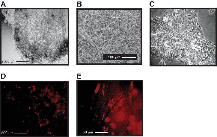
Scanning electron microscopy observations reveal the fine structure of these nonwovens (Figs. 2A, B, and 3B). Silk microfibers display a random arrangement forming a network limited by many interconnected voids. At first sight, the pictures appear similar to those of the FA cross-linked nonwovens.7,8 However, at closer inspection, the present nonwoven SF microfibers are thinner (diameter, 5–10 μm), and no film-like structures join them at cross points as no FA-mediated partial solution of the SF microfibers did occur. Besides, the void spaces among the thinner SF microfibers are roomier (Fig. 3A, B).

Physical features of 3D silk-based carded-needled nonwovens.
Adhesion and growth of keratinocyte-like C4-I human cervical carcinoma cells on silk-based nonwovens.
Notably, the preparative procedure is clean and easy to perform. The starting material is silk noils and the final textural features of the carded-needled nonwovens depend on the settings of the machines used. The present nonwovens are thin and light, as showed by the thickness and mass per unit area values reported in Table 1 (132 μm and 37 g sqm−1, respectively). The wide voids among the SF microfibers make it soft and elastic. Once wetted, it becomes even softer because of the lubricating and plasticizing action of water (just as would happen once grafted in vivo). Notably, the individual silk microfibers forming the nonwoven network display a smooth and clean surface.
No traces of sericin, the silk gum protein, surrounding the fibroin core of native microfibers or other contaminants remain, thus confirming the high level of purity of the starting material used to make these nonwovens.
A typical set of stress–strain curves of carded-needled nonwovens measured under wet conditions is shown in Figure 2C, whereas Table 1 summarizes its tensile properties. Nonwoven specimens showed a reproducible mechanical behavior under measuring conditions. The observed curves variability relates to changes in the local density of nonwovens, which is attributable to a slightly worse performance of the carding machine when short silk fibers are processed as the small microfiber neps characteristic of silk noils are difficult to open.
Under the measuring conditions, which is at a gauge length greater than fiber length, the stress–strain curve reflects the mechanical contribution of the texture of the nonwovens (i.e., the strength of the fiber entanglement), rather than of the individual silk microfibers, whose mean stress values are two orders of magnitude larger than those measured on the nonwovens. 94
The mechanical response of silk-based nonwovens to the applied stress is worth noting (Fig. 2C). The raising part of the curve, until it reaches the maximum value of stress, displays three distinct parts. A first phase where a large deformation exists at rather low stress matches the extending crimped silk fibers. A second intermediate region, where the silk fibers lining up in the stretching direction change the curve slope because the load augments the locally increasing fiber density, and makes the material stiffer and stiffer. A third high-tensile stress region where straight and compacted silk fibers slide relative to one another until complete separation. Finally, the stress–strain curve collapses suddenly when complete disentanglement of fibers occurs.
This mechanical behavior is remarkably alike to that reported for soft tissues, such as human skin, subjected to unidirectional tensile force. 82 Even more strikingly, the stress and strain values of the 3D carded-needled nonwovens (Table 1) fall in the range of the mechanical properties reported for human skin, which are 1–20 MPa for tensile strength and 30–70% for tensile strain. 82
These results are of great interest in view of the envisaged clinical application of the 3D carded-needled nonwovens. The mechanical response under wet conditions mimics that of the skin, thus ensuring a good biomechanical compliance at the implantation site. Concurrently, strength is enough to favor making and packing the nonwoven sheets, as well as handling and shaping the scaffold by the surgeon before and during implantation. All these features likely lead to high levels of mechanical and physiological integration of the scaffold, thanks to the recognized biological properties of SF fibers as a biomaterial. 10
Therefore, the present nonwovens represent a significant advance on the previous FA-crosslinked nonwovens.7,8 In fact, the latter are stronger (value of stress is double), less elastic (value of strain is less than half), and much stiffer (value of initial modulus is fourfold greater) than the novel carded-needled nonwovens. And stiffness, that is, the low tendency to deform under a small applied load, makes FA-crosslinked nonwovens7,8 much less compliant with the mechanical behavior of native human skin. 82
In vitro studies
A previous quantitative transcriptomics study carried out on nine human cervical carcinoma cell lines defined the human keratinocyte-like C4-I cells (Fig. 3C) as those most closely mimicking late-stage invasive in vivo cervical cancer. 83 To assess seeding efficiency, C4-I cells were seeded onto sterilized samples of the 3D carded-needled SF nonwovens in vitro and in plastic flasks. Four hours later, 58% ± 4% (n = 5) of the seeded cells had adhered to the SF microfibers as compared with 27% ± 3% (n = 5) that had stuck to the plastic surfaces (fibroin vs. plastic, +115%, p < 0.0003). Therefore, the adhesion of C4-I cells to the 3D SF nonwovens was more intense than to plastic surfaces. These findings agree with observations that various forms of SF exert proattachment effects on rodent and human cells.8,42,95,96
Being nearly spherical when just attached to SF microfibers (Fig. 3D), C4-I cells soon acquired a flatter morphology and by 24 h started proliferating to colonize the nonwovens (Fig. 3E). On these scaffolds, C4-I cells displayed an active metabolism consuming significant amounts of glutamine (and of glucose: not shown) (Fig. 4, top panel). As energy source glutamine is an important alternative to glucose 97 in human cervical carcinoma cells, such as HeLa cells 98 and C4-I cells, as well as in human fibroblasts and pluripotent stem cells.42,99

Metabolism of keratinocyte-like human C4-I cells cultured on 3D silk-based carded-needled nonwovens.
Concurrently, SF nonwoven-attached C4-I cells discharged significant amounts of lactate into the growth medium (Fig. 4, middle panel). Fibroblasts are able to metabolize both glutamine and glucose to lactate, which they next release.
100
C4-I carcinoma cells share the same metabolic ductility, the upshot of which is a favorable extracellular acidic environment.
101
In fact, plasmalemmal lactate receptor GPR81 is essential for cancer cell survival.
102
Besides,
Notably, inflammatory stimuli in the genital mucosal tract in vivo can induce an increased IL-1β expression in cervical intraepithelial carcinoma. 104 Therefore, our findings further confirm previous suggestions that SF is biocompatible toward human cells and not at all inflammogenic.7,8 Conversely, on 3D SF nonwovens or on plastic surfaces, C4-I cells similarly released TNF-α in discrete amounts (Fig. 4, bottom panel). Notably, this is a constitutive autocrine activity of these cells that is independent of the substrate they stick to and as such has no proinflammatory implication. In fact, once secreted, TNF-α induces the autocrine production of amphiregulin, a member of the epidermal growth factor family, in papillomavirus-transformed human cervical epithelial cells. 105
In vivo implants
To assess in vivo biocompatibility, we surgically grafted 10 × 10 × 0.13 mm samples of the silk-based 3D carded-needled nonwovens into the interscapular subcutaneous tissue of C57/BL6 mice, using sham-operated mice as parallel controls. As expected, both mice groups healed similarly within a few days with no unwanted side effects. The implants and the surrounding tissues were sampled 1 and 3 months after surgery to examine their histological features (Figs. 5 and 6C–H). Sham-operated mice at 1 and 3 months after surgery showed no local residual scar but only a slight increase in cellularity at the previous wound site versus nonoperated skin (Fig. 6A, B).

Endothelial cells and macrophages 1 and 3 months after grafting of silk-based nonwovens in vivo.
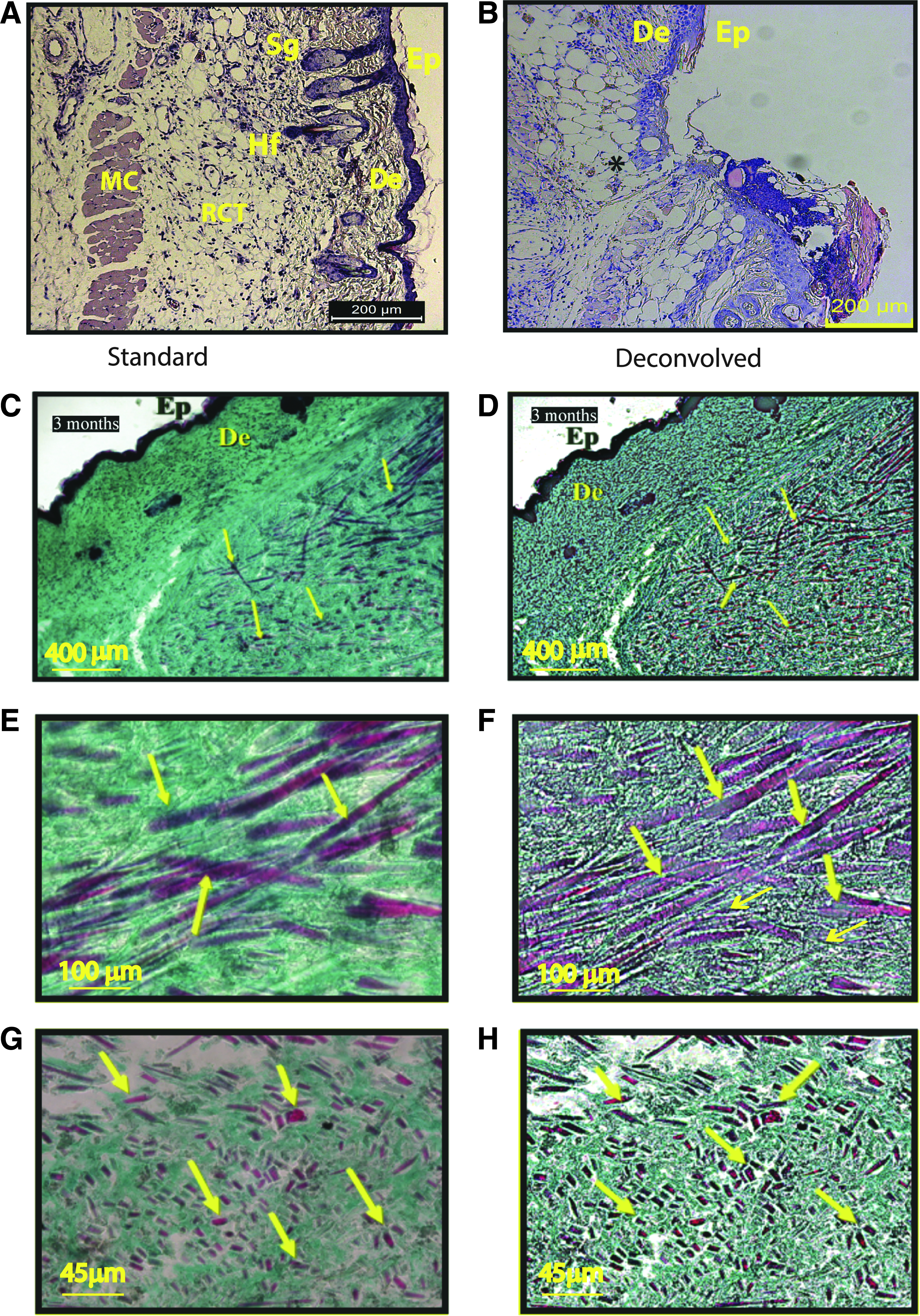
Normal, sham-operated, and nonwoven-grafted mouse skin histology.
One-month-old grafts showed rows of ET-1-positive endothelial cells lining the surfaces of the SF microfibers, which thus acted as guides for the neoangiogenesis (Fig. 5A, B). Also, a robust ingrowth of fibroblasts together with a discrete number of mononucleated macrophages and polynucleated giant cells (PGCs) resulting from the fusion of mononucleated macrophages had taken place into the intervening voids and on the surface of the SF microfibers (Fig. 5D, E).
Three months after grafting, the SF microfibers had become an integral part of the connective tissue they had helped engineer. Table 3 is a summary of the histological features at the 3D carded-needled nonwovens-grafted sites at 3 months after surgery. Fibroblasts, a few mononucleated macrophages and sporadic PGCs, an all-enveloping extracellular matrix, and newly formed vessels filled the voids besides lining the surfaces of the SF microfibers (Fig. 5C, F). As previously observed with FA-treated SF nonwovens, 7 in the silk-based nonwovens presently studied, T4 and B lymphocytes and plasmacells occurred only sporadically, and the number of T8 lymphocytes was never greater than that of the sham-operated samples (not shown).
—, absence; ±, sporadic observation; +, discrete incidence; ++, more than discrete but not exceedingly abundant presence.
PGCs, polynucleated giant cells.
Hence, no infiltrates of leukocytes or clusters could be detected (Fig. 6C–H), suggesting that the grafted nonwovens induced no inflammatory/allergic reaction of any degree in the hosts. 7 The newly produced thin collagen fibrils, but never packed fibrous masses, were visible within the implants in deconvolved pictures of Gomori-stained sections (Fig. 6C, E, G). The regular organization of the engineered connective tissue filling the voids and the absence of packed collagen fiber bundles were clear in sections cut on planes both parallel and orthogonal to the SF microfibers (Fig. 6G, H). Besides, no fibrous capsule surrounded the implants periphery, marking the absence of any FBR (Fig. 6C, D). Finally, the epidermis covering the grafted sites displayed a normal morphology, suggesting the physiological functioning of the underlying silk-based nonwovens with neoformed connective tissue (Fig. 6A, B). Thus, only the presence of the SF microfibers distinguished the novel engineered connective tissue from a normal one.
Conclusions and Future Perspectives
First, silk-based 3D carded-needled nonwovens displayed a good biocompatibility toward human keratinocyte-like C4-I cells that quickly adhered to the microfibers, and grew on them, metabolizing glutamine and releasing lactate but no detectable amounts of proinflammatory IL-1β in vitro. Moreover, like on plastic, nonwoven-attached C4-I cells kept unaltered their constitutive secretion of TNF-α which mediated their own autocrine production of mitogenic amphiregulin. 105
Second, once grafted in vivo the 3D cardled-needled nonwovens guided the de novo engineering of a connective tissue in vivo endowed with a vascular network and populated by fibroblasts, which produced thin collagen fibrils and an extracellular matrix, and, at the end of the third month, by a few sporadic PGCs. At the same endpoint, there were no detectable signs of any inflammatory, immunological, fibrotic, or encapsulating reaction in keeping with the absence of any FBR at the graft site.
Third, and most important, the biophysical properties of the novel carded-needled nonwovens closely resemble those proper of the human skin. 82 This finding suggests that they can integrate into a newly engineered connective tissue without altering its normal consistency and pliability while hindering, because of the presence of the supporting SF microfibers, any scar hypertrophy and retraction.
Therefore, silk-based 3D carded-needled nonwovens are promising tools for skin and more in general connective tissue engineering, regeneration, and repair in human clinical settings and in veterinarian medicine too. The striking number of human proteins sharing amino acid sequences with SF, a sound biological reason of its good immunological tolerability, strengthens our view. 106 So, our results warrant translating such scaffolds into clinical settings.
Footnotes
Disclosure Statement
No competing financial interests exist.