
Abstract
The management of tendon tissue injury presents a significant clinical challenge due to the unique properties of tendons. Cell-based therapy provides a new alternative for regenerating functional tendons, such as in tendon rupture repair, but largely remains at the preclinical research stage. A cell source for graft preparation is essential for successful clinic application. In this study, a novel cell coculture system of bone marrow mesenchymal stem cells (BMSCs) and tendon-derived stem cells (TDSCs) was developed and investigated. BMSCs and TDSCs were cultured separately or in combination at ratios of 20:1, 10:1, 5:1, and 1:1 in vitro, and the cocultured cells showed an enhanced proliferation and collagenous protein production. The coculture system promoted tenogenic differentiation with enhanced tenogenic marker gene expression and collagen matrix production, particularly in the groups with a ratio of 1:1. Using a rat patellar tendon window injury model, we demonstrated that the cell sheets formed by cocultured cells promoted tendon healing significantly, compared to those with a single-cell source. Our study suggests that BMSCs and TDSCs cocultured at the 1:1 ratio may be an improved cell source/preparation for tendon tissue engineering.
Introduction
A
Mesenchymal stem cells (MSCs) were first isolated from bone marrow by Friedenstein et al in 1970. 5 MSCs have been isolated from many other tissues,6–9 and cells from different tissue sources share common characteristics, including the ability for self-renewal, proliferation, and multidifferentiation potential, while also exhibiting tissue-specific characteristics. 10 Although MSCs are suggested cell source for tissue engineering applications, the method of preparation for specific tissue regeneration remains controversial. 11 Adult bone marrow-derived mesenchymal stem cell (BMSC)-based tissue engineering is a potential treatment option that promotes musculoskeletal tissue repair,12,13 but poor tenogenic differentiation ability and the risk of ectopic bone formation limit BMSCs in tendon repair. 14 Tendon-derived stem cells (TDSCs) have higher expression of tenogenic markers and therapeutic efficiency in tendon injury animal models than BMSCs, and have emerged as a more promising candidate cell source for tendon repair.9,15,16
In the management of acute tendon injury, damaged tendon tissues should be removed and repaired through surgery. It is possible to obtain small pieces of normal tendon tissues adjacent to the damaged tendon areas for the preparation of TDSCs, but it is often difficult to harvest enough normal tissue for TDSC culture within a limited time.
Recent studies have shown that cocultures of different cells can affect the fate and functions of both cells in the culture.16–18 MSCs have regulatory functions and the ability to facilitate other cell growth or differentiation in direct or indirect coculture systems.19–21 The coculture system produces specific microenvironments/niches that support cell proliferation and differentiation, without the need for supplementing growth factors, at a relatively low cost.
Cell sheet engineering permits tissue regeneration either through the direct transplanting of cell sheets to host tissues or by creating a three-dimensional (3D) structure via the layering of individual cell sheets.22,23 Complications associated with traditional tissue engineering approaches, such as host inflammatory responses to implanted polymer materials, can be avoided if no additional materials such as carrier substrates or scaffolds are used.22,24,25 We have previously reported that tenogenically induced TDSCs can form cell sheets and enhance tendon healing. 22
We hypothesize that a coculture of BMSCs and TDSCs will enhance their tenogenic potential, and that the cell sheets formed from the coculturing can be used as bioscaffolds in tendon repair. In the current study, the optimal ratio of coculture for BMSCs and TDSCs is identified, with a coculture at a 1:1 ratio resulting in the highest tenogenic differentiation ability of the cells. The cell sheets formed by coculturing BMSCs and TDSCs promoted tendon healing in a rat central one-third patellar tendon injury model.
Materials and Methods
Isolation and culture of rat BMSCs and TDSCs
All experiments were approved by the Animal Research Ethics Committee of the Chinese University of Hong Kong. Four-week-old male green fluorescent protein (GFP)-expressing Sprague Dawley rats and wild-type Sprague Dawley rats (weighing 250–300 g) were used in this study. Both TDSCs and BMSCs were isolated and cultured as previously described.9,26 To isolate the BMSCs, bone marrow was flushed out from femoral marrow cavities with low-glucose Dulbecco's modified Eagle's medium (LG-DMEM), followed by filtration with a 70-μm cell strainer (BD Biosciences, Franklin Lakes, NJ) to remove bone debris and blood clots. After washing with phosphate-buffered saline (PBS), the collected mononuclear cells were plated directly into a 100-mm culture dish at 500 cells/cm2.
TDSCs were isolated from the rat patellar tendon by digesting with type I collagenase (3 mg/mL; Sigma-Aldrich, St Louis, MO) for 45 min at 37°C. The tissue enzyme solution was filtered with a 100-μm cell strainer. Single cells were collected from the filtrate, washed twice with PBS, and plated into a 100-mm culture dish at 500 cells/cm2. Both BMSCs and TDSCs were grown in LG-DMEM (Gibco, New York, NY) with 10% fetal bovine serum and antibiotics (penicillin 100 U/mL, streptomycin 100 g/mL; Gibco) at 37°C with 5% CO2. Both cell types were washed the next day with PBS to remove nonadherent cells and trypsinized at 70–80% confluency. Cells of passage 2 were used in this study.
Coculture assay
A direct contact coculture of BMSCs and TDSCs was performed. Two individual groups and four coculture groups were tested (Table 1), with a single-cell group for BMSCs and TDSCs. Both were cocultured at the ratios of 20:1 (20:1 group), 10:1 (10:1group), 5:1 (5:1group), and 1:1 (1:1 group). The total cell number in each group was consistent.
BMSC, bone marrow mesenchymal stem cell; TDSC, tendon-derived stem cell.
Cell proliferation assay
BMSCs, TDSCs, and cocultured cells were cultured at 2000 cells/well (cell growth area of 0.32 cm2/well) in a 96-well plate. Cell proliferation for 2 and 5 days was tested with an MTTc (3-(4,5-dimethyl-2-thiazolyl)-2,5-diphenyl-2-H-tetrazolium bromide) assay, and 20 μL of 5 mg/mL MTT (Sigma, Saint Louis, MO) was added into each well. After 4 h of incubation, the medium was removed and the formazan crystals solubilized in 200 μL dimethyl sulfoxide (DMSO). The optical density at 490 nm was measured using a spectrophotometer (PerkinElmer, Waltham, MA).
Quantification of collagenous proteins
The total amount of collagenous protein production in the cell culture was assessed with Sirius red staining. Cells were plated in 12-well plate at 5000 cells per well (cell growth area of 0.32 cm2/well). After 2 weeks of culturing, cells were washed with PBS three times and with 1 mL of saturated picric acid solution with 0.1% (w/v). Sirius red F3BA was added into each well for 30 min. The reddish stains were eluted with 1 mL of 1:1 (v/v) 0.1% NaOH and absolute methanol, and the intensity was determined at 540 nm using a spectrophotometer (PerkinElmer).
Gene expression using real-time quantitative polymerase chain reaction
After being cultured for 7 and 14 days in a normal medium, the cells were harvested for quantitative polymerase chain reaction (qPCR) analysis to identify the messenger RNA (mRNA) expression of tenogenic genes Tenomodulin (TNMD) and Scleraxis (Scx), and of the major tendon extracellular matrix (ECM) genes Tenascin C (TnC), Decorin (Dcn), Collagen type I (COL1A1), and Collagen Type III (COL3A1). The cells were harvested and homogenized for RNA extraction with the RNeasy Mini Kit (Qiagen, Dusseldorf, Germany). The mRNA (200 μg per sample) was reversely transcribed to complementary DNA (cDNA) by the First Strand cDNA Kit (Promega, Madison, WI). One microliter of total cDNA of each sample was amplified in a 10 μL reaction mix using the Platinum SYBR Green qPCR SuperMix-UDG. Custom forward and reverse gene-specific primers are shown in Table 2. The expression of the target gene was normalized to that of the GAPDH gene. Relative gene expression was calculated with the 2−ΔCT formula. The ratio of COL1A1 to COL3A1 served as an indicator of tenogenic differentiation. 17
Immunofluorescence staining
Collagen type I-positive cells were determined by immunofluorescence staining. In this study, GFP expression BMSCs and non-GFP TDSCs were cultured at different ratios for 7 and 14 days and then replated with trypsin into a new plate at 10,000 per well to enable the type I collagen expression of individual cells to be examined. After 24 h, cells were fixed with 4% paraformaldehyde for 10 min and permeabilized with 0.1% Triton-X 100 for 15 min. After blocking with 5% normal goat serum for 30 min, the cells were incubated with primary antibodies against collagen type I (sc-8784, Santa Cruz Biotechnology; MS-235-P, Thermo Scientific; 1:100) overnight at 4°C. They were then incubated with the Alexa Fluor 488-conjugated secondary antibody (Invitrogen Corporation, Carlsbad, CA) for 1 h at room temperature. Mounting solution containing 1 μg/mL DAPI (Sigma) solution was used to stain the nuclei of the cells. The percentage of collagen type I-positive and GFP expression BMSCs was counted using ImageJ software, as previously reported. 27
In vitro cell sheet formation
Cells were plated at 10,000 cells/cm2 in a 10-cm dish. The culture medium was changed every 3 days. After 3 weeks of culture, abundant cell matrix formation was observed on a cellular sheet. The sheet was treated with 1 mL 0.05% trypsin (Invitrogen Corporation) for 1 min and carefully collected with a sterile cell scraper and rolled into a 0.5 cm tendon-like structure. These cell sheets were used in the subsequent studies.
Histology and immunohistochemistry staining
The cell sheet was washed in PBS three times and quick-frozen by immediately embedding it in an optimal cutting temperature compound (Sakura Finetk USA, Inc., Torrance, CA). Samples were cut into 5-μm-thick sections by frozen serial section, mounted on 3-aminopropyl-triethoxy-silane (Sigma-Aldrich)-coated slides, and stored at −80°C. The sections were rewarmed and fixed in 10% formalin for 10 min before staining. Hematoxylin and eosin (H&E) staining was used for gross observation of the sheets. Immunohistochemistry staining was carried out as previously described. 22 The sections were fixed by 10% formalin for 10 min, quenched of endogenous peroxidase activity by 3% hydrogen peroxide for 20 min, and subjected to antigen retrieval in 100 mM of citrate buffer at 37°C for 20 min.
After blocking with 5% normal goat serum, sections were incubated with specific antibodies against collagen type I and collagen type III (sc-8784, sc-8780, Santa Cruz Biotechnology; MS-235-P, Thermo Scientific; 1:100 dilution with 5% goat serum in PBS containing 1% bovine serum albumin) at 4°C overnight. Donkey anti-goat horseradish peroxidase-conjugated secondary antibodies (sc-2020; 1:100; Santa Cruz Biotechnology) were then added for an hour at room temperature, followed by 3,3′diaminobenzidine tetrahydrochloride (K3468; DAKO, Glostrup, Denmark) in the presence of H2O2. The sections were counterstained with hematoxylin and dehydrated before being mounted with DPX. The sections were analyzed with light microscopy (Leica DM 4000 B; Leica Microsystems, Heidelberg, Germany).
Rat patellar tendon injury model
One hundred and twelve Sprague Dawley male adult rats (6–8 weeks, body weight of 250–300 g) were used in the study. The rats were kept in restraint cages under controlled temperature and humidity, with a regular 12-h light cycle over a 3-day acclimation period before surgery. They were randomly divided into six groups: BMSCs, 1:20 (B:T), 1:10 (B:T), 1:5 (B:T), 1:1 (B:T), and TDSCs. Six rats from each group were used for the histology examination and 10 from each for the mechanical test. The positive and negative controls consisted of 16 rats (8 from each group). Uninjured rats were tested as the positive control group (Normal tendon), and postoperative rats without cell sheets were tested as the negative control group (Injury only). All operations were carried out under sterile conditions. After anesthesia, the left knee was shaved and the skin disinfected with 70% ethanol, and the patellar tendon was then exposed. The central one-third patellar tendon injury and repair model were created as previously described. 22
A 1-mm-wide tendon defect was made with two stacked sharp blades from the distal apex of the patella to the insertion of the tibial tuberosity. Each rolled cell sheet from one dish at the culture ratio defined above was cut into four equal pieces, and each was placed in the defect window of one rat. The plated cell sheet was sutured to the superior and inferior extremity of the defected tendon. After 4 weeks, the rats were sacrificed and the patellar tendons, including the injury sites, were prepared for histology examination (n = 6). The samples were fixed in formalin followed by dehydration with gradient alcohol. After embedding in paraffin, the specimens were cut into 5 μm sections. Sections from each sample were stained with H&E for histology examinations. For each sample, two randomly selected histology slides were evaluated under light microscopy (LEICA DM 4000 B; Leica Microsystems).
A statistical analysis of tendon healing was then conducted, as previously described, 28 with the following grades: Grade 1, excellent (good collagen alignment and tendon continuity); Grade 2, good (tendon collagen bundles exhibited good repair but were partly interrupted by randomly organized matrix); Grade 3, fair (irregularly arranged and partly broken tendon bundles); and Grade 4, poor (failed healing or massive overgrowth of granulation tissue). This analysis was performed by two independent investigators blinded to the treatment.
Biomechanical test
The injured tendon tissues were harvested for biomechanical tests (n = 10) according to the methods described in the previous study. 29 The tendon–patellar and tendon–tibia composite was harvested and the normal tendon tissue and surrounding muscles were carefully removed. The tibia shaft and proximal patella were fixed on the testing jig with two clamps. The whole construct was then mounted onto a Hounsfield H25KS mechanical testing machine (Tinius Olsen Ltd., Salfords, United Kingdom). The strength of the samples was investigated using a standard failure test with a testing speed of 40 mm/min and a preload of 0.1 N. The cross-sectional area was measured with a Vernier caliper before the test. The ultimate stress (N/mm2) and Young's modulus (N/mm2) were then calculated.
Statistical analysis
The results are reported as the mean of three independent experiments. Differences between groups were examined for statistical significance using a one-way analysis of variance, and the results are expressed as mean ± standard deviation. SPSS (SPSS, Chicago, IL) software was used for the statistical analysis.
Results
Cell proliferation
Cell proliferation was evaluated with an MTT assay. Figure 1A shows that no significant difference between the BMSCs and the cocultured groups was found at day 2, while the TDSC group had a higher proliferation. At day 5, higher cell proliferation was found in the coculture groups than in the BMSC group, which reached a similar level of cell proliferation as the TDSC group.

Cell proliferation was evaluated by the MTT assay
Quantification of collagenous proteins
As determined by Sirius red staining, less collagenous protein production was found in the BMSC group compared to the other groups (Fig. 1B, C). More collagenous protein production occurred in the coculture groups than in the TDSC group (Fig. 1B, C). In the coculture groups, collagenous protein production depends on the ratio of BMSCs and TDSCs, and the more the TDSCs the better the collagenous protein production.
Gene expression analysis
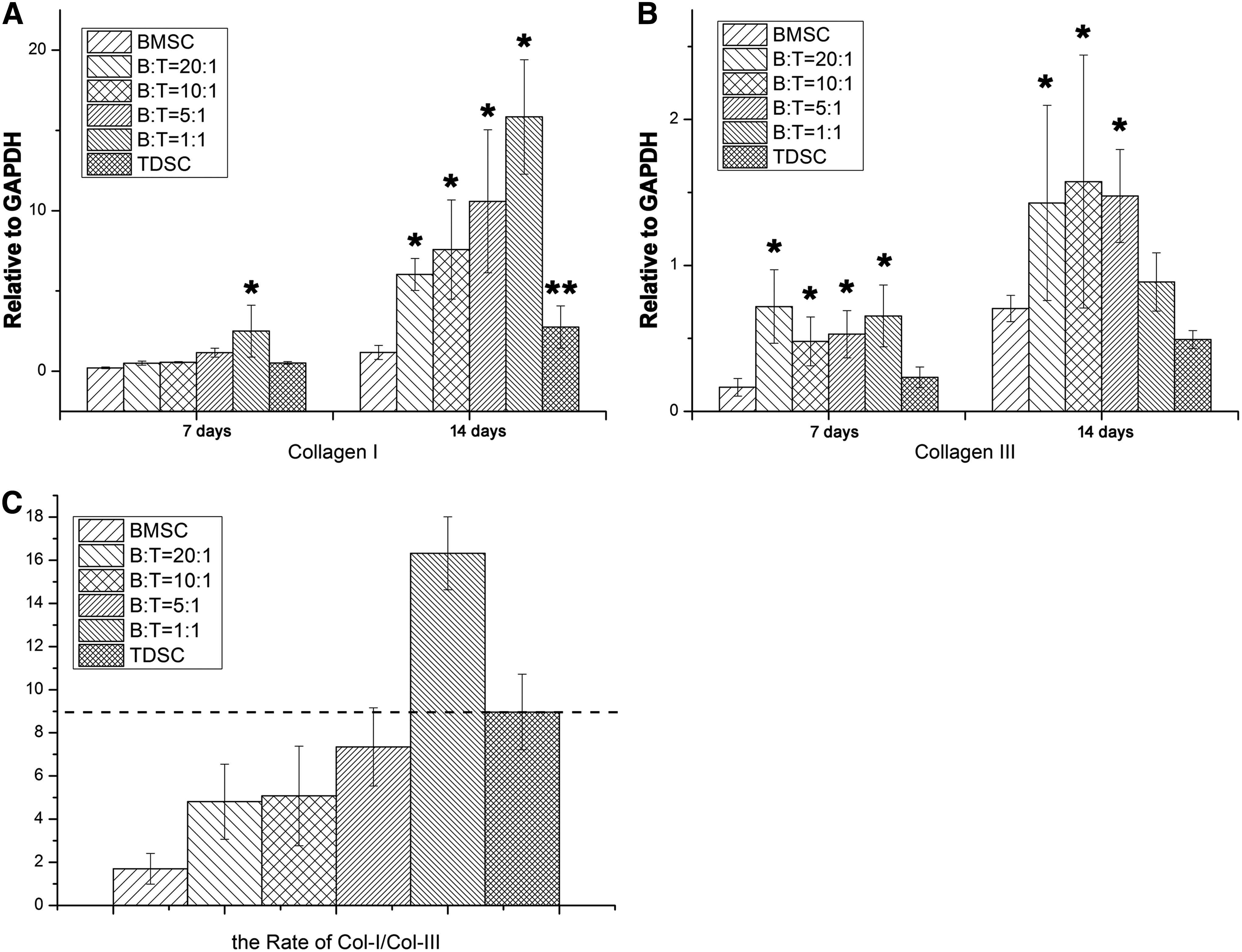
Compared to the BMSC and the TDSC groups, the tenogenic marker genes TNMD, Scx, Dcn, and TnC were all upregulated at day 14 in all of the cocultured groups, but only significantly upregulated in the 1:1 (B:T) cocultured group at day 7 (Fig. 2). The expression of ECM markers was also examined. As Figure 3A and B illustrates, the expression of COL1A1 was significantly upregulated in all of the cocultured groups at day 14, but only enhanced in the 1:1 (B:T) cocultured group at day 7. The COL3A1 was upregulated at day 7 and 14 in all of the cocultured groups. The ratio of COL1A1 to COL3A1 provides a new marker for tenogenic potential. 17 As shown in Figure 3C, the highest ratio was found in the 1:1 (B:T) cocultured group, although the TDSC group's ratio was higher than that of the BMSC group. Taken together, the cocultured system at 1:1 (B:T) provided the best ratio for tenogenic differentiation.
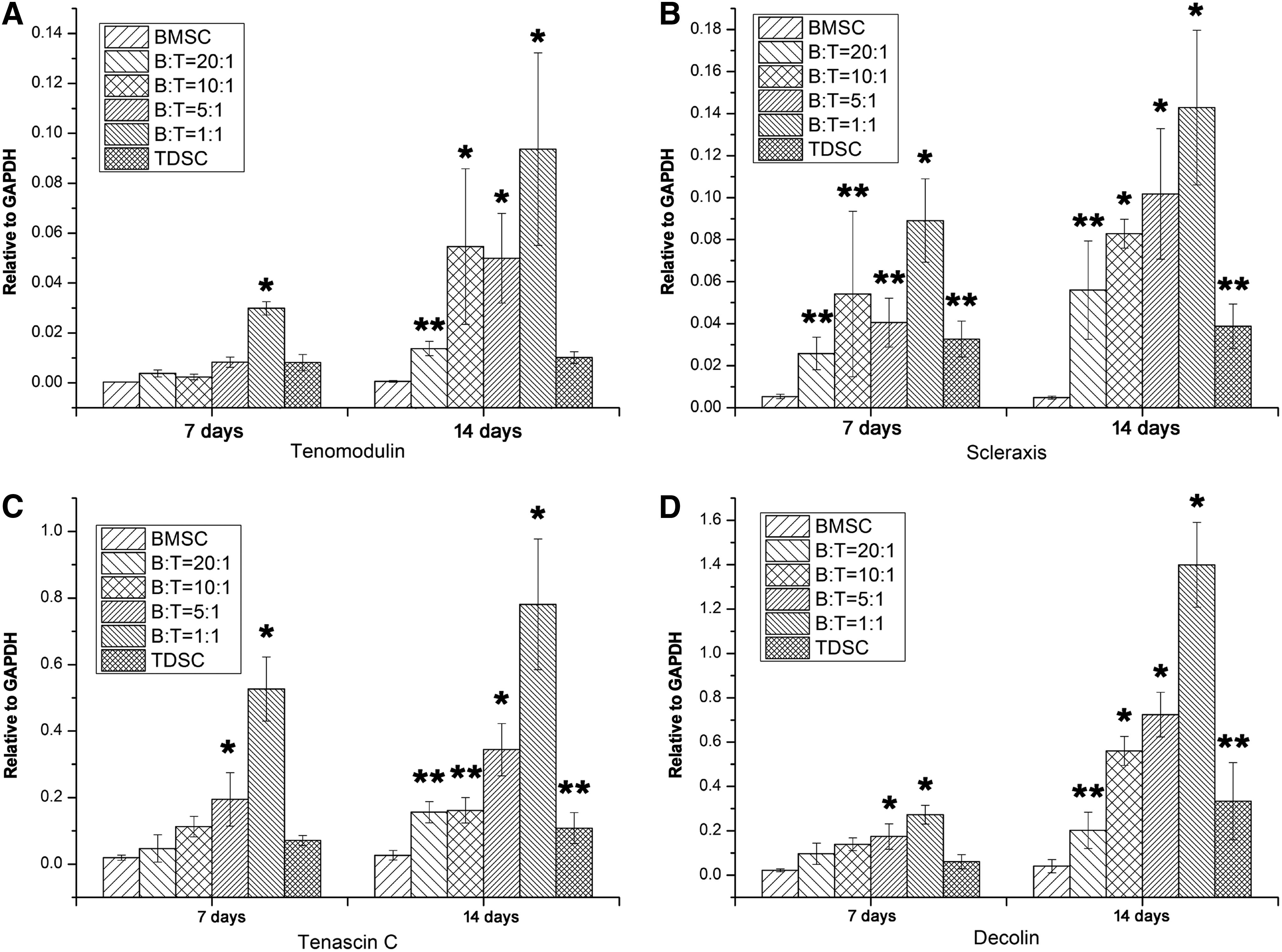
The gene level of tenogenic markers Tenomodulin
The gene level of tendon extracellular matrix markers collagen type I
Immunofluorescence staining of collagen type I-positive cells
Type I collagen expression was examined at day 7 and 14 using immunofluorescence staining. Figure 4 shows that the TDSCs expressed type I collagen (Fig. 4A-f, B-f), while most BMSCs did not (Fig. 4A-a, B-a). An increase in the type I collagen expression in the coculture system was dependent on the ratio of BMSC:TDSC. The highest number of BMSCs expressing type I collagen (Fig. 4A-e, B-e) was found at a BMSC:TDSC ratio of 1:1.
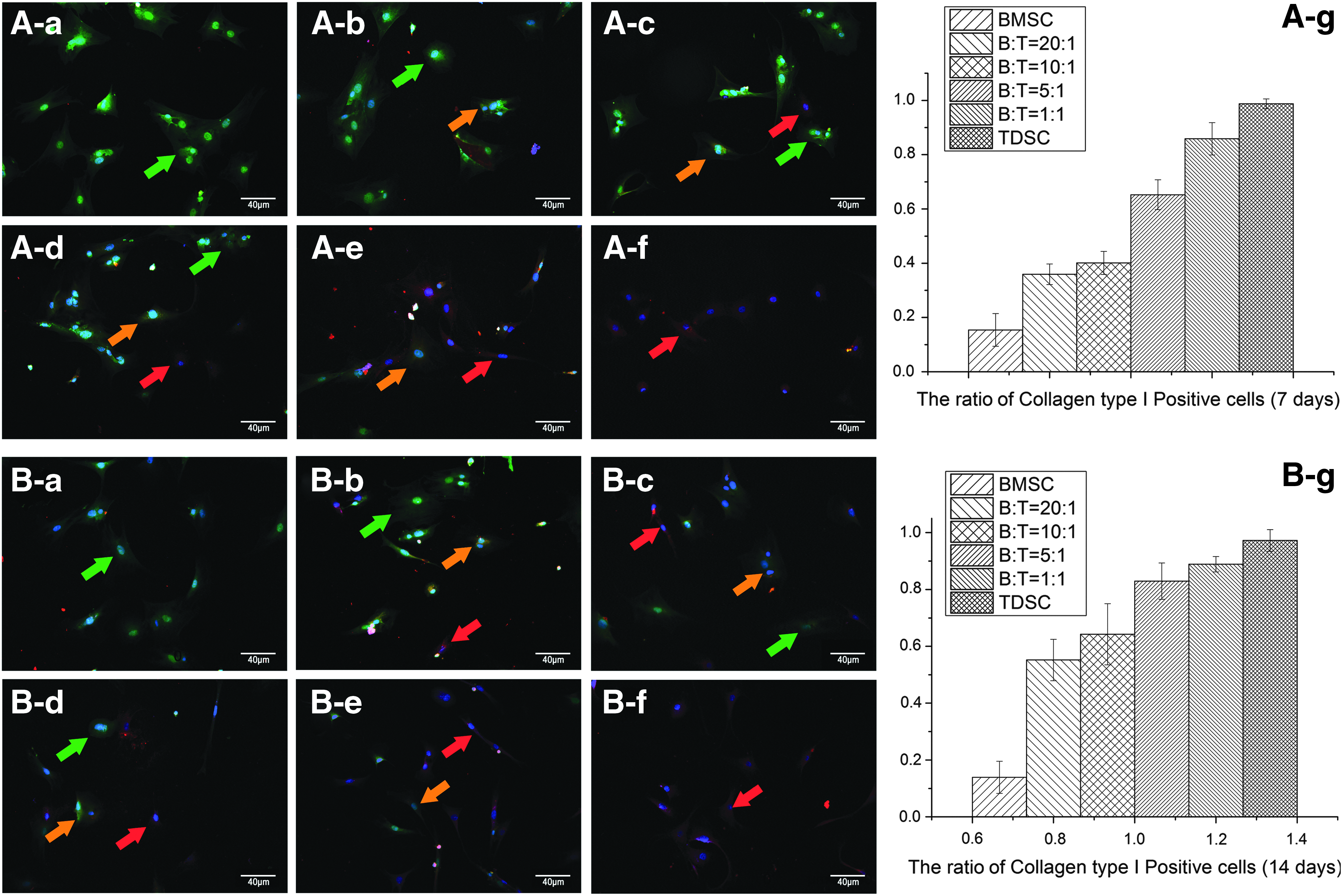
Collagen type I-positive cells at 7 days
Histological analysis of the cell sheets
H&E staining of the cell sheets revealed a disorganized matrix structure and a high level of cellularity in all six groups. Cells were round and randomly distributed within the extracellular matrices (Fig. 5A–F). No tendon-like structures were found in these cell sheets. Immunohistochemistry staining for COL1A1 (Fig. 5G–L) and COL3A1 (Fig. 5M–R) was used to determine the collagen quality. The increased expression of collagen type I may improve the strength of tendons, while collagen type III is found in fibrous tissues. Compared to the BMSC group (Fig. 5G) and the 20:1 (B:T) group (Fig. 5H), stronger staining of COL1A1 was observed in the 5:1 (B:T) group (Fig. 5J), the 1:1 (B:T) group (Fig. 5K), and in the TDSC group (Fig. 5L). The 1:1 (B:T) group (Fig. 5K) showed the strongest staining of COL1A1, while the collagen type III staining was similar in all six groups, which showed weak staining of COL3A1 (Fig. 5M–R).
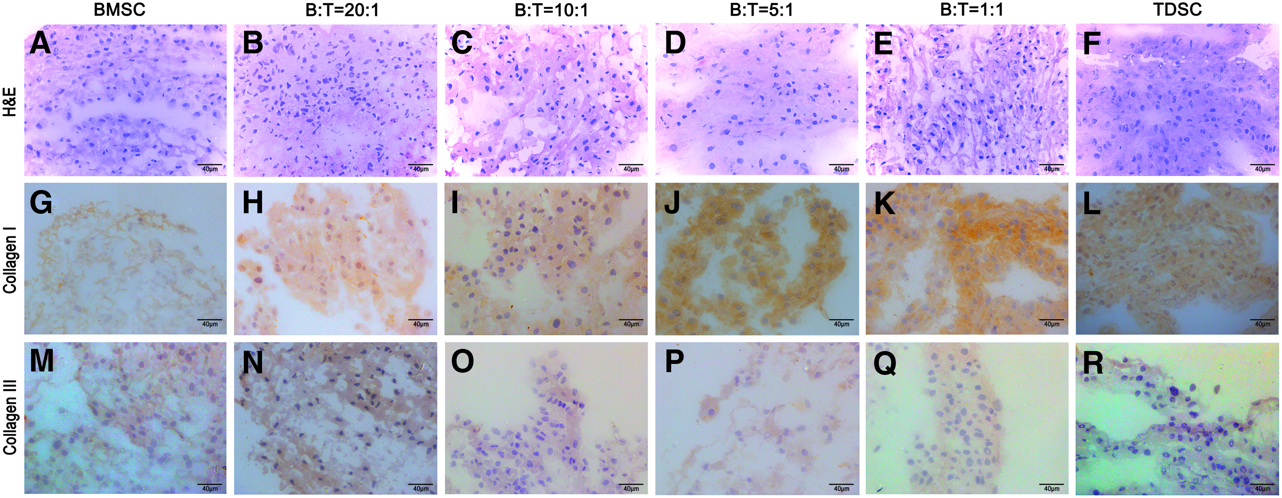
Formed cell sheets were immediately frozen and embed in an optimal cutting temperature compound. Both H&E staining
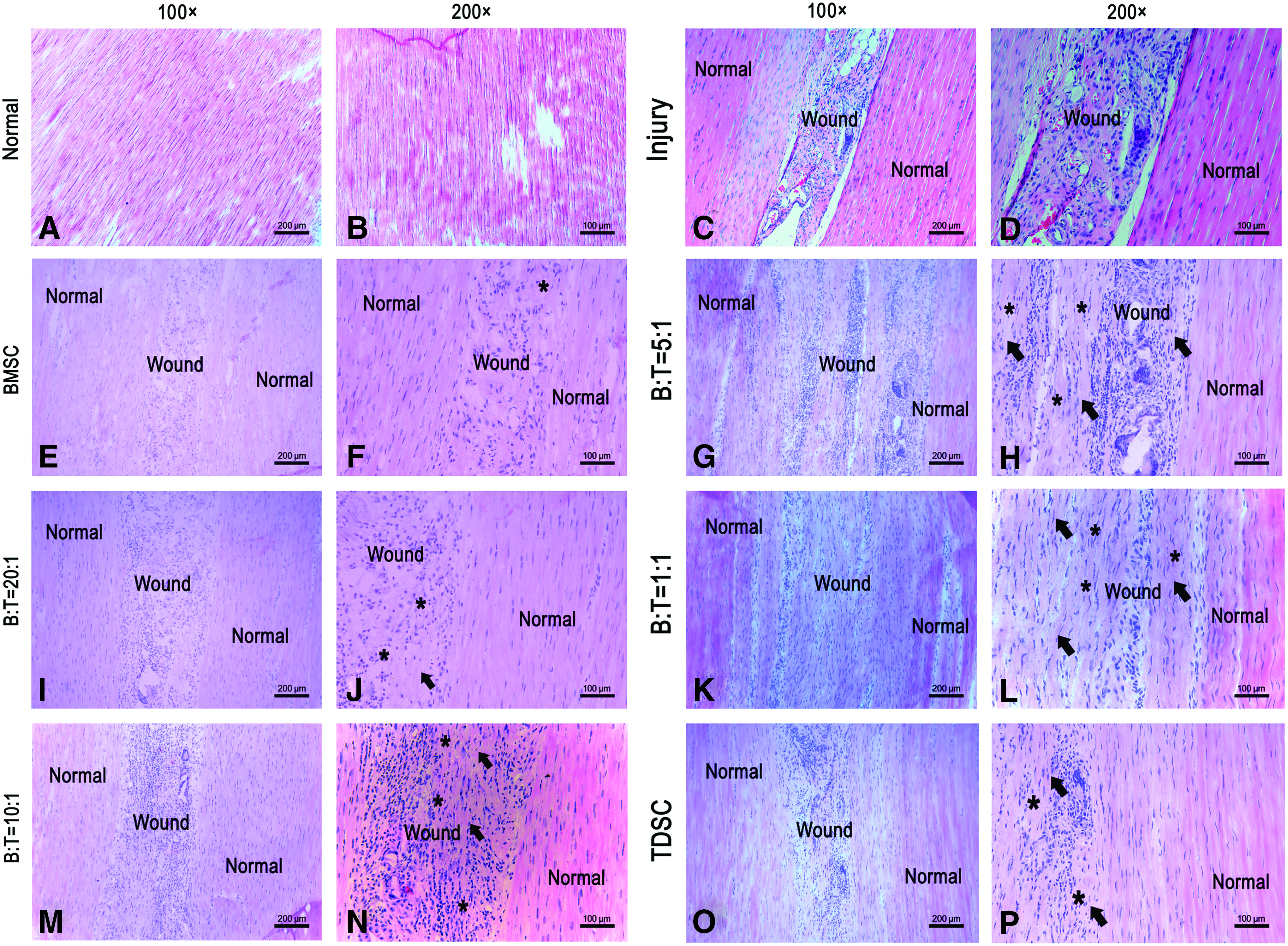
Histological analysis of patellar tendon injury healing samples
Four weeks after surgery, the patellar tendon was examined with H&E staining (Fig. 6). Normal tendon is composed of a parallel array of collagen fibers and spindle-shaped tenocytes (Fig. 6A, B). After 4 weeks of healing, the injured window was still clearly seen. The wound area was filled with randomly oriented cells and fewer collagen fibrils without any typical tendon structures (Fig. 6C, D). The injured area had a high level of cellularity, except in the 1:1 (B:T) group (Fig. 6K, L). Both the BMSC (Fig. 6E, F) and the 20:1 (B:T) groups (Fig. 6I, J) had more randomly oriented collagen fibrils and fewer healing tendon cells (Fig. 6, asterisk [*]) in the wound area than those of the other groups. We found a better collagen fiber alignment (Fig. 6, arrow) in the 10:1 (B:T) group (Fig. 6M, N), the 5:1 (B:T) group (Fig. 6G, H), the 1:1 (B:T) group (Fig. 6K, L), and the TDSC group (Fig. 6O, P) with more elongated healing cells (Fig. 6, asterisk [*]). Healing cells are defined as those that actively proliferate during tendon repair (also known as healing tendon cells). Cells in the injured area were round and randomly oriented in the early phase, then became elongated and spindle shaped. More healing tendon cells embedded between the parallel collagen fibers indicated better tendon healing. More typical tendon structures were found in the 5:1 (B:T) and 1:1 (B:T) groups, but not in the other groups. No fibrocartilage or ectopic bone was found in any of the groups during the 4-week healing period. The evaluation of tendon healing is summarized in Figure 7A. The 1:1 (B:T) group had the lowest tendon healing grade of the six groups, indicating that the healing tissue had the best collagen alignment and tendon continuity. The tendon healing grades in the normal tendon, 10:1 (B:T) group, 5:1 (B:T) group, 1:1 (B:T) group, and the TDSC group were significantly lower (better) than in the BMSC group (p < 0.05). The normal tendon and 1:1 (B:T) group had significantly lower (better) tendon healing grades than those of the BMSC and TDSC groups (p < 0.05).
After 4 weeks of healing, the healing of patellar tendon was tested by H&E staining. Normal tendon tissue

Evaluation of tendon healing was summarized in
Biomechanical testing
The ultimate load of failure and Young's modulus of the normal tendons were higher in the coculture groups. At 4 weeks, the ultimate load of failure was higher than for the BMSC group (Fig. 7B), and only the 1:1 ratio group showed any significant difference among the groups (p < 0.05). The ultimate load of failure was also significantly higher in the 1:1 (B:T) group and normal tendons compared to the other groups. The Young's modulus was also significantly higher in the 1:5 (B:T), 1:1 (B:T), and TDSC groups compared to the BMSC group (Fig. 7C).
Discussion
The success of tissue engineering depends on the source of cells. MSCs have been found to be able to differentiate toward specific lineages after coculturing with chondrocytes, nerve cells, and muscle cells.20,30 The cell coculture system provides a new platform for studying cellular function through direct interaction with stem cells. 21 BMSCs cocultured with fibroblasts indicated a significantly enhanced mRNA expression of ECM genes. 31 A similarly instructive interaction was found when BMSCs were cocultured with native tendon tissue. 32 The coculture technique has enhanced tenogenic differentiation in vitro, but its application in vivo has not been studied. The optimal ratio of cocultured cell source remains unclear. In this study, the tenogenic effect of direct cocultures on different TDSC/BMSC ratios was investigated and the cell sheets formed were found to promote tendon healing in a rat patellar tendon window injury model.
Tenogenic differentiation is a complex process and includes the expression of tenogenic transcription factors and the production of tendon extracellular matrices. The genes of TNMD, Scx, and TnC have been regarded as markers of tenogenic differentiation. 33 COL1A1 and COL3A1 are the main collagens found in tendon and ligaments, and act as tendon extracellular matrices.34–36 In the coordination of collagen formation and tendon maturation, decorin has a primary role. 37 Our in vitro results showed that the cocultured TDSCs and BMSCs led to an increase in the mRNA expression of tenogenic markers (TNMD and Scx) and tendon ECM markers (COL1A1, Dcn, and TnC). The increased tendon ECM production in the coculture groups, particularly in the 1:1 (B:T) group, was further confirmed by the immunohistochemistry results, suggesting that the tenogenic properties of TDSCs were enhanced in the presence of BMSCs. Therefore, the abundant ECM production creates a suitable microenvironment for tenogenic differentiation in cells.
Tissue engineering has been successfully applied to tissue repair and regeneration in recent years. Traditional tissue-engineered scaffolds for tendon healing included nanofiber scaffolds and hydrogel,38–41 but these usually have poor biocompatibility and biodegradability. In contrast, cell sheets can create an ideal microenvironment to modulate both cell shape and intracellular signals. 42 We have previously shown that cell sheets produced by tenogenic TDSCs could be used for tendon healing.22,29 Cell-based therapy usually requires a large number of cells and possibly a minimum of a 20-fold expansion of the harvested cells.43–45 To gain enough cells, at least four population doublings are required. A continuous culture of cells may result in a significant loss of specific tissue phenotype and can be extremely time-consuming. Our study demonstrated that a better cell proliferation occurs in the presence of TDSCs rather than solely BMSCs. Therefore, enhancing cell proliferation is helpful in achieving the required numbers of cells in a shorter time period. The enhanced cell proliferation also promotes the production of cell matrices and the associated collagen synthesis. Cell sheet technology has been used in tissue engineering and recognized as a promising alternative bioscaffold. 29 The 3D culture microenvironment provides better cell–cell and cell–matrix interactions. 46 The microenvironment created by extracellular matrices is crucial to cell survival. 47 When using cell sheets as tendon grafts, the extracellular matrices produced by tenogenic cells are biocompatible, bioactive, biodegradable, and nontoxic. 48 Cell sheets can form a preliminary specific tissue structure and gain certain mechanical properties necessary for their physiological function. Our results demonstrated that cocultured BMSCs/TDSCs enhance the production of collagenous proteins and tenogenic differentiation.
Collagen type I is predominantly present in the extracellular matrices of normal tendons, accounting for almost 90% of total tendon extracellular matrices. 35 Increased expression of collagen type I can improve the strength of tendons during repair. 41 TDSCs in the cocultured cell sheet exhibited more collagen type I-positive cells compared to BMSCs or TDSCs alone. Collagen type III, another main component of normal tendon tissues, increases during tendon repair progress and in scar tissue. 36 The ratio of collagen types I to III in normal tendon tissue is around 9:1. 17 The coculture 1:1 (B:T) group exhibited the highest collagen type I to type III ratio, indicating that this coculture may produce the best ECM production for tendon regeneration. In our study, after 2 weeks, the cocultured cells produced enough extracellular matrices to form cell sheets. The newly formed sheets were structured with randomly oriented collagen fibrils and round cells, and there was no difference among the groups. However, a higher expression of collagen type I was found in the cocultured cell sheets, particularly in the 5:1 (B:T) and 1:1 (B:T) cocultured groups.
The central one-third patellar tendon injury model is a standard model used in previous studies for assessing tendon and ligament healing.22,29,49 After the cell sheets were transplanted into the rat patella tendon defect site, we found that cocultured sheets significantly enhanced tendon healing, with an increased production of extracellular matrices, a better alignment of collagen fibers, and a larger number of tendon healing cells at 4 weeks. After injury, cells proliferate in large amounts in the damage region immediately. The morphology of the cells appears to be roundish and irregular. When early inflammatory reaction disappears, cell proliferation follows in damage region. In the ideal condition, healed tissue would form the proliferating cells gradually turning from roundish to elongated spindle-shaped-like tendon cells and surrounded by collagen composition aligning. Spindle shape and parallel collagen fibers are standard to judge regeneration of tendon. Cell sheets from the coculture with a ratio of 1:1 (B:T) exhibited the most promising effect on tendon healing, with a typical tendon structure after 4 weeks of healing. The tendon repair quality in the cocultured cell sheet groups was found to be overwhelmingly better than that of the solely TDSC or BMSC groups, with improved ultimate loading stress in mechanical tests, suggesting that there are syngeneic effects of BMSCs on TDSCs. The mechanical parameter of ultimate loading stress is an important indicator of the functional restoration of tendon structures. 48 Our data showed that the ultimate loading stress of healed tendon significantly decreased after injury. After using 1:1 ratio cell sheet, the ultimate loading stress of healed tendon improved compared to the injured tendon, which indicated a great effect on tendon function recovery. So, BMSC and TDSC cocultured cell preparation may be a novel approach in preparing cells for tendon repair.
Regarding the cells in tendon tissue engineering, their differentiation into tenocytes is the key. Our previous studies had demonstrated that comparing to BMSCs, TDSCs are a better cell source for tendon regeneration with greater tenogenic marker expression and tenogenic differentiation ability. As shown in our data, the tenogenic marker expression and tendon matrix formation were greater in the TDSC group or TDSC/BMSC mixture groups than those in the BMSC group, which is further confirmed in the rat patellar tendon window defect model. BMSCs had regulatory functions on other cells through paracrine function; hence, the presence of BMSCs may enhance the tenogenic differentiation of TDSCs. Our data suggested that 1:1 is the best ratio according to cell proliferation, tenogenic differentiation, and tendon-specific matrix formation data. In the rat patellar tendon window defect model, cell sheet from the 1:1 ratio group significantly improved tendon healing and shortened the healing time.
The limitations of the current study are as follows. (1) Our mechanistic studies into the observed positive effects on tenogenic differentiation of coculturing BMSCs and TDSCs were limited, and the exact underlying mechanisms require further careful investigations. (2) We only tested the in vivo effects of the cocultured cell sheets in one patellar tendon injury model, other models such as the Achilles tendon injury model should be tested in future studies. (3) Even though we successfully reduced the required TDSC number after cocultured with the 1:1 ratio, we still need to harvest a large number of TDSCs, which may limit their clinical application. How to further reduce the cell number of TDSCs is one of our main objects in our future study, such as the use of allogenic TDSC coculture with autologous BMSCs as one of the possible solutions.
In conclusion, our data showed that cell sheets produced by coculturing BMSCs/TDSCs at a ratio of 1:1 possess similar physiological characteristics to normal tendons, and may be used as an alternative bioscaffold for tendon tissue engineering. The promising therapeutic outcome of using BMSC/TDSC cocultured cell sheets in tendon repair may be further explored in animal investigations and clinical trials.
Footnotes
Acknowledgments
This work was supported by a grant from the National Natural Science Foundation of China (NSFC No. 81371946, 31400834) to G.L. and L.X.; and from the Hong Kong Government Research Grants Council, General Research Fund (CUHK471110 and CUHK470813) to G.L. The work was also supported, in part, by SMART program seed funding to G.L., and the Lui Che Woo Institute of Innovation Medicine of the Chinese University of Hong Kong.
Disclosure Statement
No competing financial interests exist.