
Abstract
In this study, we successfully fabricated a novel drug and plasmid DNA (pDNA) dual delivery system by electrospinning the dispersion composed of polyethyleneimine-carboxymethyl chitosan/pDNA-angiogenin (ANG) nanoparticles, curcumin (Cur), poly (D, L-lactic-co-glycolic acid) (PLGA), and cellulose nanocrystals (CNCs). In vitro release studies showed that the bioactivity of Cur and ANG was preserved in the nanofibers, and a sequential release pattern was achieved in which nearly 90% of the Cur was released in ca. 6 days and the ANG release lasted up to about 20 days. In vitro cell culture results suggested that the composite nanofibers exhibit excellent biocompatibility. To evaluate the in vivo angiogenesis and anti-infection properties, the PLGA/CNC/Cur/pDNA-ANG composite nanofibers were transplanted into the infected full-thickness burn wounds. Biopsy specimens were harvested for histology, immunohistochemistry, immunofluorescence, real-time quantitative PCR, and Western blotting analyses. The results indicated that the PLGA/CNC/Cur/pDNA-ANG composite nanofibers not only prevented local infection but also promoted skin regeneration.
Background
W
There has been growing interest in gene-activated matrices (GAMs) because they provide platforms for gene delivery, hold plasmid DNA (pDNA) in situ until endogenous repair cells arrive, and sustain expression of these growth factors. 6 Nevertheless, direct addition of naked pDNA in GAMs is associated with low transfection efficiency. 7 Incorporating vector/pDNA complexes rather than naked pDNA into a scaffold is desirable to improve the efficiency of gene expression. Guo et al. developed N, N, N-trimethyl chitosan chloride nanoparticles as a gene vector to load and deliver pDNA encoding human vascular endothelial growth factor-165 over long term and thereby efficiently promote skin regeneration. 6
Infection is one of the most common and serious complications of acute trauma. 8 It is a constant threat to human health because bacterial infections can delay or prevent wound healing or even lead to death. 9 The remedy for burns involves developing a novel biomimetic scaffold to prevent burn-wound infection. Lan et al. explored the applications of gentamycin sulfate-impregnated gelatin microsphere/silk fibroin scaffolds for the treatment of full-thickness infected burns and found that the scaffolds were effective. 10
Curcumin (Cur) displays multiple pharmacological activities, including antioxidant, antitumor, and anti-inflammatory behavior, which may be used as a promising agent for wound healing. 11 Currently, development of a suitable material as drug carrier for curcumin has positive results. Yang et al. prepared electrospun composite nanofibers composed of Cur-loaded micelles and doxorubicin hydrochloride (Dox). The results from this study indicated that Cur could be sustainably released from Cur-loaded micelles/Dox nanofibers and possesses great potential in improving the efficiency of cancer chemotherapy. 12
Many kinds of bioactive factors can be encapsulated into polymeric nanofibers and maintain their biomedical functional properties. Yang et al. incorporated polyplexes of basic fibroblast growth factor-encoding plasmid with poly(ethyleneimine) into electrospun fibers; in vivo results showed that electrospun fibrous mats rapidly restored the structural and functional properties of wounded skin. 13 A potential way to improve the performance of nanofibers is to incorporate one or more nanophase materials into polymer matrices. 14 Cellulose nanocrystals (CNCs) obtained by sulfuric acid hydrolysis of native cellulose have good mechanical properties (ca. 7 GPa), high surface area, and biocompatibility. Many CNC-based functional biomaterials have been developed, including membranes, 15 hierarchical three-dimensional porous constructs, 16 and hydrogels, 17 and have been widely used in drug delivery and tissue engineering. 18 We previously designed a fully collagen-based composite material in which CNCs were dispersed within a collagen matrix, 19 exhibited a stable mechanical functionality, and good biocompatibility. We showed that CNCs introduced into PLGA nanofibers had better mechanical strength and biocompatibility. 20
In this study, we fabricated PLGA/CNC composite nanofibers loaded with polyethyleneimine (PEI)-carboxymethyl chitosan (CMCS)/pDNA-ANG and Cur complexes. The PEI-CMCS/pDNA-ANG nanoparticles and Cur were then encapsulated into polymeric nanofibers by electrospinning. The general procedure employed for the preparation of the composite nanofibers is shown in Figure 1. The angiogenesis and anti-infection activity of the composite nanofibers were systematically investigated both in vitro and in vivo.
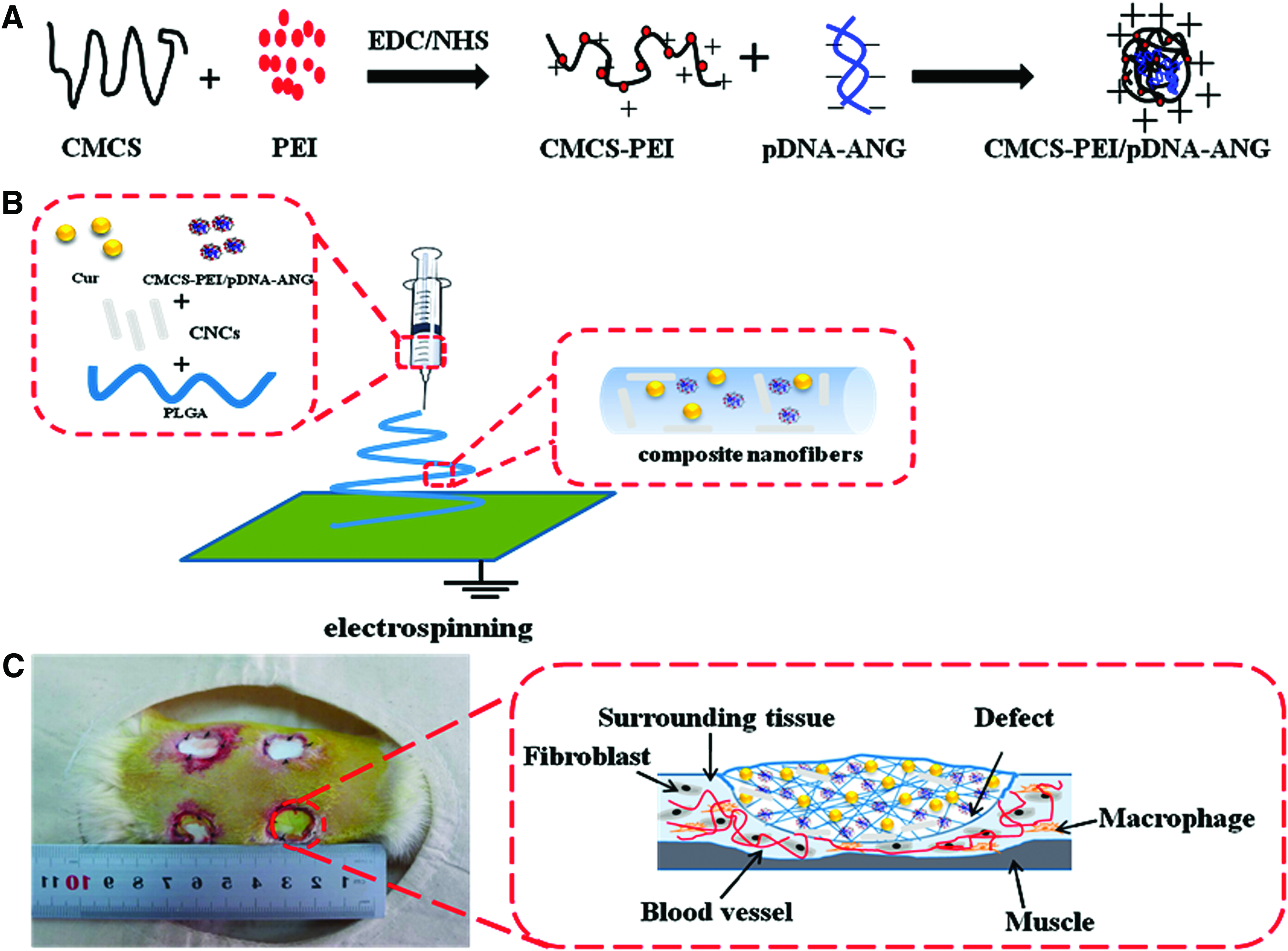
Schematic illustration of
Materials and Methods
Materials
Plasmid DNA encoding recombinant human ANG was kindly donated by Dr. X. Wang, Zhejiang University, China. Chitosan (CS, medium molecular weight, deacetylation degree 75–85%), branched polyethyleneimine (PEI, Mw = 1.8 kDa and 25 kDa), 1-ethyl-3-(3-dimethylaminopropyl) carbodiimide (EDC), N-hydroxysuccinimide (NHS), and Cur were purchased from Sigma. CNCs were obtained by acid hydrolysis of microcrystalline cellulose (50 μm). 20 PLGA having a lactic acid:glycolic acid ratio of 75:25 and a molecular weight of 97,000 Da was obtained from Polysciences, Inc. (Warrington, PA). The cell culture medium composed 89% 1640 Dulbecco's modified Eagle's medium, 10% fetal calf serum from Life Technologies (Carlsbad, CA), 1% Penicillin (100 U/mL), and 100 mg/mL streptomycin. Human umbilical vein endothelial cells (HUVECs) were maintained at 37°C in an atmosphere containing 5% carbon dioxide (CO2). The culture medium was changed every other day. All other reagents were of analytical grade and used without further processing. Triple-distilled water was used throughout the experiments.
Preparation of PEI-CMCS copolymer and PEI-CMCS/pDNA complexes
CMCS was prepared according to procedures described previously. 21 Chitosan (10 g) was suspended in 40% aqueous NaOH solution (100 mL) and isopropyl alcohol (50 mL) mixture solution and kept stirring at 30°C for 12 h. After the excess alkali solution was extracted, monochloroacetic acid (24 g) in isopropyl alcohol (50 mL) was added to the alkali-treated chitosan at 65°C for 5 h. Then, the 40% aqueous NaOH solution (100 mL) was added and stirred for another 5 h. Finally, the remaining precipitate in mixture solution was removed by centrifugation and excess anhydrous ethanol was added to the solution to precipitate out the CMCS.
PEI-CMCS copolymer was performed using EDC and NHS as coupling reagents. 21 CMCS (110 mg) and PEI (1.6 g) were dissolved in distilled water (20 mL) and kept stirring at 25°C for 30 min. Then, the pH of the mixture solution was adjusted to 5.5 by adding 1M HCl solution. Subsequently, 10 mL of an aqueous solution containing 200 mg of EDC and 110 mg of NHS was added dropwise to the mixture at room temperature over 1 h. Stirring at room temperature was maintained for 24 h. Finally, the product was dialyzed (MWCO = 10,000; Spectra/Por, Laguna Hills, CA, USA) against distilled water and lyophilized.
Generally, CMCS-PEI/pDNA nanoparticles were freshly prepared. First of all, freshly prepared CMCS-PEI was dissolved in distilled water (controlled at a concentration of 1 mg/mL). Then, the solution was filtered through 0.45-μm filter paper. CMCS-PEI/pDNA complexes at 25N/P ratio were formulated by adding corresponding volumes of the CMCS-PEI solution to an equal volume of the pDNA solution, put under vortex movement for 30 s, and then incubated for 30 min at room temperature.
In this study, the N/P ratio was fixed at 25, because previous research showed that those PEI-CMCS/pDNA complexes were associated with higher transfection efficiency in vitro. 22
Fabrication of composite nanofibrous scaffolds
The CNCs were dispersed in a 15% (w/v) PLGA solution (HFIP solvent) at 7 wt% loading (based on the PLGA weight). One milliliter sterilized phosphate-buffered saline (PBS) containing PEI-CMCS/pDNA complexes (1 μg/mL ANG) was added to 10 mL of the PLGA/CNC electrospinning solution at room temperature. The PLGA/CNC/Cur/pDNA-ANG solution was produced by adding 10 mg Cur to 10 mL of the PLGA/CNC/pDNA-ANG electrospinning solution in the dark. The obtained composite electrospun nanofibers were found to be between 100 and 200 mm in thickness. All electrospinning experiments were carried out at room temperature.
Characterization of composite nanofibers
Transmission electron microscopy
The morphological examination of the CMCS-PEI/pDNA complexes and CNCs was performed by field emission transmission electron microscopy (JEM–2010HR; JEOL, Tokyo, Japan) at an electron acceleration voltage of 140 kV.
Scanning electron microscopy
The nanofibers (on the aluminum foils) were then gold coated using sputter coating to observe the surface topographies by scanning electron microscope (SEM, LEO1530 VP; Philips, Amsterdam, The Netherlands) at an accelerating voltage of 25.0 kV.
In vitro Cur and pDNA release
The fabricated composite nanofibers were immersed in 10 mL of PBS (pH = 7.4). The samples were incubated at 37°C under continuous agitation. At the preset time points, 1.0 mL of the release medium solution was removed from each vial and replenished with an equal volume of fresh PBS. The amount of Cur in the supernatant was determined spectrophotometrically at 428 nm using an ultraviolet/visible (UV/Vis) spectrophotometer (UV-2550; Shimadzu, Otsu, Japan) and calculated using a Cur standard curve (Supplementary Fig. S1; Supplementary Data are available online at www.liebertpub.com/tea). 23
To evaluate the release kinetics of pDNA-ANG from the composite nanofibers, the sample was immersed in 10 mL sterile PBS at 37°C under continuous agitation. At the preset time points, 1.0 mL of the release medium solution was removed from each vial and replenished with an equal volume of fresh PBS. The quantity of the released pDNA was tested by a fluorometer with Hoechst 33258.
In vitro ANG expression and viability of HUVECs and transfection
ANG expression of HUVECs cultured in vitro in the composite nanofibers was evaluated by the human ANG Elisa Kit (Abcam, Cambridge, UK). Generally, samples seeded with HUVECs at a density of 1 × 10⁵ cells per well were incubated at 37°C in a CO2 incubator. At the preset time points, the nanofibers were washed thrice with PBS and then homogenized in the lysis buffer (0.1 M tris-HCl, 2 mM EDTA, 0.1% Triton X-100). To collect the supernatant, the lysate (2 mL) was centrifuged at 15,000 rpm and 4°C for 3 min.
HUVEC proliferation was measured using the Cell Counting Kit-8 (CCK-8; Dojindo, Tokyo, Japan). Before cell seeding, the composite nanofibers were cut into sections with diameters of 1.5 cm, placed into a 24-well culture plate, and sterilized with an UV lamp for 1 h. Then, the samples were soaked with 1 mL medium overnight. The next day, 2 × 10⁴ HUVECs were seeded in each well on the sample with 1 mL culture medium, which was changed every other day. For the CCK-8 assay, after the cells were cultured for 1, 3, 5 and 7 days, 100 μL of medium from each sample was transferred into separate wells of a 96-well plate and CCK-8 reagent (10 μL) was subsequently added to each 96 well, followed by another 2 h of incubation at 37°C. The optical density (OD) value at 450 nm was then measured using a microplate absorbance reader (Model 680; Bio-Rad, Hercules, CA).
The PEI-CMCS and pDNA were isolated according to the method described previously.6,24 The transfection capacity of the released pDNA complexes was evaluated by using pDNA-ANG and HUVECs. After the cells were seeded at a density of 1 × 10⁴ cells per well and cultured for 24 h, the pDNA complexes released at 7, 14, and 21 days were added with a concentration of 1 mg pDNA per 104 cells, respectively. The transfection efficiency was analyzed by a FACS scan instrument (BD, Mountain View, CA).
In vivo animal experimental and surgical procedures
All of the animal studies were conducted according to the guidelines of the Jinan University and the U.S. National Institutes of Health. Male Sprague-Dawley (SD) rats weighing 150–250 g were used in the study. Food and water were supplied ad libitum.
25
The rats were anaesthetized by intraperitoneal injection of pentobarbital sodium (Sigma) at 30 mg kg−1. Once anaesthetized, the dorsal hair of the rats was removed. The dorsal skin area was then burned with a hot circular copper billet ( = 15 mm, 95°C, 40 s) to induce a full-thickness burn. To mimic the clinical treatment of burns, the burns were excised to the level of the panniculus carnosus 24 h later.
10
A total of four full-thickness burns (20-mm in diameter) were created on the dorsum of each rat. Then, the wounds were covered with PLGA/CNC, PLGA/CNC/Cur, PLGA/CNC/pDNA-ANG, and PLGA/CNC/Cur/pDNA-ANG composite nanofibers. The entire wound area was then wrapped with sterile gauze and fixed with an elastic bandage. The tissue samples were harvested at 7, 14, 21, and 28 days postsurgery.
= 15 mm, 95°C, 40 s) to induce a full-thickness burn. To mimic the clinical treatment of burns, the burns were excised to the level of the panniculus carnosus 24 h later.
10
A total of four full-thickness burns (20-mm in diameter) were created on the dorsum of each rat. Then, the wounds were covered with PLGA/CNC, PLGA/CNC/Cur, PLGA/CNC/pDNA-ANG, and PLGA/CNC/Cur/pDNA-ANG composite nanofibers. The entire wound area was then wrapped with sterile gauze and fixed with an elastic bandage. The tissue samples were harvested at 7, 14, 21, and 28 days postsurgery.
Histology evaluation
For histological analyses, the harvested samples were fixed with paraformaldehyde (4% in PBS, 0.01 M, pH 7.4) and after 24 h, the samples were transferred to PBS buffer at 4°C, dehydrated in a graded series of ethanol, and then embedded in paraffin for routine hematoxylin and eosin (H&E) staining and Masson's trichrome staining for collagen fibers. 26
The slides were then observed with a light microscope (Axio Scope A1 FL; Carl Zeiss, Wetzlar, Germany). All histological analyses were performed on at least three wounds per group per time point; images presented are representative of all replicates.
Immunohistochemistry and immunofluorescence
For the immunohistochemical staining, the paraffin sections (5 μm) were deparaffinized, washed thrice in PBS for 5 min, and then blocked with 5% serum for 30 min. The slides were subsequently incubated with primary antibodies against ANG (Abcam), tumor necrosis factor-α (TNF-α) (Abcam), interleukin-1β (IL-1β) (Abcam), and interleukin-6 (IL-6) (Abcam) at 4°C overnight. After rinsing thrice with PBS, the slides were incubated with secondary antibodies at 37°C for 20–30 min, further developed with 3,3′-diaminobenzidine tetrahydrochloride solution, and then finally counterstained with hematoxylin. Positive staining was indicated by a brown color observed under the optical microscope. The number of newly formed blood vessels was counted by CD31-positive staining per area.
Immunofluorescence was performed following the procedures previously described. 27 Briefly, paraffin sections (5 mm) were deparaffinized, washed thrice in PBS for 5 min, blocked with 5% serum for 30 min, and incubated overnight at 4°C with rabbit anti-CD31 primary antibody (1:100; Abbiotec, San Diego, CA) and mouse anti-a-smooth muscle cells (SMA) primary antibody (1:50; Abcam). After rinsing thrice with PBS, the slides were incubated with rhodamine-conjugated goat-anti-rabbit secondary antibody and fluorescein isothiocyanate (FITC)-conjugated goat-anti-mouse secondary antibody (1:50; Dako, Carpinteria, CA) for 30 min. After washing thrice in PBS, the cell nuclei were stained by DAPI for 10 min at room temperature. Images were acquired with a fluorescence microscope (IX 81; Olympus, Tokyo, Japan). The number of mature blood vessels was counted by dually SMA-positive and CD31-positive vascular structures per area.
Real-time quantitative PCR analysis
Total RNAs of the tissues were extracted using TRIzol Reagent (Invitrogen, Carlsbad, CA). 28 Total RNA (1 mg) was used for reverse transcription into complementary DNA (cDNA) by an M-MLV Reverse Transcriptase cDNA synthesis kit (Promega, Madison, WI). Gene primers for ANG, CD31, and α-SMA, as well as the calibrator reference gene β-actin, are summarized in Supplementary Table S1.The real-time PCR was performed using iQ SYBR green PCR master mix (Bio-Rad). The samples were subject to the following conditions in a One iCycler iQ5 (Bio-Rad): initial denaturation at 95°C for 10 min followed by 45 cycles of 94°C for 5 s and 62°C for 20 s. Ct (threshold cycle) values were calculated using the iQ5 optical system software (version 2.0).
Western blotting analysis
The frozen tissue samples were homogenized in RIPA lysis buffer having protease inhibitors. The lysates were then clarified by centrifugation at 12,000 rpm for 15 min at 4°C and separated on sodium dodecyl sulfate-polyacrylamide gel electrophoresis (SDS-PAGE). After transferring to a polyvinylidene fluoride (PVDF) membrane (Millipore, Billerica, MA), the proteins were incubated overnight with antibodies and detected with the enhanced chemiluminescence (ECL) (Abcam) system following treatment with 5% milk powder in triethanolamine buffered saline solution (TBS) to prevent nonspecific reaction. The specific antibodies used for this experiment were anti-ANG primary antibody (Abcam), anti-IL-6 primary antibody (Abcam), anti-IL-1β primary antibody (Abcam), anti TNF-α primary antibody (Abbiotec), and anti-NF-κB primary antibody (Abcam).
Statistical analysis
All statistical computations were performed using the SPSS for Windows software (version 16.0; SPSS, Inc., Chicago, IL). Differences with a value of p < 0.05 were considered statistically significant. All experiments were performed in triplicate and the results are presented as the mean ± standard deviation; statistical significance was calculated by Student's t-test.
Results
Characterization of the PEI-CMCS/pDNA complexes and composite nanofibers
The migration capability of DNA was completely retarded at a weight ratio of 0.1, which indicated that all of the pDNA was trapped inside the particles (Supplementary Fig. S2). PEI-CMCS/pDNA complexes were formed at weight ratios of 25:1. PEI-CMCS condenses pDNA-ANG into spherical nanosized particles with diameters ranging from 100 to 200 nm. The mean values of the length and diameter of the prepared rod-like CNCs were 180 ± 25 and 5 ± 2.1 nm, respectively (Supplementary Fig. S3).
The PLGA/CNC nanofibers had a smoother surface and a larger diameter of 400 ± 25 nm (Fig. 2A). The composite nanofibers had rougher surfaces, which was attributed to the partial dissolution of Cur or PEI-CMCS from the nanofibers. The nanofibers were homogeneous and their diameters were all in the nanoscale range; a smaller fiber size was noted for the composite reduced with the addition of Cur or PEI-CMCS/pDNA-ANG. 29

Morphological characterization of electrospun nanofibers. SEM images of electrospun nanofibers:
In vitro Cur and pDNA release
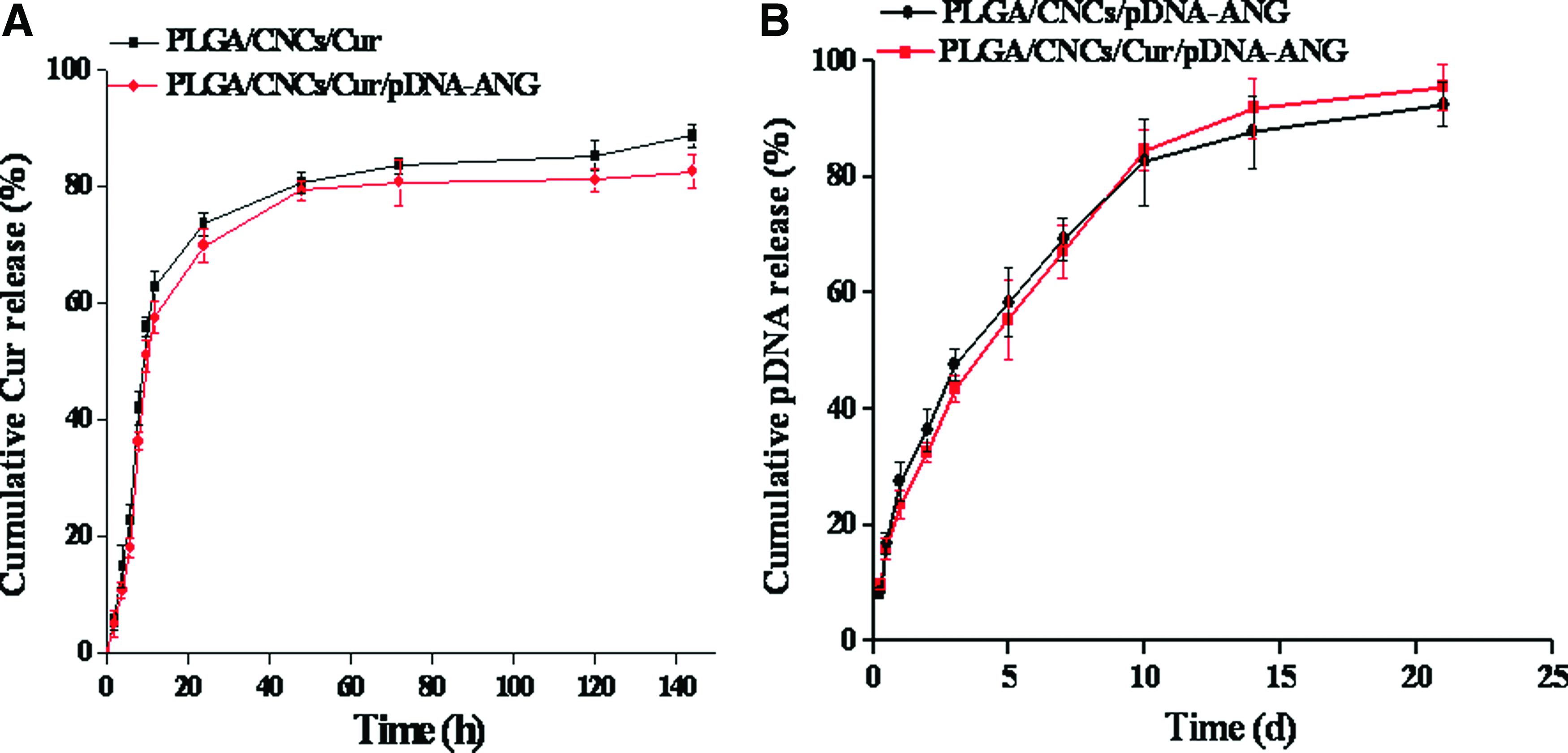
The results of the release behaviors of Cur from composite nanofibers in vitro are illustrated in Fig. 3A. The release curve showed a biphasic pattern. An initial burst release was observed within the first 24 h. Within the first 24 h, the percentages of released drug reached ca. 73.6% and 69.8% with the PLGA/CNC/Cur and PLGA/CNC/Cur/pDNA-ANG nanofibers, respectively. After this initial burst release, the remaining drug in the composite nanofibers was released at a slightly slower rate during the following 144 h. When measured over the release time of 144 h, the percentage of released Cur reached ca. 88.7% and 82.5% with the PLGA/CNC/Cur and PLGA/CNC/Cur/pDNA-ANG nanofibers, respectively.
Cumulative release of
The incorporated pDNA-ANG complexes were released in a sustained manner for ca. 21 days, with a gradually decreasing release rate with increasing time (Fig. 3B). The loaded pDNA was released at a rate of nearly 50% over the first 3 days, which increased to more than 90% over the following 21 days.
In vitro ANG expression, cell viability, and transfection
The ANG expression of the PLGA/CNC/pDNA-ANG and PLGA/CNC/pDNA-ANG composite nanofibers was significantly higher than for the other samples, that is, 1.6- and 2.8-fold higher compared with PLGA/CNCs (as control) at days 3 and 7, respectively (Fig. 4A).
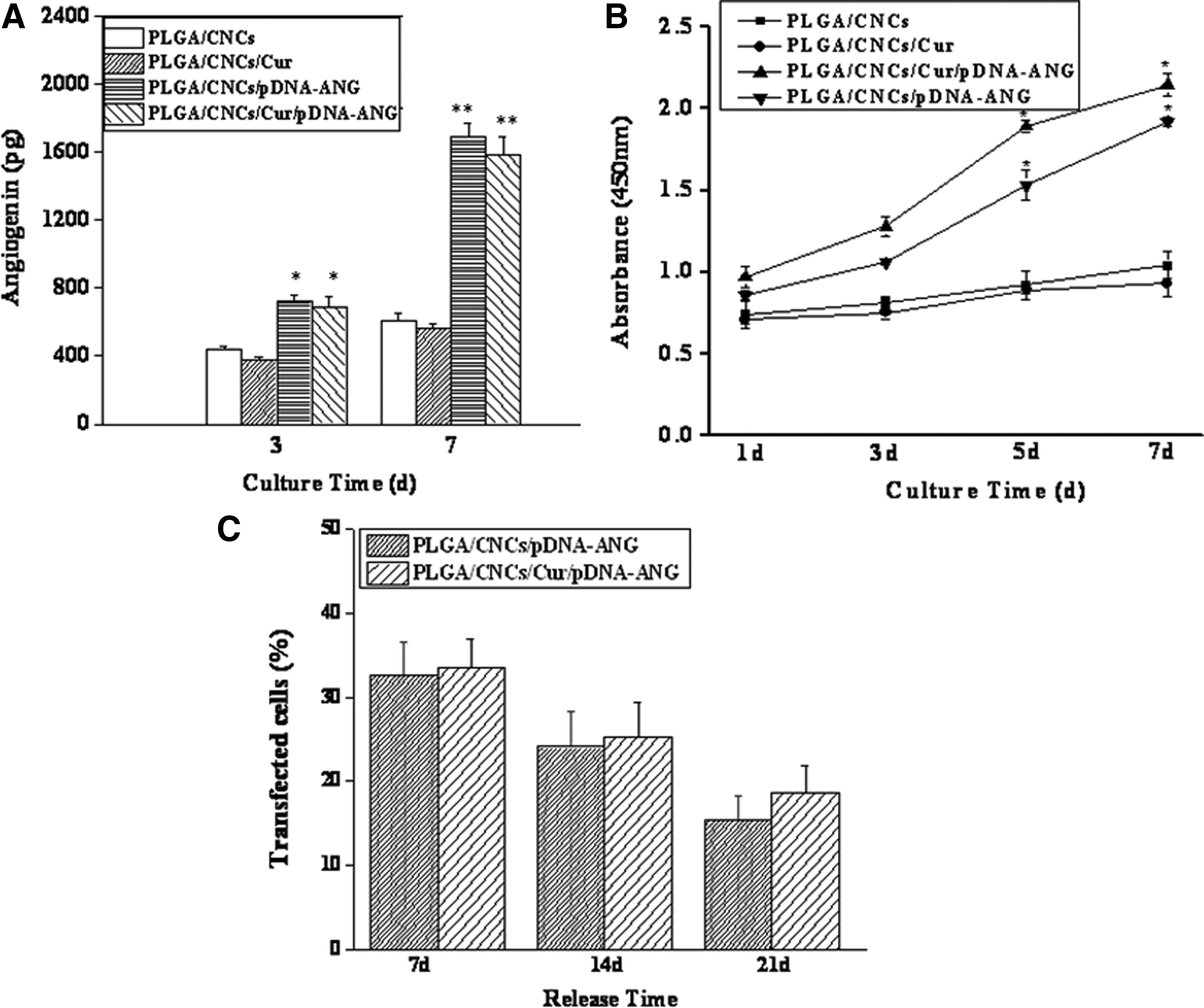
Biocompatibility of the composite nanofibers is essential for their application in tissue engineering. Thus, cell viabilities of the composite nanofibers were evaluated using the CCK-8 assay. The increased OD values on days 1, 3, 5, and 7 exhibited good cytobiocompatibility (Fig. 4B).
The transfection ability of the released pDNA from composite nanofibers indicated that the transfection efficiency of the released pDNA was decreased along with the release time (Fig. 4C). However, it could remain 15% until 21 days. This is due to most of the plasmids being released at earlier times.
In vivo wound healing
Figure 5 shows the general observation of the burn wounds on different days. Inflammation was still detected in all rats at 7 days postoperation. At 14 and 21 days postoperation, the wound area was still exposed in all groups, while for the PLGA/CNC/Cur/pDNA-ANG group, the extensive brownish red granulation was smaller compared with the other groups. At 28 days postoperation, the flat surface of the wounds and some degree of reepithelialization were observed for the PLGA/CNC/Cur, PLGA/CNC/pDNA-ANG or PLGA/CNC/Cur/pDNA-ANG groups. The wound images were quantified to show the unhealed areas of each group at different time points (Supplementary Fig. S4). The differences in the area of wounds among all the groups were not significant at 7 days postoperation, but gradually became obvious after treatment for 14, 21, and 28 days postoperation. In addition, the PLGA/CNC/Cur/pDNA-ANG group owned the minimal mean area of the wound in all groups.
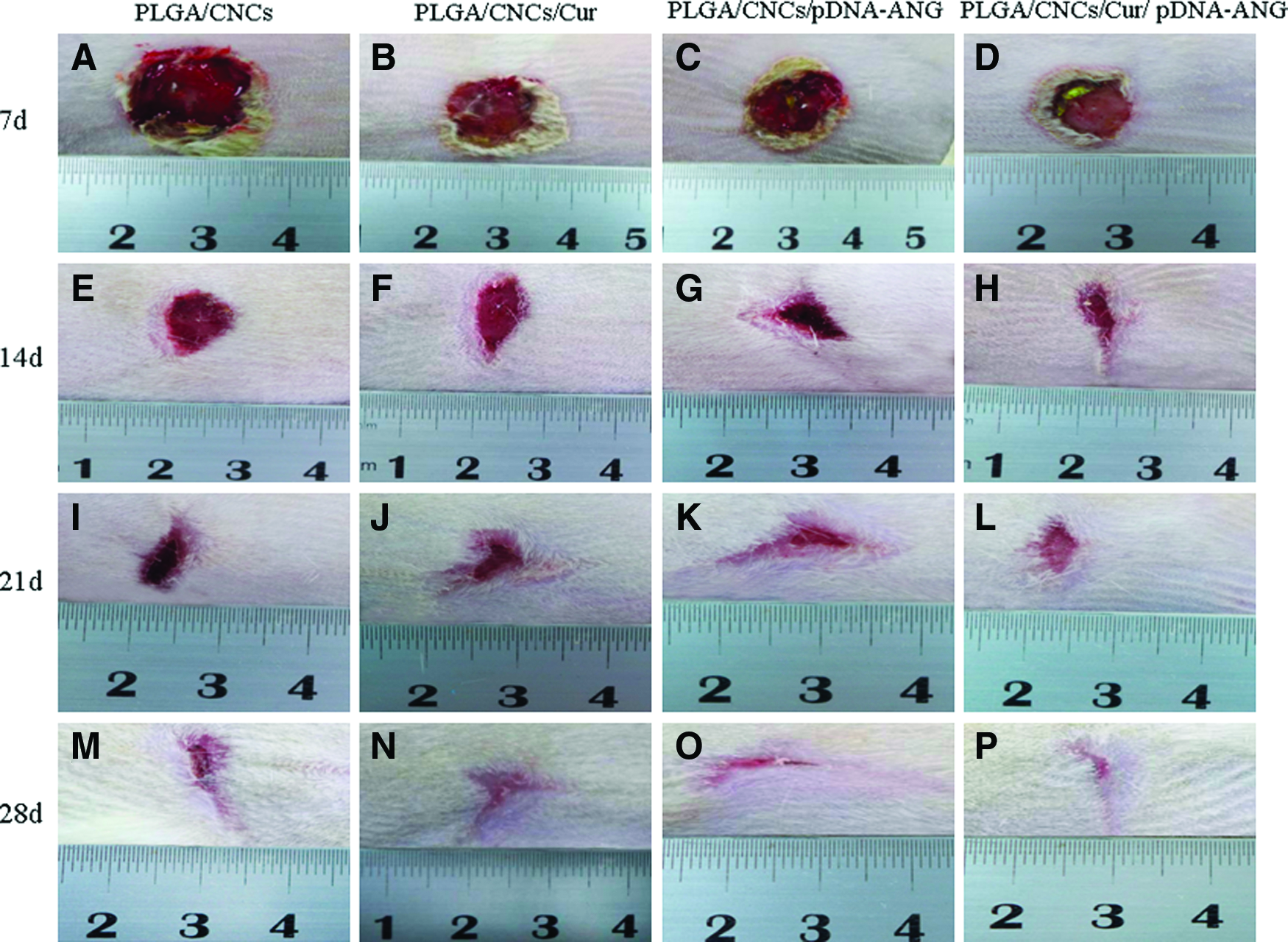
Change in appearance of wounds dressed with PLGA/CNC
Histological observation
To investigate these microchanges in a full-thickness burn, histological analysis was carried out by observation of H&E (Fig. 6) and Masson's trichrome-stained sections (Supplementary Fig. S5). At predetermined time points, the tissue samples were removed from the rats. On day 7 and 14, granulocytes and lymphocytes were still evident in wounds treated only with the PLGA/CNC, PLGA/CNC/Cur, and PLGA/CNC/pDNA-ANG groups. On day 21, the epidermis layer reached a certain thickness on the edge of wounds in the group treated with PLGA/CNC/Cur/pDNA-ANG nanofibers. On day 28, hair follicle cells and papillary structures were observed in the PLGA/CNC/Cur/pDNA-ANG groups (red arrows indicated in Fig. 6P).
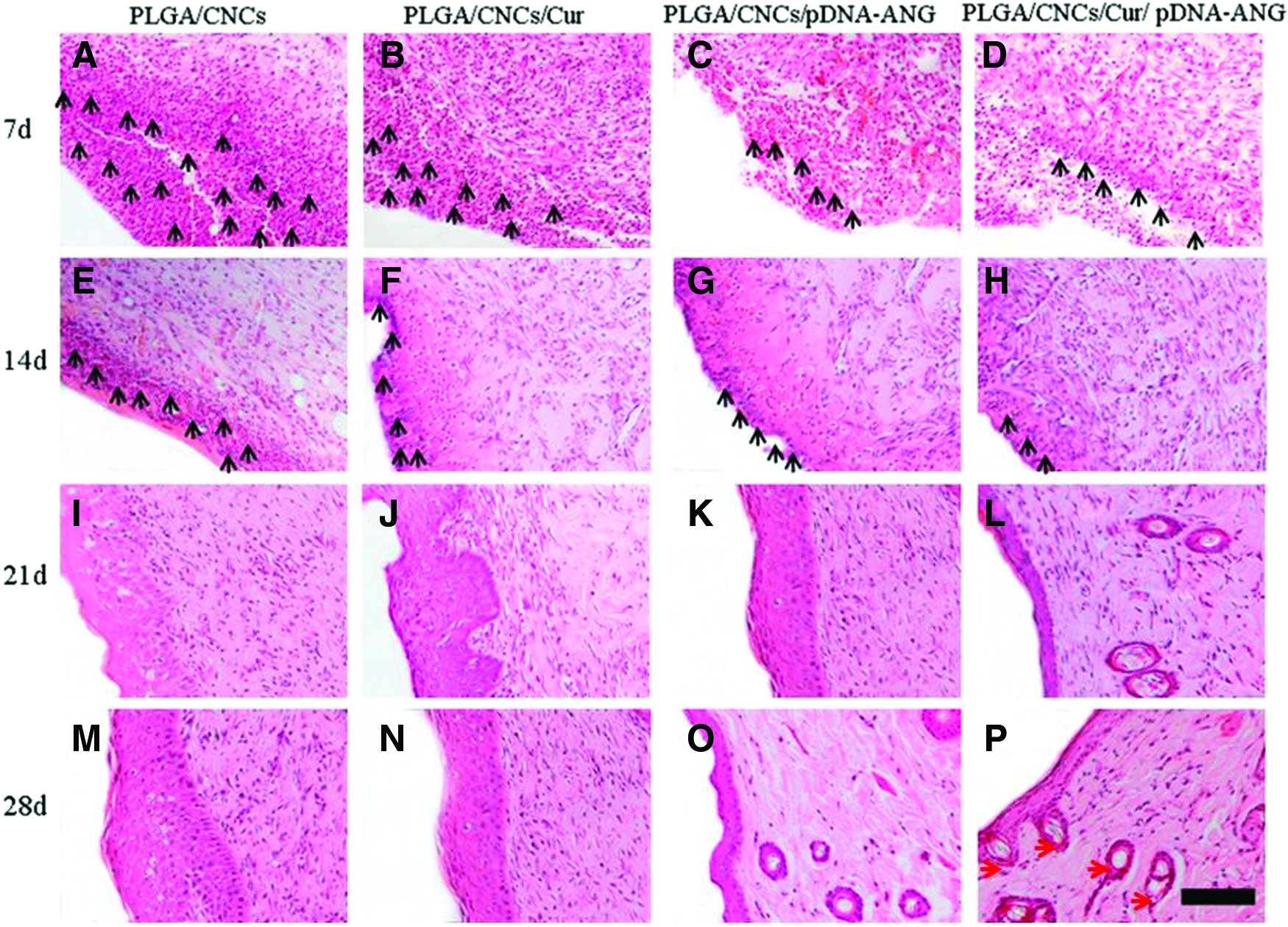
Histological analyses of dorsal skin stained with haematoxylin and eosin after full-thickness burn infection treated with PLGA/CNCs
Similarly, the deposition of collagen was observed using Masson's trichrome stain on samples from the various groups at the indicated time intervals (Supplementary Fig. S5). On day 28, deposition of collagen in the PLGA/CNC/Cur/pDNA-ANG group was greater and more regular than in the other groups and the structure of regenerated skin was similar to that of normal skin (Supplementary Fig. S6).
Immunohistochemistry analysis
ANG is one of cytokines that is involved in the infection response and is significantly elevated in the plasma of patients with thermal injury compared to control. Throughout the experiment points, expression levels of ANG were measured in the full-thickness burn infection of rat dorsal back skins at the indicated time points (Fig. 7). In all groups, there were no significant differences in the levels of ANG on day 7. However, a significant decrease in the levels of ANG was observed between days 14 and 21 in the PLGA/CNC/Cur/ANG group. This same trend was observed in the PLGA/CNC/ANG group. In the control group PLGA/CNCs, there were no significant differences in the levels of ANG before day 28.
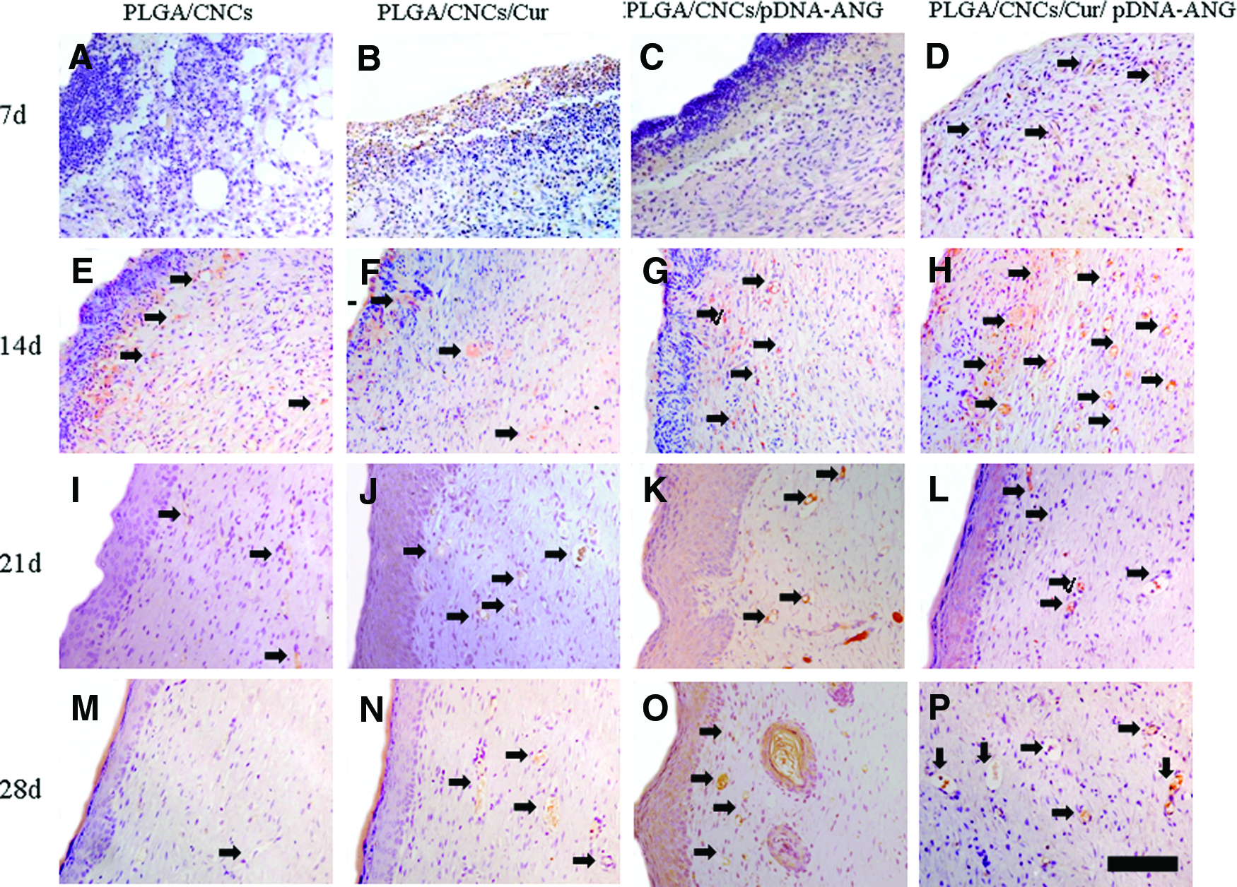
Expression of ANG in dorsal skin after full-thickness burn infection treated with PLGA/CNCs
Immunofluorescence
The mature vessels were further characterized by immunofluorescence, by costaining CD31 and α-SMA (Fig. 8). 30 The number of mature blood vessels is summarized quantitatively in Supplementary Figure S7. The density of the mature blood vessels increased monotonously with increasing implantation time for all the groups. The PLGA/CNC/Cur/pDNA-ANG group always had a significantly higher density of mature vessels. No significant difference was found among the other three groups. Blood vessels with thicker walls and round shape were also present in the PLGA/CNC/Cur/pDNA-ANG group on days 21 and 28 (Fig. 8L, P).
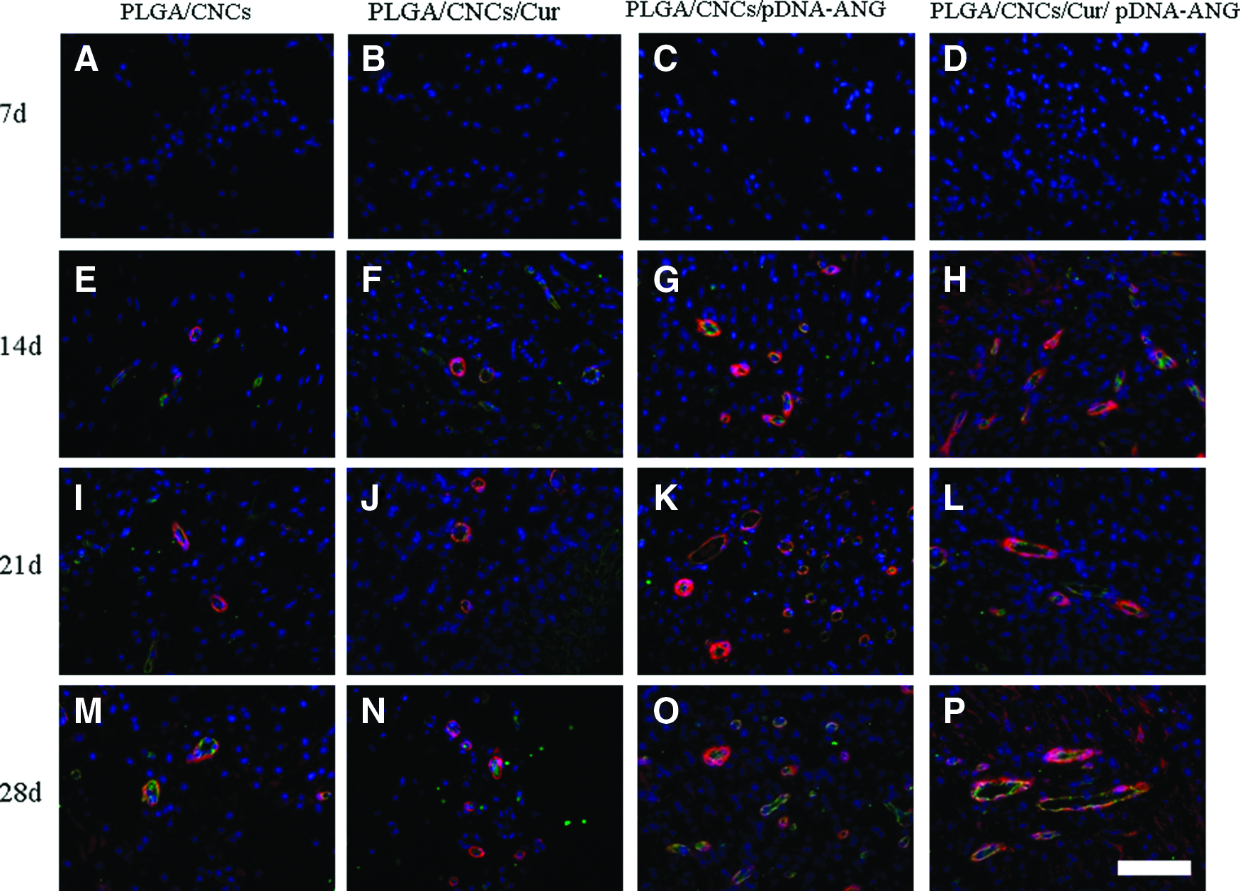
Fluorescence triple staining of sections of burn wounds treated with PLGA/CNCs
Real-time quantitative PCR and Western blotting analysis
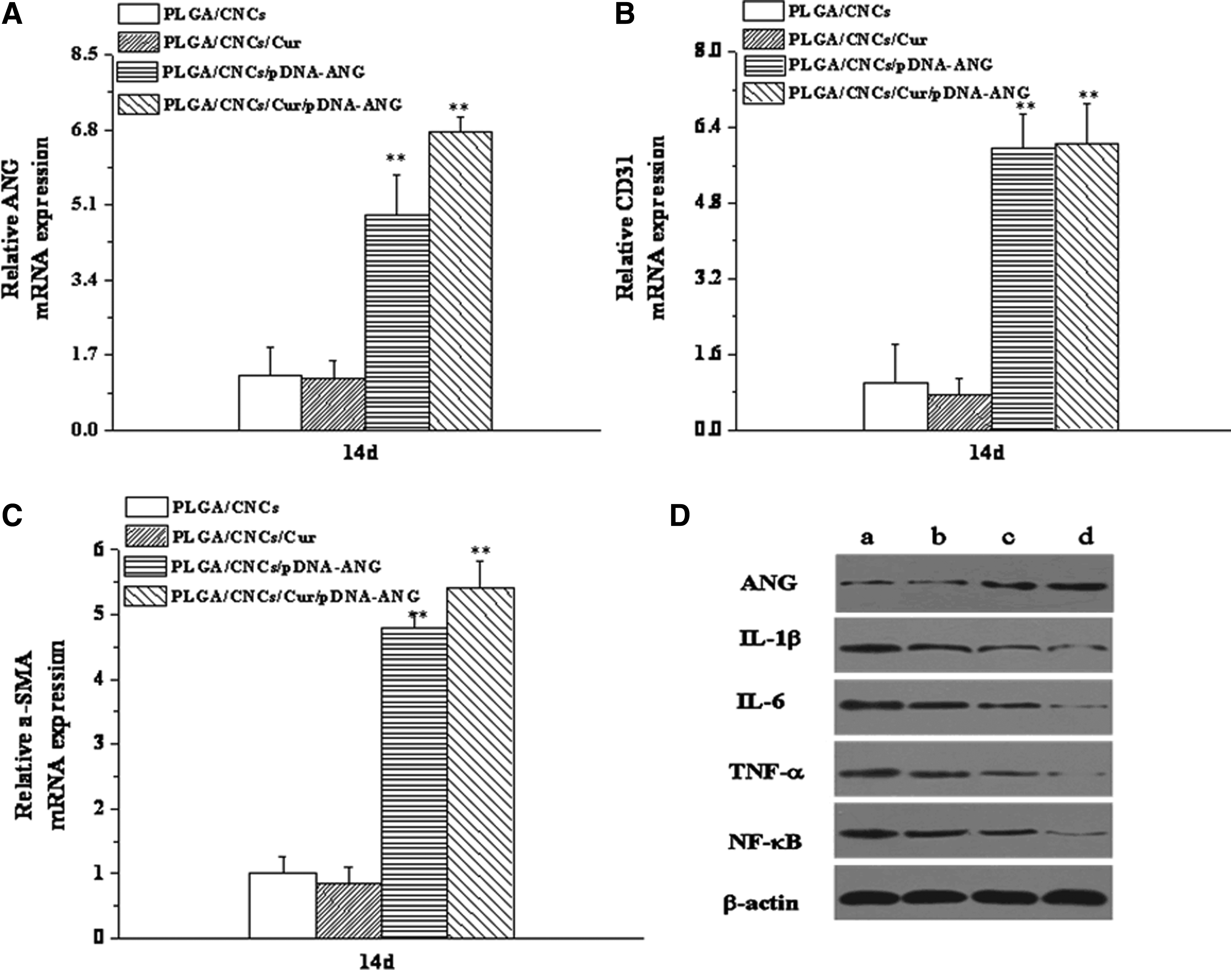
Real-time quantitative PCR (RT-qPCR) was used to quantify the in vivo expression of ANG, CD31, and α-SMA at 14 days after the excision (Fig. 9A–C). It is obvious that the expression of ANG, CD31, and α-SMA in the PLGA/CNC/Cur/pDNA-ANG group is higher than the other groups. In addition, Western blotting analysis of the tissue extracts was conducted to directly detect IL-lβ, IL-6, TNF-α, NF-κB, and ANG in the regenerated tissues at the same time point (Fig. 9D). The ANG band of PLGA/CNCs/Cur/pDNA-ANG was the darkest in all groups. That means the expression level of ANG was the highest. Correspondingly, IL-lβ, IL-6, TNF-α, and NF-κB bands of PLGA/CNCs/Cur/pDNA-ANG were the lightest compared to other groups, which indicates that the expression of IL-lβ, IL-6, TNF-α, and NF-κB in the PLGA/CNC/Cur/pDNA-ANG group is lower than the other groups. Also, expression levels of IL-lβ, IL-6, and TNF-α being measured in the PLGA/CNC/Cur/pDNA-ANG group were lower at the indicated time points (Supplementary Figs. S8–S10). In all groups, there were no significant differences in the levels of IL-lβ, IL-6, and TNF-α on day 7. A significant decrease in the levels of IL-lβ, IL-6 and TNF-α was observed between days 14 and 21 in the PLGA/CNC/Cur/pDNA-ANG group.
Real-time quantitative PCR analyses of mRNA for
Discussion
ANG is a potent stimulator of angiogenesis in skin regeneration and has been identified as the main component involved in the induction of early-stage neovascularisation. Despite the vast interest in ANG and its potential for wound healing, clinical trials have, in most cases, been disappointing because ANG proteins with very short half-lives in vivo may be easily denatured by the loading processes used to protect them from degradation and allow for prolonged release. Therefore, constructing a delivery carrier that can solve these problems is very important. PEI-CMCS has recently been used as a growth factor or DNA delivery carrier. 21
In this study, we attempted to prepare novel electrospun PLGA/CNC composite nanofibers for the controlled dual delivery of pDNA and Cur. SEM characterization showed that both the pDNA-ANG- and the Cur-loaded nanofibers had rougher surfaces compared with the smooth PLGA/CNC composite nanofibers. It was attributed to the fact that part of Cur dissolves out from the nanofibers or the occurrence of phase separation during the electrospinning of the PLGA/CNCs/Cur and PLGA/CNCs/Cur/pDNA-ANG. In addition, pDNA-ANG- and the Cur-loaded nanofibers looked thinner compared to PLGA/CNC nanofibers, which may have resulted from an enhanced solution conductivity with the addition of Cur or PEI-CMCS/pDNA-ANG nanoparticles. 31
An obvious initial burst release was observed within the first 24 h for the composite nanofibers. 32 This initial burst may have resulted from dissolution of Cur from the surface of the nanofibers during electrospinning. In contrast, because of the protection provided by the multibarrier structure (PLGA/CNC matrix), the Cur release from the Cur-loaded composite nanofibers followed a successive biphasic release profile. 33 This combination of dissociation pathways resulted in a slower Cur diffusion rate, thereby achieving a sustained release profile. These results demonstrate that a controlled delivery of Cur can be achieved by electrospinning to prepare nanofibers, and indicated that this system may be suitable to load drug for wound healing.
The multibarrier structure of the nanoparticle-embedded electrospun nanofibers was mainly aimed at prolonging the half-life. 34 The result of the in vitro DNA release analysis revealed that the release time could last up to 21 days, and after this time point, below 5% of the loaded pDNA remained in the scaffold. Furthermore, the release results of drug and DNA also illustrated that the PLGA/CNC/Cur/pDNA-ANG nanofibers had superior sustained-release function and the PEI-CMCS nanoparticles were the key determining factor in the controlled release of pDNA. 22
The incorporation of pDNA-ANG, with the protection of PEI-CMCS, showed some positive effects on ANG expression. As a result of this overexpression, the HUVECs showed good viability in composite nanofibers loaded with PEI-CMCS/pDNA-ANG complexes (Fig. 4B). At most time points, the PLGA/CNC/Cur/pDNA-ANG composite nanofibers showed a lower inflammatory response and presented more new vessels; these features would be favorable to skin regeneration. 10 It is supposed that under the condensation and protection of PEI-CMCS, pDNA-ANG are readily endocytosed to perform transfection more easily and consequently stimulating cells to secrete relevant ANG. 35 TNF-α is a major cytokine mediator of the acute inflammatory response following skin injury. 36 There is evidence that TNF-α can increase IL-1β and IL-6 and the release of somatostatin and inhibit the release of growth hormone. 37 Growth hormone increases protein synthesis and is beneficial to wound healing. 38 It is quite clear that Cur release from composite nanofibers can inhibit the expression of TNF-α, which is beneficial to wound healing.
Expressed as a transmembrane protein, CD31 can serve as a label to assess the formation of new blood vessels in the early stage of angiogenesis. 39 To become mature blood vessels, the whole endothelial cell layer should be surrounded by smooth muscle cells, which can be marked by a plasma protein named α-SMA. 40 The presence of endothelial cells and smooth muscle cells meanwhile manifests mature blood vessels. The PLGA/CNC/Cur/pDNA-ANG group had significantly higher numbers of mature blood vessels than the other groups throughout the experiment.
RT-qPCR analyses confirmed that the PLGA/CNC/Cur/pDNA-ANG group had the highest messenger RNA expression of those critical factors ANG, CD31, and α-SMA related to vascular formation in vivo (Fig. 9A–C). Meanwhile, Western blotting analysis of the tissue extracts was conducted to detect ANG, IL-lβ, IL-6, TNF-α, and NF-κB in the regenerated tissues on day 14 (Fig. 9D). The densitometric analysis of ANG, IL-1β, IL-6, TNF-α, and NF-κB expression was shown in Supplementary Table S2. NF-κB has been revealed to originate the transcription procedures of many genes, which correlated to the immune response, the expression of inflammatory molecules, as well as antiapoptosis and proliferation of cells. 41 Moreover, there is evidence to show that NF-κB is a key transcription factor that is capable of activating IL-lβ and TNF-α. 42 Therefore, restraining effectively the viability of NF-κB is crucial to treatment of burns. In this study, inhibition of NF-κB was one of the major functions attributed to interaction between Cur and ANG. Cur can inhibit the NF-κB activity and decrease inflammatory factors such as IL-lβ and IL-6. 43 Meanwhile, pDNA-ANG can enhance the expression of ANG. Thus, the PLGA/CNC/Cur/pDNA-ANG composite nanofiber group greatly inhibited the NF-κB activity. On the contrary, the PLGA/CNC group showed no significant changes in NF-κB activation. Hence, Cur and ANG releasing from composite nanofibers had a relatively longer half-life and increased bioavailability. Moreover, PLGA/CNC/Cur/pDNA-ANG nanofibers could effectually improve the bioavailability of Cur and thereby accelerate burn wound healing.
In conclusion, composite electrospun nanofibers loaded with drug and growth factor have great potential for the treatment of severely burned patients.
Footnotes
Acknowledgments
This study was supported financially by the Natural Science Foundation of China (Grant Nos. 51303064, 81171812, and 81272105), the National Basic Science and Development Program (973 Program, 2012CB518105), Health and Medical Treatment Collaborative Innovation Major Special Projects of Guangzhou (Grant No. 201508020253), the Science and Technology Program of Guangzhou (Grant Nos. 201601010270 and 2017010160489), the Science and Technology Project of Guangdong province (Grant Nos. 2011B031300006 and 2015A010101313), and the Science and Technology key Project of Guangdong province (Grant No. 2014B020212010).
Disclosure Statement
No competing financial interests exist.
References
Supplementary Material
Please find the following supplemental material available below.
For Open Access articles published under a Creative Commons License, all supplemental material carries the same license as the article it is associated with.
For non-Open Access articles published, all supplemental material carries a non-exclusive license, and permission requests for re-use of supplemental material or any part of supplemental material shall be sent directly to the copyright owner as specified in the copyright notice associated with the article.